
Special Article - Oral Squamous Cell Carcinoma
J Dent & Oral Disord. 2016; 2(5): 1026.
Basaloid Squamous Cell Carcinoma arising in the Buccal Mucosa: A Rare Location and p16-Positive Patient
Harada Y1,2,*, Yamada S³, Noguchi H¹, Kimura S¹, Miyawaki A², Oya R² and Nakayama T¹
¹Department of Pathology, School of Medicine, University of Occupational and Environmental Health, Japan
²Department of Dentistry and Oral Surgery, University Hospital of Occupational and Environmental Health, Japan
³Department of Pathology, Field of Oncology, Graduate School of Medical and Dental Sciences, Kagoshima University, Japan
*Corresponding author: Harada Y, Department of Pathology & Department of Dentistry and Oral Surgery, School of Medicine, University of Occupational and Environmental Health, Japan
Received: June 14, 2016; Accepted: July 11, 2016; Published: July 13, 2016
Abstract
Basaloid Squamous Cell Carcinoma (BSCC) is a rare, histologically, aggresive variant of Squamous Cell Carcinoma (SCC), commonly localized in the upper aerodigestive tract, but extremely rare in the buccal mucosa. Herein, we present a female patient, who was diagnosed with BSCC involving the buccal mucosa, and was disease free for more than 5 years after surgical treatment. Microscopic examination of the small, exophytic nodule revealed numerous atypical epithelial cells with hyperchromatic nuclei and scant cytoplasm, arranged in a lobular configuration, and partially embedded in hyalinised stroma. Immunohistochemically, the tumour cells were positive for the Human Papillomavirus (HPV) marker, p16. This report presents an exceptional case of a HPV-positive patient with BSCC of the buccal mucosa, who had a good prognosis.
Keywords: Basaloid Squamous Cell Carcinoma (BSCC); Buccal mucosa; Human Papillomavirus (HPV)
Introduction
Basaloid Squamous Cell Carcinoma (BSCC) is a rare, histologically aggressive variant of Squamous Cell Carcinoma (SCC), first characterized in the head and neck region, and defined as a variant of SCC [1-3]. BSCC usually arises in the aerodigestive tract and encompasses 2-5% of all head and neck SCC [4]. In the oral cavity, the tumour has a strong predilection for the tongue (61%) [3], whereas less than 0.9% of oral BSCC occur in the buccal mucosa [5,6]. BSCCs usually present as Stage III and Stage IV tumours with high overall mortality rates (60%) due to frequent lymph node (64%) and distant (44%) metastases [2,4,7]. Interestingly, recent studies have shown that Human Papillomavirus (HPV) might play a role in the pathogenesis of BSCC [3], whereas HPV-positive patients have better survival than HPV-negative patients [4,6].
BSCC is composed of basaloid and squamous components; the basaloid cells are small with scant cytoplasm and hyperchromatic nuclei that are devoid of nucleoli. They are generally arranged in a solid growth pattern and present with a lobular configuration and occasional cystic spaces filled with mucin-like material. Accessory findings include foci of coagulative necrosis and hyalinised stroma within the tumour lobules. BSCC typically coexists with dysplastic epithelium or invasive SCC components [1,2,8]. Although the solid variant of adenoid cystic carcinoma (ACC) is considered as the main differential diagnosis, it generally lacks squamous differentiation and other SCC components [2,8]. Immunohistochemistry has been used to confirm the epithelial origin of these tumours. High-molecular weight cytokeratin (CK, clone 34βE12) is most sensitive for the detection of basaloid cells and most useful marker for this tumour [5,8]. The basaloid cells were positive for p63, but not CK7 which is marker for salivary gland origin [3,9]. Whereas, 34βE12 and p63 are abundantly expressed by basaloid cells in BSCC, the ACC cells show strong immunoreaction for CK7 [3,4,9]. Herein, we present a case of an HPV-positive patient with BSCC in the buccal mucosa.
Case Presentation
A 43-year old female with an unremarkable previous medical history presented with a chief complaint of a gradually growing nodule on the left buccal mucosa. Intraoral examination identified a small (size 11 × 8 mm), poorly demarcated, exophytic growth with an irregular surface on the left buccal mucosa (Figure 1). Histopathological examinations indicated conventional SCC. No regional lymph node metastases were identified on the CT scans. The tumour, which did not involve the cheek muscle or skin was clinically staged as T1N0M0, and resected under general anaesthesia. Microscopically, the tumour was predominantly composed of numerous atypical epithelial cells with hyperchromatic nuclei and scant cytoplasm in a lobular configuration, partially embedded in hyalinised stroma, and arranged in sheets or solid nests (Figure 2a). The superficial areas of the tumour demonstrated features of conventional SCC beneath moderately dysplastic mucosa (Figure 2b), whereas the central area presented with foci of coagulative necrosis (Figure 2c). Periodic acid-Schiff staining revealed mucin-like material within several small cystic spaces. Immunohistochemically, the tumour cells were positive for 34βE12 (Figure 3a), p63 (Figure 3b), and p16 (Figure 3c), but negative for CK7, synaptophysin and α-smooth muscle actin. Based on these findings, we finally reached a diagnosis of BSCC. More than 5 years after surgery, the patient remains well with no further recurrences or metastases.
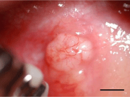
    Figure 1: The intraoral findings on initial visit. The exophytic, poorly
demarcated, white-coloured nodule was identified in the left buccal mucosa,
presenting with an irregular surface (Scale bar, 5 mm).
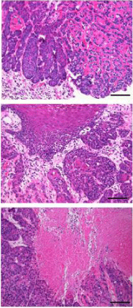
    Figure 2: Histopathological findings of the surgical specimen.
(a) The tumour was predominantly composed of numerous atypical epithelial
cells with hyperchromatic nuclei and scant cytoplasm, arranged in a solid
growth pattern, with lobular configuration, and micro cyst formation. The
cells were arranged in small to medium sized nests, and partially embedded
in hyalinised stroma. (b) The conventional SCC was recognized beneath
moderately dysplastic mucosa. (c) The foci of necrosis within the central area.
(haematoxylin and eosin stain; ×200; Scale bar, 100 μm).

    Figure 3: Immunohistological findings of the surgical specimen. The tumour
cells were specifically positive for (a) 34βE12 (×200; Scale bar, 100 μm), (b)
p63 (×400; Scale bar, 50 μm), and (c) the HPV marker p16. (×200; Scale bar,
100 μm). (Inset: negative control).
Discussion
BSCC of the buccal mucosa is a very rare entity, and its prognosis in HPV-positive patients has not been reported so far. In this study, earlier and easier detection of the lesion was possible due to its exophytic nature arising from buccal mucosa than the other head and neck sites. Furthermore, the tumour cells exhibited positive immunoreaction for the HPV marker, p16 [4]. Interestingly, HPVinfection is a major risk factor for the development of head and neck SCC [10], whereas HPV-positive patients with BSCC of the head and neck have significantly better 5-year survival rates (75%) than the HPV-negative patients (45%) [4]. This reason might be elaborated with lower ability to proliferate or metastasize for HPVpositive patients. Indeed, the stage of the present case was T1N0M0. Nevertheless, further analyses, including real-time quantitative polymerase chain reaction (qRT-PCR) and in situ hybridization, may be needed in order to verify HPV infection. In addition, we may have to accumulate more samples to provide convincing evidence for accurate prediction of this disease in the near future. Therefore, HPVpositive patients with BSCC of the buccal mucosa might potentially have much better prognosis than those arising in other regions of the head and neck. Nevertheless, careful follow-up is mandatory.
Conclusion
This case highlights an extremely rare case of BSCC arising from the buccal mucosa in an HPV-positive patient, and potentially leading to a good prognosis.
References
- Wain SL, Kier R, Vollmer RT, Bossen EH. Basaloid-squamous carcinoma of the tongue, hypopharynx, and larynx: report of 10 cases. Hum Pathol. 1986; 17: 1158-1166.
- Pieter J, Slootweg, Mary Richardson. Basaloid Squamous Cell Carcinoma. Douglas R, Gnepp, editors. In: Diagnostic Surgical Pathology of the Head and Neck, 2nd edn: SAUNDERS. 2009; 82-86.
- Shivakumar B, Dash B, Sahu A, Nayak B. Basaloid squamous cell carcimoma: A rare case report with review of literature. J Oral Maxillofac Pathol. 2014; 18: 291-294.
- Thariat J, Badoual C, Faure C, Butori C, Marcy PY, Righini CA. Basaloid squamous cell carcinoma of the head and neck: role of HPV and implication in treatment and prognosis. J Clin Pathol. 2010; 63: 857-866.
- Ereño C, Gaafar A, Garmendia M, Etxezarraga C, Bilbao FJ, López JI. Basaloid squamous cell carcinoma of the head and neck: a clinicopathological and follow-up study of 40 cases and review of the literature. Head Neck Pathol. 2008; 2: 83-91.
- Jayasooriya PR, Tilakaratne WM, Mendis BR, Lombardi T. A literature review on oral basaloid squamous cell carcinomas, with special emphasis on etiology. Ann Diagn Pathol. 2013; 17: 547-551.
- Raslan WF, Barnes L, Krause JR, Contis L, Killeen R, Kapadia SB. Basaloid squamous cell carcinoma of the head and neck: a clinicopathologic and flow cytometric study of 10 new cases with review of the English literature. Am J Otolaryngol. 1994; 15: 204-211.
- Cardesa A, Zidar N, Ereño C. Basaloid squamous carcinoma. Barnes L, Eveson JW, Reichat P, Sidransky D, editors. In: WHO Classification of Tumours, Pathology and Genetics of Head and Neck Tumours. Lyon, IARC Press. 2005; 124-125.
- Coletta RD, Cotrim P, Almeida OP, Alves VA, Wakamatsu A, Vargas PA. Basaloid squamous carcinoma of oral cavity: a histologic and immunohistochemical study. Oral Oncol. 2002; 38: 723-729.
- Lindel K, Beer KT, Laissue J, Greiner RH, Aebersold DM. Human papillomavirus positive squamous cell carcinoma of the oropharynx: a radiosensitive subgroup of head and neck carcinoma. Cancer. 2001; 92: 805-813.