
Special Article - Gingivitis
J Dent & Oral Disord. 2016; 2(6): 1029.
An Eagle’s View in Case Analysis Unveils Diagnostic Dilemmas: A Case Report
Satyanarayana KV1*, Anuradha BR1, Priyanka M1 and Praveena CH2
1Department. of Periodontics, MNR Dental College and Hospital, India
2Department of Prosthodontics, Sibar Institute of Dental Sciences, India
*Corresponding author: Satyanarayana KV, Department. of Periodontics, MNR Dental College and Hospital, Sangareddy, Hyderabad, India
Received: June 28, 2016; Accepted: July 29, 2016; Published: August 01, 2016
Abstract
Chronic and aggressive periodontitis are differentiated mainly by progression of the disease; local factors, age of onset and neither of the diseases are having pathognomonic features to diagnose specifically. Sometimes overlapping features like patterns of bone loss, pathological migration, pocket formation leads to diagnostic dilemma. This ambiguity can be overcome by thorough clinical examination and case analysis. Here, one such case is reported with spacing between teeth, radio graphically bilateral mirror images and pathological migration mimicking features of aggressive periodontitis which later on cast analysis revealed other clinical features like traumatic occlusion with reversal of occlusal phenomenon, plunger cusps causing pocket formation, related to site specificity of chronic periodontitis and finally alignment of teeth which decide plane of interdental septum and giving mirror images radio graphically in this case. Hence a thorough clinical history and careful analysis of the patient is mandatory prior to proper diagnosis and treatment planning.
Keywords: Periodontitis; Chronic; Aggressive; Occlusion
Introduction
Chronic and aggressive periodontitis can be differentiated in few cases and in some other cases overlapped features makes diagnosis difficult but not impossible. Bilateral mirror images of alveolar bone loss around first molars tend to conclude aggressive periodontitis but angulated plane of interdental septum is determined by alignment of teeth and cementoenamel junction of approximating teeth. Many such cases are improperly diagnosed which finally results in erroneous prognosis and treatment plan. Here a unique case is reported which exemplifies the ambiguous nature of periodontitis in diagnosing it precisely.
Case Presentation
Case report
A twenty two years old healthy female patient reported to the Department of Periodontics, MNR Dental College and Hospital, Sangareddy, with the chief complaint of spacing between upper front teeth region since 3 months, and space was gradually progressing. No relevant systemic and family history noticed.
Intra oral examination revealed pale pink color of the gingiva with discrete melanin pigmentation and erythematous marginal gingiva in relation to upper left central incisor. The consistency was soft and edematous with loss of scalloping in relation to upper and lower anterior region. Generalized bleeding on probing was observed. Oral hygiene status was fair with gingival and plaque index with a score of one. Periodontal examination revealed probing depth of five to seven millimeters in relation to first molars and right and left central incisors. Grade II mobility in relation to upper central incisors and grade I in relation to lower central incisors was observed. Tongue thrusting habit with open contacts was noticed anteriorly between 11&21; 11&12; 21&22; 31&32; 41&42 and posteriorly between 16&17; 26&27. Plunger cusps were seen in relation to 37 and 47, causing food impaction interdentally. Pathological migration was seen in relation to upper central incisors. The extruded right and left central incisors resulted in uneven gingival margins [1] (Figure 1).
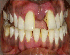
Figure 1: Uneven gingival margins.
Occlusion of the patient was class II malocclusion, with increased anterior vertical overbite (Figure 2). Habitual rest position of the patient is an edge-edge relation which is different from physiological rest position (Figure 3). Reversal of occlusal plane was observed in which palatal cusps of maxilla and buccal cusps of mandible were attrited (Figure 4 & Figure 5).
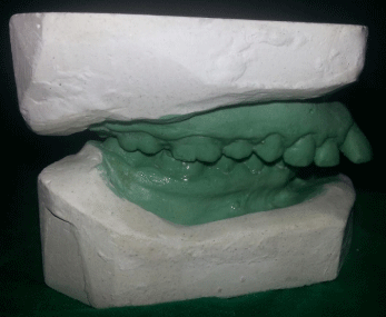
Figure 2: Class II malocclusion with increased overjet and overbite.
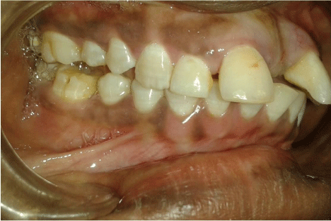
Figure 3: Habitual rest position.
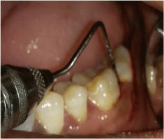
    Figure 4:  Change of occlusal plane with lower second molar mesial
inclination.
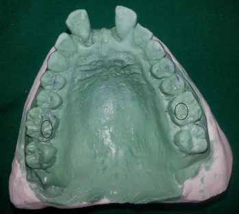
Figure 5: Attrition of supporting cusps.
Based on above clinical findings, provisionally diagnosed as localized aggressive periodontitis, differentially diagnosed as chronic localized periodontitis [2].
Radiographic investigation with Ortho Pantomograph (OPG) (Figure 6). Revealed angular bone defects involving both mesial and distal aspects of coronal one third of radicular portion of first molars.
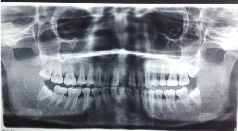
    Figure 6:  OPG revealing bilateral mirror images involving molars: Line drawn
between the CEJs of approximating teeth.
Horizontal bone defects involving the coronal one third of roots of premolars and right and left central and lateral incisors.
No abnormality detected in biochemical investigation (Complete blood picture and fasting blood sugar levels) and viral screening (Hepatitis B and HIV).
Case analysis
A twenty two year old female patient presented with soft, erythematous marginal gingiva in relation to maxillary left central incisor, and extruded upper incisors with uneven gingival margins. Clinical examination revealed generalized bleeding on probing, periodontal pockets, class II malocclusion with reversal of occlusal plane in relation to posterior teeth and plunger cusps. In this case, pockets formation was observed in relation to posterior teeth where patient complains of food impaction which is associated with plunger cusps. Prominent mesiolingual inclination of lower second molars and slight buccally placed lowerfirst molars creating plunger cusp and wedging of food into the corresponding interproximal embrasures. Reversal of occlusal plane was observed with attrition of supporting cusps of maxillary and mandibular molars.
Radiographic examination (Figure 6) revealed angular bone defects involving both mesial and distal aspects of coronal one third of radicular portion of first molars. At this instance, plane of interdental septum was evaluated according to Ritchey B and Orban [3]. criteria. The mesiodistal angulation of the crest of interdental septum usually parallels a line drawn between the cemento-enamel junctions of the approximating teeth. It was revealed that plane of interdental septum in relation to all first molars was slightly slanted i.e. apically from first molars to coronal aspect of adjacent teeth. So here point of discussion is before initiation of the disease, interdental septum is in oblique direction to the long axis of first molar, later on with disease progression it would or wouldn’t alter the pattern.
Presence of traumatic bite along with food impactions leading to angular type of bony destruction involving the molars with open proximal contacts contributing to periodontal pocket formation. Pathological migration was seen as extrusion in relation to upper incisors.
In the present case report, patient is systemically healthy with minimal local factors and no relevant environmental factors.
Though presence of angular defects involving the first permanent molars and absence of local factors mimics the aggressive nature of the disease, thorough clinical history and careful analysis of patient reveals the role of occlusion and plunger cusps aggravating the periodontal destruction.
Discussion
Periodontitis is characterized by extension of inflammation to the supporting structures of the tooth which may be modified by the host response or microbial challenge [4]. Presence of local factors, systemic factors, environmental and genetic factors are considered as risk factors for disease.
Chronic periodontitis has been defined as an infectious disease resulting in inflammation within the supporting tissues of the teeth, progressive attachment loss and bone loss [5].
The characteristic features of chronic periodontitis are prevalent in adults but can occur in children, amount of destruction consistent with local factors, associated with variable microbial pattern, slow to moderate rate of disease progression, sub gingival calculus frequently found and modified with systemic diseases, local factors and environmental factors. WhereasLang’s criteria [6]. for aggressive periodontitis includes affecting otherwise clinically healthy patients, rapid attachment loss and bone destruction, amount of microbial deposits inconsistent with disease severity and familial aggregation of diseased individuals.
Variations in aggressive periodontitis according to Baer, [7]. (atypical form of aggressive periodontitis) include occurrence of a pattern of bone loss localized to one proximal surface of the first molar inmultiple arches as an initial stage in the development of the disease, or anincipient localized case and alveolar bone loss progresses only to a certain point and then may remain quiescent for many years.
In this case, age of the patient, involvement of molars and incisors, mirror image pattern of bone loss, fair oral hygiene status suggest aggressive periodontitis. At the same time moderate pocket depth, site specificity of pockets where plunger cusps were observed, reversal of occlusal phenomenon in both arches, traumatic occlusion suggest chronic periodontitis.
Gould and Picton [8]. found that teeth with opener poorly shaped contacts had significantly higher periodontal index scores when compared with teeth that had sound proximal contacts. In this case, in relation to upper anteriors and molars high periodontal index scores with food impaction were observed. Hirchfeld [9]. has defined food impaction as the ‘forceful wedging of food through occlusal pressure into interproximal spaces’.
Greater probing depth and loss of attachment observed in this patient around first molars and incisors was also in accordance with Jernberg GR et al [10]. who had reported that greater probing depth and loss of attachment for sites that exhibited open contacts and food impaction compared with contralateral sites without open contacts or food impaction.
Various theories have been proposed to explain the cause of vertical bone resorption in interproximal intrabony lesions which include occlusal traumatism with local irritation, degeneration of the periodontium and food impaction. The relationship between occlusion and subgingival plaque is co-destruction. The concept of occlusion as a co destructive factor was questioned by Waerhaug [11]. But in a study conducted by Nunn ME and Harrel SK, [12]. There was strong association between initial occlusal discrepancies and various clinical parameters indicative of periodontal disease. Evidence suggests that occlusal discrepancy could be considered as an independent risk factor contributing to periodontal disease. The presence of angular osseous defects observed in OPG was due to chronic periodontitis, aggravated by trauma from occlusion and food impaction.
A diagnosis of chronic periodontitis is made (AAP International Workshop for classification of periodontal diseases, 1999) [13]. based on historical data, clinical findings like occlusal considerations and subgingival plaque accumulation.
As periodontitis is a multifactorial disease, relying on the single factor is not advisable. Since chronic periodontitis is considered as a site specific disease, the clinical signs of periodontitis are caused by the direct, site specific effects of trauma from occlusion and sub gingival plaque accumulation.
Conclusion
Diagnosis in periodontics is subjected to tantalizing towards a misdiagnosis if it is done irrationally. Every aspect of case history including clinical findings, investigations, case analysis always guide towards accurate diagnosis and hence apposite prognosis and treatment plan.
References
- Rufenacht C. Structural esthetic rules. Fundamentals of esthetics. Chicago: Quintessence publishing Co Inc. 1992; 67-134.
- Armitage GC. Periodontal diseases: diagnosis. Ann Periodontol. 1996; 37-215.
- Ritchey B, Orban B. The crests of the interdental alveolar septa. J Periodontol. 1953; 24: 75-87.
- John Novak M, Karen F Novak, Carranza FA. Chronic Periodontitis. Textbook of clinical periodontology, New Delhi: Elsevier Publishers. 2007; 10: 494-499.
- Fleming TF. Periodontitis. Ann Periodontol. 1999; 32-38.
- Lang N, Bartold PM, Cullinan M, Jeffcoat M, Mombelli A, Murakami S, et al. Consensus report-aggressive periodontitis. Ann Periodontol. 1999; 39-53.
- Baer PN. The case for periodontosis as a clinical entity. J Periodontol. 1971; 42: 516-520.
- Gould MS, Picton DC. The relation between irregularities of the teeth and periodontal disease. Br Dent J. 1966; 12: 20-23.
- Hirschfeld I. Food impaction. J Am Dent Assoc. 1930; 1504-1528.
- Jernberg GR, Bakdash MB, Keenan KM. Relationship between proximal tooth open contacts and periodontal disease. J Periodontol. 1983; 529-533.
- Waerhaug J. The angular bone defect and its relationship to trauma from occlusion and down growth of subgingival plaque. J Clin Periodontol. 1979; 61-82.
- Harrel SK, Numm ME. The effect of occlusal discrepancies on Periodontitis. II. Relationship of occlusal treatment to the progression of periodontal disease. J Periodontol. 2001; 72: 495-505.
- Armitage GC: Development of classification system for periodontal diseases and conditions. Ann Periodontol. 1999; 4: 1-6.