
Special Article - Dental Implants
J Dent & Oral Disord. 2016; 2(6): 1030.
Safety Zone to Mandibular Canal for Posterior Mandible Implant Surgeries
Garcia Blanco M* and Puia SA
Department of Oral and Maxillofacial Surgery, University of Buenos Aires, Argentina
*Corresponding author: Garcia Blanco M, Department of Oral and Maxillofacial Surgery, School of Dentistry, University of Buenos Aires, Buenos Aires, Argentina
Received: July 04, 2016; Accepted: August 04, 2016; Published: August 05, 2016
Abstract
Dental implant surgeries are widely used in dentistry due to their numerous advantages. Many articles have focused on local accidents and complications of this treatment. One of the major complications is the injury of the inferior alveolar nerve, which could potentially be damaged permanently. In order to protect clinicians and patients, some authors recommend leaving a safety zone around this nerve. The aim of this article is to review guidelines proposed to avoid this complication, and discuss critical clinical situations when this recommendation could be reappraised.
Keywords: Dental implants; Diagnosis; Nerve injury; Safety zone; Short implants
Abbreviations
IAN: Inferior Alveolar Nerve; MC: Mandibular Canal; CT: Computed Tomography; CBCT: Cone Beam CT
Introduction
Nerve injuries and safety zone
Replacement of teeth through dental implants is a widely accepted technique due to its numerous advantages [1-3]. Although it has many benefits, there may be some unfavorable outcomes. Damage to the Inferior Alveolar Nerve (IAN) is a potential major complication in mandibular dental implant surgeries. Physical harm can occur during anesthesia, flap elevation or advancement, bone graft harvest, osteotomy preparation or implant placement [4-7]. The prevalence of nerve damage that results in altered lip sensations ranges from 0% to 40% in old literature [8-10]. Currently, midcrestal incisions and Computed Tomography (CT) help prevent this type of injury, and recent studies report prevalence below 3% [11,12]. Patients’ symptoms include complete absence of sensation, diminished or increased sensitivity, abnormal sensations which may not be unpleasant and spontaneous or mechanically evoked painful symptoms [13-15]. The IAN enters the mandible in the internal side of the ramus, lies beside the lingual plate and makes a sudden turn in direction towards the buccal plate in the first molar area to the mental foramen, which size and frequency are still controversial [16-19]. Although Cone Beam CT (CBCT) seems to have the best potential efficiency in the identification of the Mandibular Canal (MC) [20], nerve detection maybe difficult in some cases [21].
Recommendations have been proposed to avoid injuring the inferior alveolar nerve. It is especially important to set guidelines in the dental community about how to act prudently to avoid nerve damage, and it is essential for beginners in the discipline. Lack of experience can cause injuries due to not recognizing drill length marks, confusing these marks through poor visualization, extending drilling time and overheating the bone, and other deficiencies in surgical technique (such as vertical incision next to mandibular foramen, or excessive traction of the flap). As some manufacturing companies make implant drills slightly longer to improve drilling efficiency [22,23] drill stops could be used to prevent over-drilling [4,22,24]. It would also be useful to take intra-operative radiographs during implant bed preparation in atrophic mandibles to confirm distance to MC [22,24].
The safety zone to the MC that most authors have proposed is 2 mm [25,26]. Implant length is chosen by making vertical linear measurements, from the top of the alveolar crest to the upper border of the mandibular canal, subtracting 2 mm as a safety zone to the MC. Since the advent of CT, clinicians have begun to reduce this limit, setting a safety zone in the range of 1 to 2 mm to MC [27-29]. This is especially important in some clinical situations, when bone height is reduced, and the safety zone needs to be reappraised. The aim of this article is to review the guidelines proposed to avoid nerve injuries and discuss different clinical situations.
Literature Review
Bone height 12 mm or greater
When patient’s bone height is 12 mm, the clinical situation has a simple, predictable resolution by placing a 10mm long dental implant, maintaining a 2 mm safety zone to the nerve (Figures 1-6). If there is greater bone height, implant length could be increased, improving bone implant contact, although some authors recommend not placing implants longer than 12 mm in posterior mandible, because it would increase the possibility of complications [30].
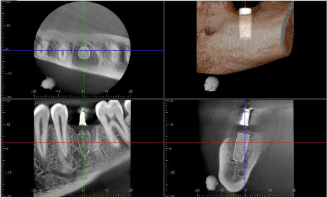
    Figure 1:  Screenshot of treatment planning for implant length and position in
good clinical conditions.
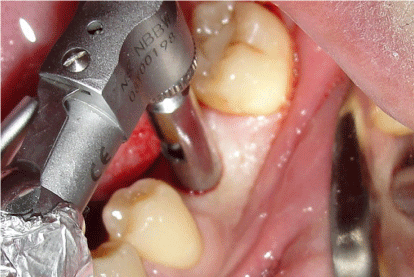
Figure 2: Posterior mandible flapless approach.
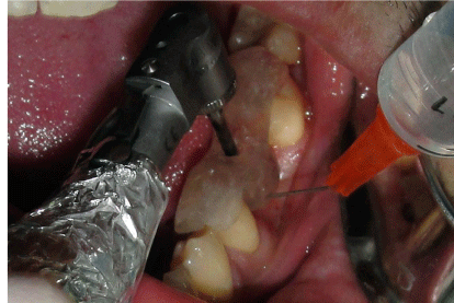
Figure 3: Cortical bone drilling in left first molar area.
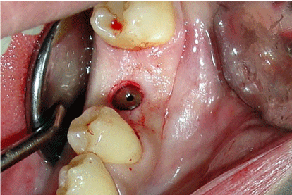
Figure 4: Implant placement leaving 2 mm of safety zone or more.
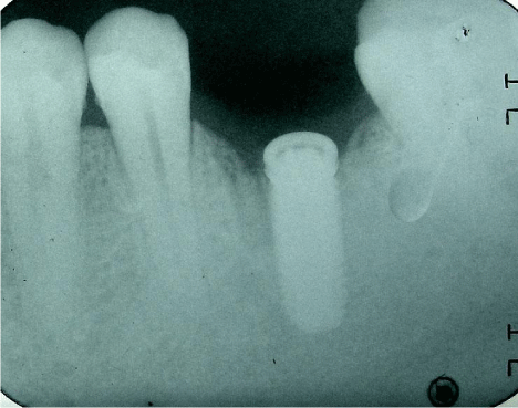
Figure 5: Periapicalpost-surgical radiograph.
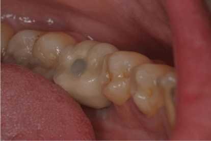
Figure 6: Screw-retained single crown in good clinical condition.
Bone height 11 mm or less
Problems begin when bone height is 11 mm or less, as the clinician may decide to use bone regeneration, IAN lateralization, placing the implant buccal or lingual to the IAN, placing short implants (less than 10 mm) [31], or reducing the safety zone by placing a standard 10 mm long implant.
In this situation, an experienced surgeon may consider drilling the bone with controlled drill pressure to 10 mm and placing a 10 mm long implant (Figures 7-10). This resolution would provide greater predictability over time than a shorter implant would. The safety zone set to MC is a general guideline to avoid nerve injuries, but sometimes 1 or 2 mm can make the difference between performing a short, predictable surgery with low morbidity or a long, complex, less predictable bone regeneration surgery.
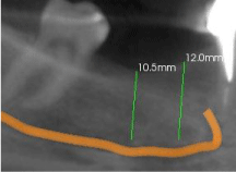
Figure 7: 10.5 mm bone height for right second molar implant placement.
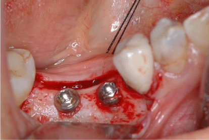
    Figure 8:  Intra operative picture leaving external hex dental implant platform
located slightly above the bone limit.
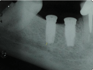
    Figure 9:  Less than 2 mm safety zone for replacement of a right second
molar.
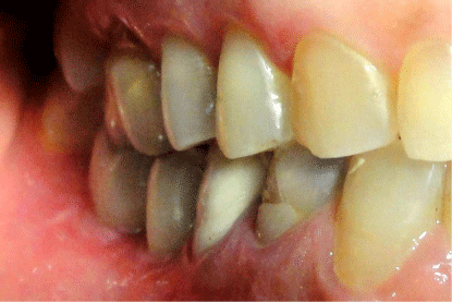
    Figure 10:  Prosthetic rehabilitation of standard length implant in 10.5 mm
bone height.
IAN lateralization is a complex surgery that requires more research to determine whether the technique can be recommended or not, as postoperative permanent nerve injury of 22% has been reported [32].
Placement of an implant buccal or lingual to the MC is a highly sensitive and blinded surgery. From the prosthetic point of view, the implant emergence profile is usually far from the occlusal surface. Currently, there is no long-term evidence to support its use [33].
Bone height less than 10 mm
The most challenging situation is to have less than 10mm, as the clinician may decide either to place a short implant or to perform bone regeneration.
Mandibular bone regeneration includes procedures ranging from simple guided bone regeneration to microvascular free flaps in extreme situations. Other procedures as distraction osteogenesis, onlay and inlay grafts may also be performed. Guided bone regeneration is the most frequently used regeneration technique due to its simplicity and predictable results for horizontal regenerations of exposed implant threads, and some articles report vertical regenerations of 3 to 6 mm, especially when titanium membranes were used. The key factor in these surgeries is to avoid exposure of the membrane, which my lead to infection and resorption of the graft [34]. Onlay bone grafts are unlikely to cause IAN injury, involve relatively simple surgery and provide immediate bone regeneration. Their main disadvantage is that the resorption rate is often high, with reports of 17 to 41% of the total bone regeneration achieved. Alternatively, regeneration by inter positional or inlay grafts have been proposed. The main advantage of this technique is the lower bone resorption because of its better vascularization. Its disadvantages are the complexity of the surgery, increased possibility of IAN injury, and the requirement of at least 4 mm of basal bone [35]. Osteogenic distraction is similar to inlay grafts, but regeneration performed also gradually recovers soft tissue. Its main disadvantage is the complexity of surgery for posterior mandible and discomfort of the device for the patient, though the latest intraosseous distractors may reduce these problems [36]. Microvascular bone grafts are highly sensitive surgeries performed in extreme cases. Their main advantage is immediate internal vascularization of the graft resulting in an almost complete absence of loss of graft volume [37].
When augmentation procedures were compared with the placement of short implants, the latter were considered more successful, since the treatment is faster, cheaper and associated to less morbidity [38]. Reducing this safety zone carefully, and considering the simplicity, speed, and predictable placement of short dental implants (shorter than 10 mm) an alternative to vertical bone regeneration in posterior mandible could be proposed (Figures 11- 14). Although many papers have highlighted the advantages of short implants in the posterior mandible; the studies do not exceed 10 years of clinical follow-up, so these implants should still be used with some caution.
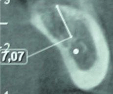
    Figure 11:  Pre surgical CT with 7.07 mm bone height for a left second molar
replacement.
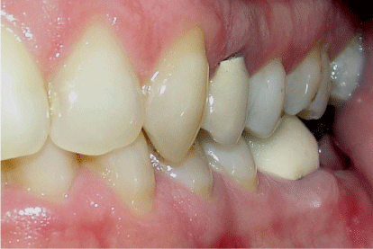
Figure 12: Reduced interocclusal space for placing a crown.
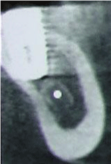
    Figure 13:  Less than 1 mm safety zone for replacement of a left second
molar.
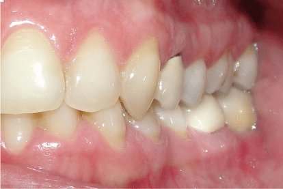
Figure 14: Prosthetic rehabilitation of short implant in 7 mm bone height.
Discussion
Dental implant surgeries in posterior mandible need to be planned carefully to avoid any permanent undesirable complications. When an acute dental intervention is performed, the clinician must clarify all possible outcomes and specify them in the informed consent form. Any high risk surgeries should be evaluated in accordance with patient’s decision. Reducing the safety zone carefully, and considering the placement of short dental implants (evaluating occlusal loads and crown to implant ratio), appears to be a straightforward, quick, predictable method for resolving intermediate atrophied mandibles. Extremely reabsorbed mandibles (less than 6 mm) should still be treated with bone regeneration surgeries, using the procedures described above.
Many research papers on morse taper implants currently report better stability and less bone resorption, recommending an infrabone position for implantation [39]. This treatment leads to more dangerous situations through closer approach to the MC in the posterior region of the mandible. As this technique still does not position the implant in the apical-occlusal dimension [40] (1 or 2 mm below crest) and no randomized controlled clinical trial has been conducted to compare it to conventional implant placement, it should be used with some caution.
Finally, if a nerve injury develop, it could be treated physiologically or pharmacologically. Physiological treatment includes removal of the implant within 36 hours post-surgery when it is in contact with or causing pressure on the MC, in order to prevent permanent damage [24]. If the implant causing the problem is already osseointegrated, it can be removed using trephine drill; or alternatively, an apicoectomy of the implant can be performed. Pharmacologic therapies for acute nerve injuries include the use of corticosteroids (administered in high doses within 1 week of the injury) and non-steroidal antiinflammatory drugs. To control inflammatory reactions in the injured nerve after the first week, a course of oral steroids can be prescribed. Oral prednisolone 1 mg per kg per day (maximum 80 mg) for the first week, stepping down by 10 mg daily over the following week, can be prescribed. As an alternative or adjunct therapy, non-steroidal antiinflammatory drugs can be prescribed (800 mg of ibuprofen 3 times daily for 3 weeks). If the situation improves, the clinician can prescribe another course of anti-inflammatory drugs [13]. The combination of high-dose steroids and non-steroidal anti- inflammatory medication can be associated with significant complications including upper gastrointestinal ulceration if prescribed long term. Even for a few weeks, prescription of these drugs should be undertaken with consideration to the patient’s medical history and caution [24]. In some complicated cases, additional pharmacologic agents can be prescribed, such as antidepressants, anticonvulsants, antisympathetic agents, and topical medications [13].
Conclusion
When placing implants in the posterior mandible, a safety zone is recommended to avoid IAN injuries. A standard implant length of 10 mm provides predictable rehabilitation, and implants longer than 12 mm are not recommended because of the greater likelihood of complications. As a general rule, when working with panoramic radiographies, a distance of 2 mm to the MC would be recommended; and this safety zone could be reduced to 1 mm with high quality CT o CBCT. Short implants appear to be a reasonable alternative to avoid performing bone regeneration surgeries which may have major complications. Each clinical situation should be analyzed individually to choose the safest, most predictable resolution for each patient.
References
- Dierens M, Vandeweghe S, Kisch J, Nilner K, De Bruyn H. Long-term follow-up of turned single implants placed in periodontally healthy patients after 16-22 years: radiographic and peri-implant outcome. Clin Oral Implants Res. 2012; 23: 197-204.
- Levin L, Laviv A, Schwartz-Arad D. Long-term success of implants replacing a single molar. J Periodontol. 2006; 77: 1528-1532.
- Noack N, Willer J, Hoffmann J. Long-term results after placement of dental implants: longitudinal study of 1,964 implants over 16 years. Int J Oral Maxillofac Implants. 1999; 14: 748-755.
- Greenstein G, Carpentieri JR, Cavallaro J. Nerve damage related to implant dentistry: incidence, diagnosis, and management. Compend Contin Educ Dent. 2015; 36: 652-659.
- Palma-Carrió C, Balaguer-Martínez J, Pearrocha-Oltra D, Pearrocha-Diago M. Irritative and sensory disturbances in oral implantology. Literature review. Med Oral Patol Oral Cir Bucal. 2011; 16: 1043-1046.
- Tay AB, Zuniga JR. Clinical characteristics of trigeminal nerve injury referrals to a university centre. Int J Oral Maxillofac Surg. 2007; 36: 922-927.
- Hillerup S, Jensen R. Nerve injury caused by mandibular block analgesia. Int J Oral Maxillofac Surg. 2006; 35: 437-443.
- Bartling R, Freeman K, Kraut RA. The incidence of altered sensation of the mental nerve after mandibular implant placement. J Oral Maxillofac Surg. 1999; 57: 1408-1410.
- Wismeijer D, van Waas MA, Vermeeren JI, Kalk W. Patients’ perception of sensory disturbances of the mental nerve before and after implant surgery: a prospective study of 110 patients. Br J Oral Maxillofac Surg. 1997; 35: 254-259.
- Ellies L, Hawker P. The prevalence of altered sensation associated with implant surgery. Int J Oral Maxillofac Implants. 1993; 8: 674-679
- Dannan A, Alkattan F, Jackowski J. Altered sensations of the inferior alveolar nerve after dental implant surgery: a retrospective study. Dentistry. 2013; 13: 1-5.
- Deppe H, Mücke T, Wagenpfeil S, Kesting M, Linsenmeyer E, Tölle T. Trigeminal nerve injuries after mandibular oral surgery in a university outpatient setting—a retrospective analysis of 1,559 cases. Clin Oral Investig. 2015; 19: 149-157.
- Juodzbalys G, Wang HL, Sabalys G. Injury of the Inferior Alveolar Nerve during Implant Placement: a Literature Review. J Oral Maxillofac Res. 2011; 2: 1.
- Hillerup S. Iatrogenic injury to oral branches of the trigeminal nerve: records of 449 cases. Clin Oral Investig. 2007; 11: 133-142.
- Khawaja N, Renton T. Case studies on implant removal influencing the resolution of inferior alveolar nerve injury. Br Dent J. 2009; 206: 365-370.
- Ozturk A, Potluri A, Vieira AR. Position and course of the mandibular canal in skulls. Oral Surg Oral Med Oral Pathol Oral Radiol. 2012; 113: 453-458.
- Ritter L, Neugebauer J, Mischkowski RA, Dreiseidler T, Rothamel D, Richter U, Zinser MJ, Zoller JE. Evaluation of the course of the inferior alveolar nerve in the mental foramen by cone beam computed tomography. Int J Oral Maxillofac Implants. 2012; 27: 1014-1021.
- von Arx T, Friedli M, Sendi P, Lozanoff S, Bornstein MM. Location and dimensions of the mental foramen: a radiographic analysis by using cone-beam computed tomography. J Endod. 2013; 39: 1522-1528.
- Greenstein G, Tarnow D. The mental foramen and nerve: clinical and anatomical factors related to dental implant placement: a literature review. J Periodontol. 2006; 77: 1933-1943.
- Juodzbalys G, Wang HL. Guidelines for the Identification of the Mandibular Vital Structures: Practical Clinical Applications of Anatomy and Radiological Examination Methods. J Oral Maxillofac Res 2010; 1: 1
- Orechovsky B, Bryk C and Von See C. Critical Evaluation of Implant Treatment Planning with a Radiological Undetectable Inferior Alveolar Nerve. J Dent & Oral Disord. 2016; 2: 1010.
- Alhassani AA, AlGhamdi AS. Inferior alveolar nerve injury in implant dentistry: diagnosis, causes, prevention, and management. J Oral Implantol. 2010; 36: 401-407.
- Alghamdi AS. Pain sensation and postsurgical complications in posterior mandibular implant placement using ridge mapping, panoramic radiography, and infiltration anesthesia. ISRN Dent. 2013; 2013: 134210.
- Renton T, Dawood A, Shah A, Searson L, Yilmaz Z. Post-implant neuropathy of the trigeminal nerve. A case series. Br Dent J. 2012; 212: 17.
- Misch CE. Root form surgery in the edentulous mandible: Stage I implant insertion. In: Misch CE, Contemporary Implant Dentistry. 2nd edn. St. Louis: CV Mosby; 1999: 360.
- Vazquez L, Saulacic N, Belser U, Bernard JP. Efficacy of panoramic radiographs in the preoperative planning of posterior mandibular implants: a prospective clinical study of 1527 consecutively treated patients. Clin Oral Implants Res. 2008; 19: 81-85.
- Worthington P. Injury to the inferior alveolar nerve during implant placement: a formula for protection of the patient and clinician. Int J Oral Maxillofac Implants. 2004; 19: 731-734.
- Frei C, Buser D, Dula K. Study on the necessity for cross-section imaging of the posterior mandible for treatment planning of standard cases in implant dentistry. Clin Oral Implants Res. 2004; 15: 490-497.
- Kraut RA, Chahal O. Management of patients with trigeminal nerve injuries after mandibular implant placement. J Am Dent Assoc. 2002; 133: 1351-1354.
- Chaushu G, Taicher S, Halamish-Shani T, Givol N. Medicolegal aspects of altered sensation following implant placement in the mandible. Int J Oral Maxillofac Implants. 2002; 17: 413-415.
- Mezzomo LA, Miller R, Triches D, Alonso F, Shinkai RS. Meta-analysis of single crowns supported by short (<10 mm) implants in the posterior region. J ClinPeriodontol. 2014; 41: 191-213.
- Vetromilla BM, Moura LB, Sonego CL, Torriani MA, Chagas OL Jr. Complications associated with inferior alveolar nerve repositioning for dental implant placement: a systematic review. Int J Oral Maxillofac Surg. 2014; 43: 1360-1366.
- Jensen O, Cottam J, Adams M, Adams S. Buccal to lingual transalveolar implant placement for all on four immediate function in posterior mandible: report of 10 cases. J Oral Maxillofac Surg. 2011; 69: 1919-1922.
- Ramel CF, Wismeijer DA, Hämmerle CH, Jung RE. A randomized, controlled clinical evaluation of a synthetic gel membrane for guided bone regeneration around dental implants: clinical and radiologic 1- and 3-year results. Int J Oral Maxillofac Implants. 2012; 27: 435-441.
- Felice P, Pistilli R, Lizio G, Pellegrino G, Nisii A, Marchetti C. Inlay versus onlay iliac bone grafting in atrophic posterior mandible: a prospective controlled clinical trial for the comparison of two techniques. Clin Implant Dent Relat Res. 2009; 11: 69-82.
- Felice P, Lizio G, Checchi L. Alveolar distraction osteogenesis in posterior atrophic mandible: a case report on a new technical approach. Implant Dent. 2013; 22: 332-328.
- Garcia Blanco M, Ostrosky MA. Implant prosthetic rehabilitation with a free fibula flap and interpositional bone grafting after a mandibulectomy: a clinical report. J Prosthet Dent. 2013; 109: 373-377.
- Felice P, Cannizzaro G, Barausse C, Pistilli R, Esposito M. Short implants versus longer implants in vertically augmented posterior mandibles: a randomised controlled trial with 5-year after loading follow-up. Eur J Oral Implantol. 2014; 7: 359-369.
- Mangano C, Iaculli F, Piattelli A, Mangano F. Fixed restorations supported by Morse-taper connection implants: a retrospective clinical study with 10-20 years of follow-up. Clin Oral Implants Res. 2015; 26: 1229-1236.
- Cassetta M, Di Mambro A, Giansanti M, Brandetti G, Calasso S. A 36-month follow-up prospective cohort study on peri-implant bone loss of Morse Taper connection implants with platform switching. J Oral Sci. 2016; 58: 49-57.