Case Report
J Dent & Oral Disord. 2016; 2(6): 1031.
An Elastofibromatous Lesion at the Tip of the Sublingual Fold
Yoshimura T1,2*, Yamada S2, Shima K3, Nakamura N1 and Tanimoto A2
1Department of Oral and Maxillofacial Surgery, Field of Oral and Maxillofacial Rehabilitation, Kagoshima University Graduate School of Medical and Dental Sciences, Japan
2Department of Pathology, Field of Oncology, Kagoshima University Graduate School of Medical and Dental Sciences, Japan
3Department of Oral Pathology, Field of Oncology, Kagoshima University Graduate School of Medical and Dental Sciences, Japan
*Corresponding author: Yoshimura T, Department of Oral and Maxillofacial Surgery, Field of Oral and Maxillofacial Rehabilitation, Kagoshima University Graduate School of Medical and Dental Sciences, Kagoshima, Japan
Received: August 01, 2016; Accepted: August 15, 2016; Published: August 17, 2016
Abstract
An elastofibromatous lesion is a slow-growing and rare pseudotumoral lesion of the soft tissues, which commonly occurs in the subscapular region, where it is known as elastofibroma dorsi. In contrast, elastofibromatous lesions of the oral cavity are extremely rare; to date, only eleven cases have been reported. Thus, the details of the clinical condition and its pathogenesis remain unclear. We here in report a case of an elastofibromatous lesion arising in the tip of the sublingual fold in an 81-year-old male patient. Although no previous case reports have described any obvious repeated irritation, it has been suggested that most oral elastofibromatous lesions probably develop as a reaction to local irritation or some type of trauma. This is the case of an oral elastofibromatous lesion might have been caused by chronic, frictional irritation by a denture.
Keywords: Oral elastofibromatous lesion; Salivary gland duct; Etiology
Introduction
Elastofibromatous lesion is a rare, slow-growing pseudotumoral lesion of the soft tissues. Most cases occur in the subscapular region [1]. Because of its location, it is often called elastofibroma dorsi. Its pathogenesis remains unclear, but the cause of this disease has been considered to be repeated, chronic trauma due to mechanical stress [2]. Elastofibromatous lesions of the oral cavity are extremely rare. Only eleven cases have been reported to date. In this paper, we describe the case of a patient with an elastofibromatous lesion in the floor of the mouth, especially at the tip of the sublingual fold, which possibly resulted from chronic irritation due to a denture.
Case Presentation
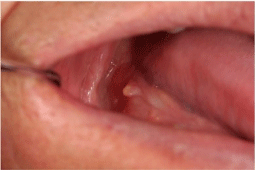
An 81-year-old Japanese man presented with a small, polypoid and yellowish-white mass on the tip of the right sublingual fold (Figure 1). It had been asymptomatic. His medical history included essential hypertension. He was a non-smoker and did not have any history of trauma in the oral cavity. He had used a partial denture of the right mandibular molar region. The lesion was located between the alveolar process and the denture on twice examinations before resection.

    Figure 1:  The intraoral findings on the initial visit.
A small, asymptomatic, polypoid, and yellowish-white mass on the tip of the
sublingual fold.
The lesion was generally soft, but the tip was hard. The lesion was measured approximately 5 mm in diameter; the hard tip was only 1 mm in diameter. The overlying epithelium was completely smooth. We clinically diagnosed it as a benign soft tissue tumor, such as fibroma. An excisional biopsy was performed under the local anesthesia.
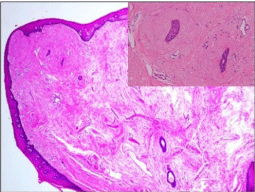
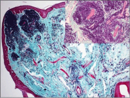
A histopathological examination revealed that the nodule was composed of partly dense and partly loose fibrous tissue, with the massive formation of eosinophilic homogeneous, amorphous elastic fibers in a small nodular fashion, intermingled with collagen fibers, reminiscent of cutaneous solar elastosis (Figure 2). The elastic fibers were distributed as variable-sized bundles in the upper subepithelial layer, and these fine fibrils were seen throughout the nodule and the perivascular to especially periductal area of the salivary gland in the nodule. It was covered with a non-disordered but parakeratotic, acanthotic squamous mucosal epithelium showing irregular-shaped reteridges. There was a minor salivary gland in the base of the lesion and slight infiltration of chronic inflammatory cells was observed in the nodule. Combined elastica-Masson staining was performed, which confirmed the presence of thickened and occasionally wavy and fragmented elastic fibers (Figure 3). Large numbers of elastic fibers were only detected in the tip of the mass (Figure 3). The lesion was diagnosed an elastofibromatous lesion based on the histopathological findings.

    Figure 2:  The histopathological findings of the surgical specimen (H&E
staining).
The nodule was composed of partly dense and partly loose fibrous tissue
showing the massive formation of eosinophilic amorphous elastic fibers,
intermingled with collagen fibers (×40; inset: ×200).

    Figure 3:  Elastica-Masson staining of the surgical specimen.
Large numbers of elastic fibers were detected only in the tip of the mass (×40;
inset: ×200).
Discussion
Only eleven cases have been reported to date [3-8]. This is the twelfth case of the elastofibromatous lesion in the oral cavity. The male-to-female ratio was 7 to 4 [3-8]. Four cases were located in mouth floor, four cases in palate (soft and hard), and in alveolar mucosa, lower lip, and tongue, each one case, respectively [3-8]. Our case was an 81-year-old male presented with a small, polypoid and yellowish-white mass on the tip of the right sublingual fold. It has been suggested that most oral elastofibromatous lesions develop as a reaction to local chronic irritation or several types of trauma [3-8]. In general, elastofibroma dorsi is caused by the mechanical friction between the scapula and the chest wall [9], and elastic degeneration of collagen or abnormal elastic fibrinogenesis may be its pathogenesis [10]. The present case is an elastofibromatous lesion might have developed due to chronic and frictional irritation by the denture, and the histopathological changes are unique in that they occur in the tip of the sublingual fold, especially around the minor salivary gland ducts in a small nodular fashion. Taken together, our case strongly supports the etiological theory that repeated frictional irritation and the stress can induce elastofibromatous lesion, and it is speculated that its oral lesion might result from abnormal proliferation of elastic fiber surrounding salivary gland duct, marked contrast to the elastic degeneration of collagen in elastofibroma dorsi and the perivascular elastosis in an oral lesion [4]. It might be difficult to diagnose oral elastofibromatous lesion correctly based merely on the haematoxylin and eosin stainings, therefore careful examination is necessary.
References
- Jarvi OH, Saxen AE. Elastofibroma dorsi. Acta Pathol Microbial Scand. 1961; 51: 83-84.
- Maldjian C, Adam RJ, Maldjian JA, Rudelli R, Bonakdarpour A. Elastofibroma of the neck. Sleletal Radiol. 2000; 29: 109-111.
- Darling MR, Kutalowski M, MacPherson DG, Jackson-Boeters L, Wysocki GP. Oral Elastofibromatous Lesions: A Review and Case Series. Head and Neck Pathol. 2011; 5: 254-258.
- Thomas Daley, Mark Darling. Elastofibroma Oralis. Head and Neck Pathol. 2011; 5: 259-260.
- Potter TJ, Summerlin DJ, Rodgers SF. Elastofibroma: the initial report in the oral mucosa. Oral Surg Oral Med Oral Pathol Oral Radiol Endod. 2004; 97: 64-67.
- Manchandu R, Foote J, Alawi F. Elastofibroma presenting as an oral soft tissue mass. J Oral Pathol Med. 2008; 37: 125-126.
- Tosios KI, Economou I, Vasilopoulos NN, Koutlas IG. Elastofibromatous changes and hyperelastosis of the oral mucosa. Head and Neck Pathol. 2010; 4: 31-36.
- Nonaka CF, Rego DM, Miguel MC, de Souza LB, Pinto LP. Elastofibromatous change of the oral mucosa: case report and literature review. J Cutan Pathol. 2010; 37: 1067-1071.
- Mark R. Wick, Virginia A. LiVolsi, John D. Pfeifer, Edward B. Stelow, Paul E. Wakely, Jr. SILVERBERG’S Principles and Practice of Surgical Pathology and Cytopathology. 5th edn. Cambridge University Press. 2015.
- Hisaoka M, Nishino J. Elastofibroma. WHO Classification of Tumours of Soft Tissue and Bone.4th edn .2013; 53-54.