Case Report
J Dent & Oral Disord. 2016; 2(8): 1042.
Interdisciplinary Conservative Approach of A Geminated Tooth
Falcón BE*
Department of Periodontics and Implantology, National University Jorge Basadre Grohmann, Perú
*Corresponding author: Falcón BE, Department of Periodontics and Implantology, National University Jorge Basadre Grohmann, Tacna, Perú
Received: October 19, 2016; Accepted: November 09, 2016; Published: November 10, 2016
Abstract
Gemination in the incisors is a rare anomaly in permanent teeth, causing malformed teeth, functional and psychosocial that leads the patient to be introverted and not to express his smile to others. It is therefore an imperative conservative interdisciplinary approach for rehabilitation. We describe a case of dental gemination, successfully treated by an interdisciplinary conservative treatment. A case study involving a 14 year old male patient, having tooth gemination causing marked dental crowding. Opting for conservative interdisciplinary treatment that included orthodontics, periodontics and aesthetic. With this conservative approach we are able to maintain the vitality of the tooth; improving the aesthetics, function and patient self-esteem.
Keywords: Gemination; Geminated; Double crown; Dental anomaly
Introduction
Dental abnormalities may be the result of many factors, including genetic and environmental, although genetic defects have higher incidence [1]. During the odontogenesis, dental anomalies can be produced by nutritional deficiency, hypervitaminosis a pregnant mother, endocrine influences, infectious/inflammatory processes, congenital diseases, local traumas and by ionizing radiation [2,3].
The correct diagnosis is very important for the success of any treatment. Dental gemination occurs when two teeth trying to develop from a single germ that leads to a larger tooth; characterized by invagination, resulting in incomplete formation of two teeth. The crown is duplicated or bifid with a slot extending from the incisal edge to the cervical region [4]. Crown halves are commonly symmetrical [4-6].
The differential diagnosis between fusion and germination is established because in gemination one root canal is shown, while in the fusion are separate root canals. Furthermore, tooth fusión results in a decrease in tooth number, whereas tooth gemination results in an increase in the number of teeth [1,6]. Fusion and dental gemination together account for 1% of dental anomalies [2].
Monitoring is important because geminated teeth often cause aesthetic problems and malocclusion, as diastema, crowding or protrusion of the teeth [6].
Several treatment methods have been described in the literature with respect to different types and morphological variations of geminated teeth, including endodontic restoration, surgical, prosthetic, periodontal and orthodontic treatment [7].
The aim of this paper is to describe a case of dental gemination treated satisfactorily by an interdisciplinary conservative treatment in an adolescent patient.
Case Presentation
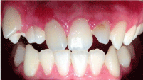
A male patient of 14 years old was presented to rate their permanent upper incisor teeth, which will cause aesthetic and chewing problems besides creating you afraid to express her smile (Figure1).

Figure 1: Initial condition of the patient.
The anamnesis does not refer significant medical or family history of dental anomalies. In the clinical evaluation the patient has permanent dentition and the normal number of teeth, gingivitis and bad position presented in the previous sector, which is more pronounced on the right side; and it notes that the right lateral incisor has a double tooth’s appearance. Thermal pulp testing, percussion and periodontal probing showed no abnormalities.
Radiographic examination to the lateral incisor with two crowns and two root canals, seen coming up the middle third radicular, which converge in a single very wide canal.
In the background you come to a diagnosis of gemination the right lateral incisor being planned an interdisciplinary conservative treatment involving the periodontal, orthodontic and dental aesthetic treatment to preserve the vitality of the tooth.
Prior to treatment informed consent was given by the family (Figure2).

Figure 2: Views. A) Buccal, B) Palatal and C) Radiographic.
In a first phase, gingivitis treated, and then begins orthodontic treatment. Once the active phase ends orthodontic treatment and aligning the teeth is achieved, a selective reduction the right lateral incisor was performed to reduce the mesial-distal width of the tooth (Figure 3).

Figure 3: A) Aligning the teeth, and B) Selective reduction piece 12.
Later the brackets are removed and gingivoplasty is performed with electrocautery (Figure 4).

Figure 4: A, B) Final phase of orthodontics, and C) Gingivoplasty.
Immediately after it proceeds to make a direct aesthetic veneer to restore the tooth, and at the same time seal the buccal and palatal face in order to prevent further decay. It controls a week and six months where the stability of the obtained result is seen and while the overall patient satisfaction (Figure 5).

    Figure 5:  Postoperative: A) Immediate, B) One week, and C) Six months
post-operative.
Discussion
Gemination is an anomaly with a hereditary tendency [8]. It is more common in primary teeth than in permanent [5,9,10], anterior teeth being the most affected [8,9]. Germination is an unusual anomaly of the hard dental tissue, with a prevalence of primary teeth of 0.5 to 0.6% and 0.1% in the permanent [9,10]. Although James, et al. A prevalence of 2.5% reported in primary teeth [7] and Nirmala, et al. reported to bilateral germination is seen in 0.01% to 0.04% in primary dentition and in 0.02% to 0.05% in permanent dentition [11]. Similar to the case reported, but in permanent dentition.
Barberio, et al. They claim that the most affected teeth are lateral incisors upper, premolars and molars [4]. However, the most affected teeth are the lower incisors (mentioned by Alves, et al.) [5]. In addition, Chen, et al. Reported what most cases occur in the deciduous maxillary lateral incisor and unilaterally [4]. Just as in this case, but here it is given in permanent lateral incisor.
It mentions that dental anomalies in the dentition are associated with their permanent successors (mentioned by Santos, et al.) [2]; however this precedent does not occur in this case because neither the patient nor the parents mentioned this background.
Aesthetic and functional implications usually requires a complex multidisciplinary approach [9,10]. The orthodontic treatment followed by complementary esthetic treatment preserved the health and restored the esthetics [9].
In cases of pulp commitment, it scans very helpful with CBCT in three dimensions before endodontic treatment and to estimate the root canal morphology, allowing a 3600 around the tooth [3,7]. To decide not perform endodontic treatment due to the not deep carious lesion, it must be done a prosthetic restoration to improve dental occlusion and aesthetic appearance [6,7,11].
In cases of pulp commitment, it scans very helpful with CBCT in three dimensions before endodontic treatment and to estimate the root canal morphology, allowing a 3600 around the tooth [3,7]. To decide not perform endodontic treatment due to the not deep carious lesion, it must be done a prosthetic restoration to improve dental occlusion and aesthetic appearance [6,7,11].
It is also suggested as one of the treatment options nowadays be the extraction and replacement of the tooth with an implant [10]. However, since the patient is too young for implant therapy and the clinical conditions are not to reach extracting the tooth perform, conservative treatment might be more appropriate.
Patient treatment must provide benefits not only functional, but must serve to your emotional and psychological state, to achieve the goal of restoring aesthetics.
Conclusion
With this conservative management employee is able to maintain the vitality of the tooth; improving the aesthetics, function and patient self-esteem, which favors social interaction in the future, because we achieved full satisfaction with the result.
References
- Guerrero F. Mandibular third molar retained and fused with a supernumerary fourth molar: a case report. Int J Dent Health Sci. 2014; 1: 443-448.
- Santos KSA, Lins CCSA, Almeida-Gomes F, Travassos RMC, Santos RA. Anatomical aspects of permanent geminate superior central incisives. Int. J. Morphol. 2009; 27: 515-517.
- TarımErtaş E, YırcalıAtıcı M, Arslan H, Yaşa B, Ertaş H. Endodontic Treatment and Esthetic Management of a Geminated Central Incisor Bearing a Talon Cusp. Case Rep Dent. 2014; 2014: 123681.
- Barbério GS, Da Costa SV, Rios D, De Oliveira TM, Machado MAAM. Rare case of bilateral gemination in deciduous teeth. Int. J. Morphol. 2013; 31: 575-577.
- Alves N, Nascimento, CMO, Patriarca, JJ. Dental Gemination in the inferior canine in both dentition. Case report. Int. J. Morphol. 2010; 28: 873-874.
- Beltran, V, Leiva C, Valdivia I,Cantên M, Fuentes R. Dental gemination in a permanent mandibular central incisor: an uncommon dental anomaly. Int. J. Odontostomat. 2003; 7: 69-72.
- James EP, Johns DA, Johnson KI, Maroli RK. Management of geminated maxillary lateral incisor using cone beam computed tomography as a diagnostic tool. J Conserv Dent. 2014; 17: 293-296.
- Bueno CE, Fontana CE, Bruno Miguita KB, Davini F, Araújo RA, Cunha RS. Tratamentoendodôntico de incisivo superior geminado. RSBO. 2011; 8: 225- 230.
- LeGall M, Philip C, Aboudharam G. Orthodontic treatment of bilateral geminated maxillary permanent incisors. Am J Orthod Dentofacial Orthop. 2011; 139: 698-703.
- Oelgiesser D, Zyc R, Evron D, Kaplansky G, Levin L. Treatment of a fused/ geminated tooth: a multidisciplinary conservative approach. Quintessence Int. 2013; 44: 531-533.
- SVSG Nirmala, Sandeep Chilamakuri, Sunny Preetham Thirupati. Bilateral Double Primary Teeth Associated with Multiple Odontogenic Anomalies in Permanent Dentition: A Case Report. EC Paediatrics.2015; 1: 54-61.