Special Article - Oral and Maxillofacial Surgery
J Dent & Oral Disord. 2016; 2(9): 1045.
Prevalence of Maxillary Lateral Incisors and Eruptive Direction of Maxillary Canine in Japanese Unilateral Cleft Lip and Alveolus and Unilateral Cleft Lip and Palate Patients
Kajii TS¹*, Takamura Y¹, Hata S¹, Nukumizu K², Tamaoki S¹, Takagi S³, Ohjimi H³ and Ishikawa H4
¹Section of Orthodontics, Department of Oral Growth & Development, Division of Clinical Dentistry, Fukuoka Dental College, Japan
²Section of Oral & Maxillofacial Surgery, Faculty of Medicine, University of Miyazaki, Japan
3Department of Plastic, Reconstructive, & Aesthetic Surgery, School of Medicine, Fukuoka University, Japan
4Fukuoka Dental College, Japan
*Corresponding author: Takashi S. Kajii, Section of Orthodontics, Department of Oral Growth & Development, Division of Clinical Dentistry, Fukuoka Dental College, Fukuoka, Japan
Received: October 26, 2016; Accepted: November 15, 2016; Published: November 17, 2016
Abstract
Purpose: The aim of present study was to confirm prevalence of the lateral incisor in Japanese unilateral cleft lip and palate or alveolus patients. The present study also hypothesized that a congenitally missing maxillary lateral incisor in the lesser segment may affect eruptive direction of the maxillary canine on the cleft side.
Methods: Participants comprised 40 Japanese oral cleft patients (23 patients with non-syndromic Unilateral Cleft Lip and Palate (UCLP), 17 patients with nonsyndromic Unilateral Cleft Lip and Alveolus (UCLA)). Panoramic radiographs taken at initial examination were used to assess the maxillary lateral incisor and measure maxillary canine angulation. Additionally, maxillary canine angulation of 20 subjects who had undergone secondary autologous bone grafting was also measured using panoramic radiographs taken after canine eruption.
Results: Frequency of a congenitally missing lateral incisor on the cleft side was 52% in UCLP patients and 24% in UCLA patients. Maxillary canine angulation during eruption on the cleft side was inclined significantly more mesially in subjects without a maxillary lateral incisor at the lesser segment than in subjects with the lateral incisor.
Conclusions: Frequency of a congenitally missing lateral incisor on the cleft side in Japanese UCLP patients was double that in UCLA patients. Presence of the maxillary lateral incisor in the lesser segment may guide the maxillary canine to a more vertical eruptive direction, although a maxillary canine located near the cleft without a lateral incisor in the lesser segment may appear to erupt at the same angle held before grafting.
Keywords: Canine; Cleft lip; Cleft palate; Lateral incisor
Abbreviations
SBG: Secondary alveolar Bone Grafting; UCLP: Unilateral Cleft Lip and Palate; UCLA: Unilateral Cleft Lip and Alveolus
Introduction
Oral clefts are one of the most common congenital craniofacial anomalies, and oral cleft patients usually require orthodontic treatment. Malpositioning of teeth adjacent to the cleft, such as rotation and tipping, and dental abnormalities such as hypodontia, malformation, abnormal eruption patterns are frequent in cleft patients [1-3]. A congenitally missing maxillary lateral incisor on the cleft side is the most common finding in cleft patients [4-7].
Bony and soft tissue cleft defect, lesser segment dimensions, alveolar bone grafting, tooth size, as well as dental anomalies including supernumerary, congenitally missing, or malformed teeth, may alter dental eruption patterns and could increase the risk of dental impaction in cleft patients [8-12]. These factors make orthodontic treatment planning difficult [13-16].
During normal eruption in non-cleft children, the canine displaces toward the occlusal plane, straightens gradually, and deviates toward a more vertical position [17-19]. Gereltzul et al. [20] reported that the maxillary canine on the cleft side tips more mesially than that on the non-cleft side in cleft patients. They also reported that a maxillary canine located near the cleft with Secondary Alveolar Bone Grafting (SBG) appears to erupt at the same angle held before grafting, although without SBG the canine erupts more vertically, guided by cortical bone. However, most patients with cleft alveolus now undergo SBG. Existence of a maxillary lateral incisor in the lesser segment may also guide the maxillary canine in a more vertical eruptive direction, but few studies have evaluated the influence of the status of the maxillary lateral incisor on eruption of the cleft-side canine.
In oral cleft patients, understanding the prevalence of the lateral incisor can help in making orthodontic treatment plans for patients. The aim of the present retrospective study was to confirm the prevalence of the lateral incisor for Unilateral Cleft Lip and Palate (UCLP) and Unilateral Cleft Lip and Alveolus (UCLA) patients in the Japanese population. The present study also hypothesized that a congenitally missing maxillary lateral incisor in the lesser segment would affect the eruptive direction of the maxillary canine on the cleft side.
Materials and Methods
Subjects
Forty Japanese oral cleft patients (23 patients with non-syndromic UCLP, 17 patients with non-syndromic UCLA) were examined (Table 1). Criteria for including UCLP or UCLA subjects in the study were: 1) intention to be treated at the Orthodontic Clinic of Fukuoka Dental College Medical and Dental Hospital between 2000 and 2015; 2) age <10 years at initial examination; 3) no eruption of canines at initial examination; and 4) no alveolar bone grafting at initial examination. Criteria for excluding a subject from the study were: 1) incomplete cleft lip and alveolus; 2) history of trauma; and 3) previous orthodontic treatment.
    #
    Cleft type
    Cleft side
    Sex
    Age at initial exam.
    Lateral inc.at cleft side
    Lateral inc. at non-cleft side


    Type
    Segment


    1
    UCLP
    Lt
    F
    8Y1M
    Missing
    -
    Normal


    2
    UCLA
    Lt
    M
    9Y8M
    Normal
    Minor
    Normal


    3
    UCLP
    Lt
    M
    9Y1M
    Missing
    -
    Normal


    4
    UCLP
    Lt
    M
    8Y5M
    Normal
    Minor
    Normal


    5
    UCLP
    Lt
    M
    7Y9M
    Missing
    -
    Normal


    6
    UCLP
    Lt
    F
    7Y6M
    Missing
    -
    Normal


    7
    UCLP
    Lt
    M
    7Y4M
    Malformed
    Minor
    Normal


    8
    UCLA
    Rt
    F
    8Y1M
    Malformed
    Minor
    Normal


    9
    UCLA
    Lt
    M
    9Y8M
    Missing
    -
    Normal


    10
    UCLP
    Lt
    F
    8Y0M
    Missing
    -
    Normal


    11
    UCLP
    Lt
    M
    8Y2M
    Missing
    -
    Normal


    12
    UCLP
    Rt
    M
    7Y11M
    Missing
    -
    Missing


    13
    UCLA
    Rt
    M
    8Y1M
    Malformed
    Minor
    Normal


    14
    UCLA
    Lt
    F
    9Y3M
    Normal
    Minor
    Normal


    15
    UCLA
    Rt
    F
    8Y2M
    Malformed
    Minor
    Normal


    16
    UCLA
    Lt
    M
    7Y6M
    Malformed
    Major
    Normal


    17
    UCLA
    Lt
    F
    7Y6M
    Missing
    -
    Normal


    18
    UCLP
    Lt
    M
    8Y9M
    Missing
    -
    Normal


    19
    UCLA
    Rt
    M
    7Y5M
    Missing
    -
    Normal


    20
    UCLA
    Lt
    F
    7Y5M
    Normal
    Major
    Normal


    21
    UCLP
    Lt
    F
    7Y8M
    Malformed
    Minor
    Normal


    22
    UCLA
    Rt
    F
    7Y1M
    Malformed
    Minor
    Normal


    23
    UCLP
    Lt
    F
    7Y2M
    Missing
    -
    Normal


    24
    UCLA
    Rt
    M
    7Y1M
    Normal
    Minor
    Normal


    25
    UCLP
    Lt
    F
    9Y6M
    Malformed
    Minor
    Normal


    26
    UCLP
    Lt
    F
    7Y0M
    Normal
    Minor
    Normal


    27
    UCLP
    Lt
    F
    6Y10M
    Missing
    -
    Normal


    28
    UCLP
    Rt
    F
    7Y5M
    Missing
    -
    Normal


    29
    UCLA
    Lt
    M
    6Y8M
    Malformed
    Minor
    Normal


    30
    UCLA
    Lt
    M
    7Y11M
    Missing
    -
    Normal


    31
    UCLP
    Lt
    M
    6Y11M
    Malformed
    Minor
    Normal


    32
    UCLP
    Lt
    F
    7Y6M
    Missing
    -
    Normal


    33
    UCLA
    Lt
    F
    9Y2M
    Normal
    Minor
    Normal


    34
    UCLA
    Rt
    F
    7Y8M
    Normal
    Minor
    Normal


    35
    UCLP
    Lt
    M
    7Y7M
    Malformed
    Minor
    Normal


    36
    UCLP
    Lt
    M
    6Y10M
    Malformed
    Minor
    Missing


    37
    UCLP
    Lt
    M
    6Y6M
    Malformed
    Minor
    Normal


    38
    UCLP
    Rt
    F
    6Y5M
    Malformed
    Minor
    Normal


    39
    UCLA
    Rt
    F
    7Y7M
    Malformed
    Minor
    Normal


    40
    UCLP
    Lt
    M
    6Y11M
    Malformed
    Minor
    Malformed


    Note: Subjects #33 and #37 showed a    supernumerary lateral incisor in the major segment on the cleft side. In    Subjects #1, #18, and #21, the canine on the cleft side was impacted at 13    years old. exam.: examination; inc.: incisor.
Table 1: Subjects in the present study.
Mean ages at initial examination for the UCLP and UCLA subjects were 7.6 ± 0.8 years and 8.1 ± 0.9 years, respectively. Ratios of females of UCLP and UCLA subjects were 11/23 and 9/17, respectively. Ratios of UCLP and UCLA subjects with alveolar cleft on the left side were 20/23 and 9/17, respectively (Table 1).
Twenty subjects underwent SBG (9 subjects with UCLP, 11 subjects with UCLA). The remaining 20 subjects underwent the initial examination after 2013 and so were too young to have undergone SBG. All SBGs were performed using bone harvested from the iliac crest before maxillary canine eruption. Mean ages at initial examination for subjects with (n=10) and without (n=10) a maxillary lateral incisor at the lesser (minor) segment were 7.9 ± 0.7 years and 8.0 ± 0.7 years, respectively. By eruption of maxillary canines, none of the 20 subjects had undergone any treatment to affect maxillary canine angulation, such as maxillary lateral expansion and/or alignment using maxillary braces.
Methods
To assess maxillary lateral incisor, panoramic radiographs and/ or periapical radiographs taken at initial examination were used. Panoramic radiographs were obtained using an AZ 3000 system (Asahi Roentgen, Kyoto, Japan), Cypher E system (Asahi Roentgen, Kyoto, Japan), or Veraviewepocs 2DE system (Morita, Tokyo, Japan). Two experienced readers who were blinded to the clinical information of subjects independently assessed the maxillary lateral incisor. Definitions for malformation of the maxillary lateral incisor were based on the study by Kim and Baek [7]. Namely, malformation of the maxillary lateral incisor was defined as a peg-like or conical shape. A =30% decrease in width of the crown compared to orthodontic models, radiographs, or intraoral photographs was also considered to represent malformation of the maxillary lateral incisor.
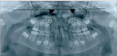
To measure maxillary canine angulation, panoramic radiographs taken at initial examination were used. As shown in Figure 1, some skeletal landmarks were defined on panoramic radiograph [19,20]. A straight line through the most inferior points of both orbital cavities was used as a horizontal reference line, then the internal angle formed by the long axis of the maxillary canine and this reference line was measured [19,20].

    Figure 1:  Landmarks and measurements used on panoramic radiographs.
Open circles: most inferior point of the orbital cavity; solid line: horizontal
reference line; dotted lines: canine long axis; closed triangles: angle between
canine long axis and horizontal reference line.
In addition, maxillary canine angulation of the 20 subjects those had undergone SBG was also measured using panoramic radiographs taken after canine eruption (without elimination of any impacted canine). The Ethics Committee of Fukuoka Dental College approved the protocols (#267) for this retrospective study. Subjects gave written informed consent for participation in this study.
Statistical methods
All tracings for measuring maxillary canine angulation were made by one observer and were repeated after at least 4 weeks. To assess the reproducibility of this method, 10 subjects were randomly selected. Measurements of maxillary canine angulation at both cleft and non-cleft sides were repeated at least 4 weeks after the first measurements. The combined error (Se) and coefficient of reliability were calculated according to Houston [21]. Se was estimated using the formula Se2=S d2/2n, where d is the difference between the first and second measurements, and n is the sample size. The coefficient of reliability was estimated using the formula 1-Se2/St2, where St is the total variance of the measurement. For both measurements, the coefficient of reliability was above 99% and was thus considered to be within acceptable limits.
The chi-squared test was used to determine the statistical significance of differences in the frequency of a congenitally missing maxillary lateral incisor between cleft and non-cleft sides, or between UCLP and UCLA. The paired t test was used to compare maxillary canine angulation between cleft and non-cleft sides. Student’s or Welch’s t test was used to compare maxillary canine angulation between absence and presence of the lateral incisor in the lesser segment. Statistical analyses were performed using the SPSS® version 20.0 statistical package (SPSS, Chicago, IL). The level of statistical significance was set at a probability level of 0.05.
Results
Prevalence of maxillary lateral incisors
Table 2 shows the prevalence of maxillary lateral incisors on the cleft and non-cleft sides for all subjects. The frequency of a congenitally missing lateral incisor was 40% on the cleft side, significantly higher than the 5% on the non-cleft side (P < 0.001). The frequency of a malformed lateral incisor on the cleft side was also 40%, again significantly higher than the 3% on the non-cleft side (P < 0.001). Subjects #33 (UCLA) and #37 (UCLP) showed a supernumerary lateral incisor in the major segment on the cleft side (Table 1). The frequency of a supernumerary lateral incisor in the major segment of the cleft side was thus 5%. No patients showed a supernumerary lateral incisor in the lesser segment of either the cleft or non-cleft side (Table 1).
    Missing
    Malformed
    Normal
    Total (n)


    Cleft side
    16
    16
    8
    40


    Non-cleft side
    2
    1
    37
    40


    Total (n)
    18
    17
    45
    80


    The chi-squared value was 42.81 (P    < 0.001).
Table 2:  Prevalence of maxillary lateral incisors on cleft and non-cleft sides of
all subjects.
For comparison of the prevalence of a maxillary lateral incisor on the cleft side between UCLP and UCLA, Table 3 shows the prevalence of a maxillary lateral incisor on the cleft side. The frequency of a congenitally missing lateral incisor on the cleft side was 52% in UCLP patients, more than double the 24% in UCLA patients, although this difference was not significant.
Eruptive direction of maxillary canine
Figure 2 shows maxillary canine angulation at initial examination on the cleft and non-cleft sides for all subjects. Mean maxillary canine angulation at initial examination was significantly smaller on the cleft side than on the non-cleft side (P < 0.001). Smaller angulation of the maxillary canine represents a more mesial inclination of the maxillary canine.
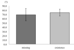
For evaluating the influence of a missing maxillary lateral incisor on maxillary canine angulation in the lesser segment, Figure 3 shows maxillary canine angulation at initial examination with or without the maxillary lateral incisor in the lesser segment on the cleft side for all subjects. Mean maxillary canine angulation at initial examination was smaller in subjects without a maxillary lateral incisor at the lesser segment than in subjects with a lateral incisor, although no significant difference in mean values was apparent between subjects with and without a lateral incisor in the lesser segment.
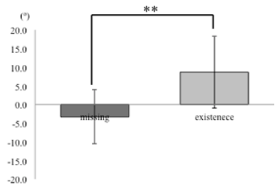
Figure 4 shows the change in maxillary canine angulation between pre-eruption (initial examination) and post-eruption on the cleft side in the 20 eligible subjects. Mean change in maxillary canine angulation was significantly negative in subjects without a maxillary lateral incisor in the lesser segment compared to subjects with a lateral incisor (P < 0.01). A negative change in maxillary canine angulation represents a more mesial inclination of the maxillary canine during eruption. In Subjects #1 (UCLP), #18 (UCLP), and #21 (UCLP), the cleft-side canine was impacted at the age of 13 years (Table 1). Frequency of an impacted canine on the cleft side was 15%. The maxillary lateral incisor in the lesser segment was congenitally absent in Subjects #1 and #18, and that in Subject #21 was malformed. No patients showed an impacted maxillary canine on the non-cleft side.
Discussion
The frequency of a congenitally missing lateral incisor was 40% on the cleft side, significantly higher than the 5% on the non-cleft side (Table 2). The frequency of a congenitally missing lateral incisor on the cleft side was 52% in UCLP patients, more than double the 24% in UCLA patients (Table 3). These results resembled those reported by Kim and Baek [7]. They evaluated 129 UCLP patients and 75 UCLA patients [7], representing a much larger cohort than in the present study. Our results for the frequency of a congenitally missing lateral incisor thus seem reliable.
    Missing
    Malformed
    Normal
    Total (n)


    UCLP
    12
    9
    2
    23


    UCLA
    4
    7
    6
    17


    Total (n)
    16
    16
    8
    40


    The chi-squared value was 5.47 (P =    0.065).
Table 3:  Prevalence of maxillary lateral incisor on the cleft side between UCLP
and UCLA.
On initial examination, the maxillary canine was significantly more mesially inclined on the cleft side than on the non-cleft side (Figure 2). This result was similar to findings reported previously [20], although the reasons remain unclear.

    Figure 2:  Maxillary canine angulation at initial examination on cleft and
non-cleft sides of all subjects. Smaller angulation of the maxillary canine
represents a more mesial inclination of the maxillary canine. *** P < 0.001.
Subject age affects the assessment of canine angulation during eruption because the canine is displaced toward the occlusal plane, gradually straightens, and deviates toward a more vertical position during normal eruption in non-cleft children [17-19]. In the present retrospective study, mean ages at initial examination for subjects with (n=10) and without (n=10) a maxillary lateral incisor in the lesser segment were 7.9 ± 0.7 years and 8.0 ± 0.7 years, respectively. While our patient cohort was not particularly large, the comparison of changes in canine angulation during eruption in the present retrospective study seemed reliable, because mean ages at initial examination for subjects with and without a maxillary lateral incisor in the lesser segment were very similar.
Maxillary canine angulation during eruption on the cleft side was inclined significantly more mesially in subjects without a maxillary lateral incisor in the lesser segment than in subjects with a lateral incisor (Figure 4). As a result, in subjects without a maxillary lateral incisor in the lesser segment, the crown of the maxillary canine on the cleft side erupted in the area of the lateral incisor, whereas the root of the maxillary canine remained in the area of the canine. The novel findings of the present study may be attributable to the presence of the maxillary lateral incisor in the lesser segment guiding the maxillary canine in a vertically eruptive direction [17], although the maxillary canine located near the cleft without a lateral incisor in the lesser segment appeared to erupt at the same angle as held before grafting.

    Figure 3:  Maxillary canine angulation at initial examination with or without
maxillary lateral incisor at the lesser segment on the cleft side for all subjects.
Smaller angulation of the maxillary canine represents a more mesial
inclination of the maxillary canine.

    Figure 4:  Change in maxillary canine angulation between pre-eruption
(initial examination) and post-eruption on the cleft side. A negative change
in maxillary canine angulation represents a more mesial inclination of the
maxillary canine during eruption. ** P < 0.01.
If the maxillary lateral incisor in the lesser segment on the cleft side is congenitally absent, SBG supports maxillary canine eruption inclined mesially through the grafted bone. Maxillary canine migration toward the occlusal plane through the grafted bone creates a good periodontal condition and maintains an interdental bone septum of good height [13,22,23]. However, as a result, SBG may affect the prevalence of impacted maxillary canine [12,13]. In the present study, the frequency of an impacted maxillary canine on the cleft side was 15%, similar to rates reported elsewhere [13,23,24]. The present findings suggest that patients with alveolar clefts have about a 20-fold higher risk of canine impaction compared to the reported 0.3- 2% frequency for impacted canine in the general population [25,26].
From a clinical perspective, the present study suggests that: 1) if orthodontists treat UCLA patients, about 80% of patients have a maxillary lateral incisor in the lesser segment on the cleft side, so orthodontists should consider aligning maxillary teeth with nonextraction for phase 2 orthodontic treatment using a fixed appliance; and 2) if orthodontists treat UCLA patients, about 90% of patients have a missing maxillary lateral incisor in the lesser segment on the cleft side, so the orthodontists should consider aligning maxillary teeth with non-extraction, uprighting the crown of maxillary canine distally, and then planning prosthetics (bridge-work or dental implant) for the space in the area of the lateral incisor after the retention phase. These suggestions are intended for patients without severe arch-length and/or skeletal discrepancies.
Not only the status of the maxillary lateral incisor, but also bony and soft-tissue cleft defects, lesser segment dimensions, alveolar bone grafting, and tooth size may influence changes in maxillary canine angulation during eruption on the cleft side among oral cleft patients [8-12]. Orthodontists and surgeons should pay attention to those influences on maxillary canine angulation during eruption.
Conclusion
In summary, frequency of a congenitally missing lateral incisor on the cleft side in Japanese UCLP patients was double that in UCLA patients. Presence of the maxillary lateral incisor in the lesser segment may guide the maxillary canine to a more vertical eruptive direction, although a maxillary canine located near the cleft without a lateral incisor in the lesser segment may appear to erupt at the same angle held before grafting.
Acknowledgment
The authors express their gratitude to Dr. Yuichiro Hata for permitting access to the patients’ records included in this study. The authors also express their gratitude to Dr. Kaoru Shimada for permitting use the software for statistics.
References
- Olin WH. Dental anomalies in cleft lip and cleft palate patients. Angle Orthod. 1964; 34: 119-123.
- Kraus BS, Jordan RE, Pruzansky S. Dental abnormalities in the deciduous and permanent dentitions of individuals with cleft lip and palate. J Dent Res. 1966; 45: 1736-1746.
- Ranta R, Tulensalo T. Symmentry and combinations of hypodontia in non-cleft and cleft palate children. Scand J Dent Res. 1988; 96: 1-8.
- Bohn A. Anomalies of the lateral incisor in cases of harelip and cleft palate. Acta Odontol Scand. 1950; 9: 41-59.
- Bohn A. The course of the premaxillary and maxillary vessels and nerves in cleft jaw. Acta Odontol Scand. 1963; 21: 463-513.
- Ranta R. A review of tooth formation in children with cleft lip/palate. Am J Orthod Dentofacial Orthop. 1986; 90: 11-18.
- Kim NY, Baek SH. Cleft sidedness and congenitally missing or malformed permanent maxillary lateral incisors in Korean patients with unilateral cleft lip and alveolus or unilateral cleft lip and palate. Am J Orthod Dentofacial Orthop. 2006; 130: 752-758.
- Kindelan J, Roberts-Harry D. A 5-year post-operative review of secondary alveolar bone grafting in the Yorkshire region. Br J Orthod. 1999; 26: 211-217.
- Dewinter G, Quirynen M, Heidbuchel K, Verdonck A, Willems G, Carels C. Dental abnormalities, bone graft quality, and periodontal conditions in patients with unilateral cleft lip and palate at different phases of orthodontic treatment. Cleft Palate Craniofac J. 2003; 40: 343-350.
- Lourenco Ribeiro L, Teixeira Das Neves L, Costa B, Ribeiro Gomide M. Dental anomalies of the permanent lateral incisors and prevalence of hypodontia outside the cleft area in complete unilateral cleft lip and palate. Cleft Palate Craniofac J. 2003; 40: 172-175.
- Aizenbud D, Camasuvi S, Peled M, Brin I. Congenitally missing teeth in the Israeli cleft population. Cleft Palate Craniofac J. 2005; 42: 314-317.
- Russell KA, McLeod CE. Canine eruption in patients with complete cleft lip and palate. Cleft Palate Craniofac J. 2008; 45: 73-80.
- da Silva Filho OG, Teles SG, Ozawa TO, Filho LC. Secondary bone graft and eruption of the permanent canine in patients with alveolar clefts: literature review and case report. Angle Orthod. 2000; 70: 174-178.
- Mitsugi M, Ito O, Alcalde RE. Maxillary bone transportation in alveolar cleft-transport distraction osteogenesis for treatment of alveolar cleft repair. Br J Plast Surg. 2005; 58: 619-625.
- Kajii TS, Alam MK, Iida J. Orthodontic treatment of cleft lip and alveolus using secondary autogenous cancellous bone grafting: a case report. World J Orthod. 2009; 10: 67-75.
- Germec-Cakan D, Canter HI, Cakan U, Demir B. Interdisciplinary treatment of a patient with bilateral cleft lip and palate and congenitally missing and transposed teeth. Am J Orthod Dentofacial Orthop. 2014; 145: 381-392.
- Broadbent BH. Ontogenic development of occlusion. Angle Orthod. 1941; 11: 223-241.
- Burdi AR, Moyers RE. Development of the dentition and the occlusion. Handbooks of orthodontics. 4th edn. Year Book Medical Publishers. 1988; 99-146.
- Fernandez E, Bravo LA, Canteras M. Eruption of the permanent upper canine: A radiologic study. Am J Orthod Dentofacial Orthop. 1998; 113: 414-420.
- Gerelzul E, Baba Y, Ohyama K. Attitude of the canine in secondary bone-grafted and nongrafted patients with cleft lip and palate. Cleft Palate Craniofac J. 2005; 42: 679-686.
- Houston WJ. The analysis of errors in orthodontic measurements. Am J Orthod. 1983; 83: 382-390.
- Boyne PJ, Sands NR. Secondary bone grafting of residual alveolar and palatal clefts. J Oral Surg. 1972; 30: 87-92.
- Paulin G, Astrand P, Rosenquist JB, Bartholdson L. Intermediate bone grafting of alveolar clefts. J Craniomaxillofac Surg. 1988; 16: 2-7.
- Bergland O, Semb G, Abyholm FE. Elimination of the residual alveolar cleft by secondary bone grafting and subsequent orthodontic treatment. Cleft Palate J. 1986; 23: 175-205.
- Bass TB. Observations on the misplaced upper canine tooth. Dent Pract Dent Res. 1967; 18: 25-33.
- Becker A, Chaushu S. Etiology of maxillary canine impaction: A review. Am J Orthod Dentofacial Orthop. 2015; 148: 557-567.
Citation:Kajii TS, Takamura Y, Hata S, Nukumizu K, Tamaoki S, Takagi S, et al. Prevalence of Maxillary Lateral Incisors and Eruptive Direction of Maxillary Canine in Japanese Unilateral Cleft Lip and Alveolus and Unilateral Cleft Lip and Palate Patients. J Dent & Oral Disord. 2016; 2(9): 1045. ISSN:2572-7710