Research Article
J Dent & Oral Disord. 2017; 3(1): 1049.
Molecular Survey of Legionella pneumophila in Dental Unit Waterlines at Najaf Dental Clinics-Iraq
Taher AA*, Alsehlawi ZS and Al-Yasiri IK
Department of Basic Science, College of Dentistry, Kufa University, Iraq
*Corresponding author: Abbas AY Taher, Department of Maxillofacial Surgery, College of Dentistry, Kufa University, Iraq
Received: January 12, 2017; Accepted: February 09, 2017; Published: February 16, 2017
Abstract
Aims: The present study was intended to determine the occurrence of Legionella spp in Dental Unit Water Lines (DUWL) among public and private dental clinics in Najaf, Iraq.
Methods and Results: Ninety-four water samples were randomly selected from various parts of the dental unit. Standardized 50ml water samples were taken from the DUWL. The processed sample was cultured on Based Charcoal Yeast Extract Medium (BCYE) plates. After incubation, the macroscopic and microscopic examinations were done to describe the bacterial morphology. Polymerase Chain Reaction (PCR) was used to examine the presence of Legionella pneumophila in tested samples. The presence of Legionella spp. was involved in 34 (36.1%) DUWL samples and detected in 28 (70.0%) of 40 bottled water samples using BCYE agar. The PCR experiment for detection of L. pneumophila revealed that 10/34 (29.4%) isolates were positive for the 16 sr RNA gene. Most of them 9/28 (32.1%) were isolated from dental unit bottles followed by one isolate that was identified in syringe water.
Conclusions: It can be concluded that L. pneumophila is moderately distributed in the DUWL of Najaf dental clinics and considered a contaminating agent that may lead to a serious risk of patient infection.
Significance and impact of study: Statistical analysis showed that the culture method had excellent correlation with PCR results (P>0.0001).
Keywords: Legiomella spp; Dental water lines; PCR assay; 16 sr RNA gene; Dental clinics
Introduction
Legionella spp. are now known to be free-living organisms surviving as natural components of freshwater ecosystems [1]. Although some phenotypic characteristics of Legionella spp. are small Gram-negative fastidious and microaerophillic rods [2]. They are encapsulated and non-spore forming. Legionella can survive in varied water conditions, in temperatures ranging from 0-63oC and pH ranging from 5.0-8.5 [3]. Biofilm formation occurs on the inner surface of the water lines as a result of water stagnation [4,5]. Hence, the water system of the hospitals with endemic Legionella may be the main source of nosocomial legionellosis responsible for mild upper respiratory tract infections or pneumonia following inhalation of contaminated water droplets from a variety of water sources [6]. Approximately 3% to 8% of all community-acquired pneumonias are caused by Legionella spp. and 85% of those pneumonias are caused by L. pneumophila [7]. The majority of the organisms in the biofilm are harmless environmental species, but some dental units may harbor opportunistic respiratory pathogens such as Legionellae [8]. Several studies have indicated that dentists and dental staff in addition to the patients admitted to the dental clinics have higher rates of respiratory infections than the general public [9]. Thus, contaminated water lines and hand pieces are believed to be at least partially responsible for these higher rates of respiratory disease. The present study intended to determine the proliferation of Legionella spp. in dental water lines at different public and private dental clinics in Najaf, Iraq.
Materials and Methods
Samples collection
During the period from Sept. 2015 to Feb. 2016, a total of 90 dental units of public and private clinics were randomly selected and screened. While, 94 diverse water samples (50 ml) were collected from bottles, Syringes and High-Speed Drills (HSD) for each dental unit, then transported to the advanced laboratory of microbiology in the Basic Science Department and kept at 8oC until used.
Preparation of sample for bacteriologic examination
Acid buffer was used to reduce the number of non-Legionella bacteria in water samples before culture according to the [1]. This buffer consisted of two solutions prepared as follows A: 0.2 M KCl (14.9 g/L in distilled water) and B: 0.2 M HCl (16.7 g/L in distilled water). Mix 18 parts of (A) with 1 part of (B) dispensed into screwcapped tubes in 1.0 ml volumes and sterilized by autoclaving. Mix 1.0 ml of the vortexed centrifuged water sample into a sterile 15 ml centrifuge tube containing 1.0 ml of sterile acid buffer. The acidified suspension mixture was incubated for 15 minutes at room temperature.
Culture and examination for Legionella
Volume 0.1 ml of the suspension was placed onto BCYE plates (previously prepared included supplement, Himedia, India) and spread with a sterile glass rod. Plates were incubated at 35oC in a candle jar with an atmosphere of 2.5% CO2 using a gas generation kit, Oxoid, UK. [1]. All cultures were examined after 3 to 4 days of incubation. Negative plates were re-incubated for seven days and then reexamined for Legionella spp. colonies. Plates were discarded after the seventh day. Macroscopic examination was conducted using a dissecting microscope to detect bacterial colonies resembling Legionella. Microscopic examination was conducted; Gram stain smear was prepared to detect stain, shape and arrangement of bacteria. Bacterial isolates with convex colonies and round edges were suspected. The center of the colony is usually bright white in color with a textured appearance that has been described as “cut-glasslike.” Cells are small Gram-negative rods according to [10,11].
DNA extraction and PCR assay
DNA was extracted from bacterial cells performed as described by Wizard Minipreps DNA kit (Promiga, USA). The DNA was used as a template for PCR amplification for the 16s rRNA gene of L. pneumophila (386 bp). PCR amplification was carried out in a gradient thermal cycler (T professional Basic Biometra, Germany) using primers (IDT, USA) according to [12]. JFP/F (5-AGGGGTTGATAGGTTAAGAGC-3’) and JFP/R (5’-CCAACAGCTAGTTGACATCG-3’). For amplification, an initial denaturation was conducted at 95oC for 20 minutes followed by 38 cycles of denaturation at 94oC for 45 seconds, annealing at 57oC for 45 seconds and elongation for 45 seconds at 72oC. The final extension step was 1 hour at 72oC. The PCR products were analyzed by agarose gel electrophoresis using 1.5% agarose. Bands were visualized with UV-transilluminator (UVP, USA) after being stained with ethidium bromide.
Statistical analysis
The (chi-square) test was used to evaluate the positivity rate of isolation and molecular identification of the Legionella spp. in DUWL samples by the proposed methods.
Results
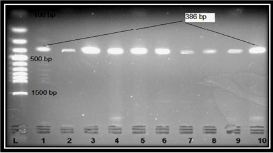
This is the first study in Iraq related to molecular detection of Legionella spp. in DUWL. Water samples were collected from various parts of dental units and investigated for the presence of Legionella spp. by culture and confirmed using PCR. Overall, there were 94 acid buffer-treated samples; 34 (36.1%) of them had heavy Legionella spp. growth on BCYE media. (Table 1) shows the number of detected Legionella spp. using BCYE and the source of the tested samples. Twenty-eight were isolated from bottled water. However, 4 were identified in syringe and 2 in HSD. Subsequently, to detect L. penumophila at the molecular level, the 16Sr RNA gene was amplified using PCR (Figure 1). Of the 34 total culture positive isolates, 10 (29.4%) were PCR positive for 16Sr RNA. Samples of bottled water mostly harbored L. pneumophila 9 (32.1%), followed by one isolate (25.0%) identified in syringe water (Table 2). This study revealed that no L. pneumophila was detected in HSD water samples by PCR. Statistical analysis showed that the culture method had excellent correlation with PCR results (P>0.0001).
    Sample source
    No. of tested Sample
    No. (%) of BCYE positive for Legionella spp.
    No. (%)of BCYE negative for Legionella spp.
    p-value**


    Bottle
    40
    28 (70.0)
    12 (30)
    0.0001


    Syringe
    30
    4 (13.3)
    26 (86.7)


    HSD*
    24
    2 (8.3)
    22 (91.7)


    Total
    94
    34 (36.1)
    60 (63.9)


    *HSD: High-Speed Drill, **Chi-Square:    34.6
Table 1: Distribution of Legionella spp. in water samples from various dental instruments using BCYE culture media.

    Figure 1:  Ethidium bromide-stained agarose gel of PCR amplified products
of extracted DNA with 16srRNA gene primers (386 bp). The electrophoresis
was performed at 70 volts for 1.5 hr. Lane (L), DNA molecular size marker
(l500-bp ladder), Lanes (1-10) positive for Legionella pneumophila isolates.
Discussion & Conclusion
Surveys of Legionella colonization in hospital water supplies have been conducted in the UK, Canada, USA, Korea and Iran [13]. The results of this study showed that Legionella was detected in DUWL in Najaf, Iraq. To the best of our knowledge, this is the first study of Legionella spp. detection in DUWL in the Middle East.
The current report provided evidence that the Legionella spp. may be moderately spread in dental clinics with a proportion of 36.1% (Table 1). This result is similar to the rate of isolation approved by some studies conducted in Iran, where the prevalence of Legionella isolation from hospitals was 36.6% and 22.7% as mentioned by [14,15] respectively. This highlighted the perceived threat to public health from DUWL contamination comes from such opportunistic and respiratory pathogens [8]. In the present study, BCYE culture media exhibit that most Legionella spp. 28 (70.0%) were isolated from dental water bottles followed by 4 (13.3%) and 2 (8.3%) identified in syringe and HSD (Table 1). Hence, the isolation of Legionela spp. from the stagnated water in the bottles may increase organisms to enter from one of the three main routes of the DUWL to form a biofilm along the length of the tubing. The biofilm acts as a reservoir for continued long-term contamination of the water line [8]. This result is compatible with the case report of [16] from the Mediated Disease Institute in Italy found that contamination of HSD with Legionella spp. related to the transmission to the patients, dentists and dental practice staff by inhalation or micro aspirations of aerosol water. Unfortunately, this may be creating a serious problem for contamination of hand pieces and transmission of Legionella spp. So, there is very little, if any, evidence of human-to-human transmission of Legionela spp. with public health relevance [17]. Moreover, there has been the successful use of artificial BCYE culture media to detect Legionella spp. Hence, these findings prompt the dental clinics to test their DUWL using water samples every month.
Many techniques have been used to detect and confirm Legionella spp., including culture, direct florescence, specific PCR and realtime PCR [18]. Culture is the gold standard method for isolation of Legionella spp. as routine work, but it has some limitations such as the growth requirements, long incubation periods, overgrowth of other bacteria and nonculturable Legionella in some samples [19]. Also, there is no molecular survey for L. penumophila in Iraqi dental clinics. For this reason, this study was designed to evaluate the culture test by PCR technique using the 16S rRNA gene.
Results revealed that of 34 culture positive isolates of Legionella spp. were recovered from DUWL. Only 10 (29.4%) were confirmed as L. penumophila using PCR, mostly (9/32.1) diagnosed in bottled water (Table 2). Increasing incidence of L. pneumophila in dental institutional clinics and hospital water supplies was observed by researchers, [20] from the USA, [21] from Italy and [13] from Iran. They demonstrated that prevalence rates of L. pneumophila were 8%, 21.8% and 7.1%, respectively. Although, the identification of L. pneumophila and non-L. pneumophila species is complex and timeconsuming, the 16S rRNA gene is present as a target gene since it exists in several copies per genome and thus allows a high sensitivity to the PCR [22]. These results suggest the presence of L. pneumophila in DUWL in Najaf dental clinics may be due to natural properties of the organism, such as a broad temperature range favoring growth, chlorine-tolerance and acid-tolerance. However, the presence of sediment, sludge and other material with biofilms in water are thought to play an important role in the persistence of Legionella spp. [23,24]. In addition, the vast majority of dentists in this survey took their water directly from the DUWL without flushing or using independent bottled water systems, anti-retraction valves on hand pieces, or any use of sterile water for minor oral surgery as the British Dental Association recommends [8].
    Sample source
    No. (%) of BCYE positive for Legionella spp.
    No. (%) of PCR positive for L.    penumophila
    No. (%) of non-L.    penumophila
    p-value**


    Bottle
    28
    9 (32.1)
    20 (67.9)
      0.436


    Syringe
    4
    1(25.0)
    3 (75.0)


    HSD*
    2
    0 (00.0)
    2 (100)


    Total
    34
    10 (29.4)
    24 (70.6)


    *HSD: High Speed Drill, **Chi-Square:    1.66
Table 2: Detection of Legionella penumophila in water sample from various dental instruments using PCR.
In conclusion, the higher recovery rates of Legionella spp. have been associated with storage and stagnated water in DUWL. Therefore, hospital tanks may act as a reservoir for repeated seeding of the Legionella spp. leading to the real threat of public health in Najaf.
Acknowledgment
The authors are very grateful to the advance research Lab of Basic Science Department faculty of dentistry. The authors thank the anonymous reviewers for their valuable comments and helpful revision suggestions.
References
- Centers for Disease Control and Prevention CDC. Procedures for the Recovery of Legionella from the Environment. Department of Health and Human Services. Public Health Service, U.S, Atlanta, GA. 2005.
- Nguyen MH, Stout JE, Yu VL. Legionellosis. Infectious Disease Clinics of North America. 1991; 5: 561-584.
- Winn WC. Legionnaires disease: historical perspective. Clin. Microbiol. Rev. 1988; 1: 60-81.
- Shearer BG. Biofilm and the dental office. J Am Dent Assoc. 1996; 127: 181-189.
- Mills SE. The dental unit waterline controversy: defusing the myths, defining the solutions. J. Am. Dent. Assoc.2000; 131: 1427- 1441.
- Cloud JL, Carroll KC, Pixton P, Erali M, Hillyard DR. Detection of legionella species in respiratory specimens using PCR with sequencing confirmation. J. Clin. Microbiol. 2000; 38:1709-1712.
- Jonas D, Rosenbaum A, Weyrich S, Bhakdi S. Enzyme-linked immunoassay for detection of PCR-amplified DNA of legionellae in bronchoalveolar fluid. J. Clin. Microbiol. 1993; 33: 1247-1252.
- Pankhurst CL. Risk Assessment of Dental Unit Waterline Contamination Primary Dental Care. 2003; 10: 5-10.
- Martin MV. The significance of the bacterial contamination of dental unit water systems. Br. Dent. J. 1987; 163: 152-154.
- Holt JG, Krieg NR, Sneath HA, Stanley JT, Williams ST. Bergeys manual of determinative bacteriology. Williams and Wilkins, USA. 1994.
- Baron EJ, Finegold SM. Baily and Scott's: Diagnostic Microbiology. Mosby Company, Missouri. 1994.
- Wojcik-Fatla A, Stojek NM, Dutkiewicz J. Efficacy of detection of Legionella in hot and cold water samples by culture and PCR. Standardization of methods. Annals of Agricultural and Environmental Medicine. 2012; 19: 289-293.
- Ghotaslou R, Sefidan FY, Akhi MT, Soroush MH, Hejazi MS. Detection of Legionella Contamination in Tabriz Hospitals by PCR Assay. Advanced Pharmaceutical Bulletin. 2013; 3: 131-134.
- Movahedian H, Shahmansouri MR, Neshat AA, Fazeli M. Identification of Legionella in the hot water supply of a general hospital in Isfahan. J. Res. Med. Sci. 2004; 6: 289-293.
- Hosseini DR, Mohabati MA, Esmailli D. Detection of Legionella in hospital water supply using mip based primers. J. Biol. Sci. 2008; 8: 930-934.
- Ricci ML, Fontana S, Pinci F, Fiumana E, et al. Pneumonia associated with a dental unit waterline. Lancet. 2012; 379: 684.
- Environmental Protection Agency EPA. Legionella: Drinking Water, Health Advisory. Office of Science and Technology, Office of Water United States, Washington, DC. 2001.
- Carvalho FS, Foronda AS, Pellizari VH. Detection of Legionella pneumophila in water and biofilm samples by culture and molecular methods from man-made systems in Sao Paulo-Brazil. J. Clin. Microbiol. 2007; 38: 743-751.
- Wellinghausen N, Frost C, Marre R. Detection of Legionellae in hospital water samples by quantitative real-time Light Cycler PCR. Appl. Environ. Microbiol. 2001; 67: 3985-3993.
- Atlas RM, Williams JF, Huntington MK. Legionella Contamination of Dental-Unit Waters. Appl. And Environment. Microbiol. 1995; 61: 1208-1213.
- Zanetti F, Stampi S, De Luca G, Fatch-Moghdam P, Antonietta M, Sabattini B, Checchi L. Water characteristics associated with the occurrence of Legionella pneumophila in dental units. Europ. J Oral Sci. 2000; 108: 22-28.
- Zhan XY, Li LQ, Hu CH, Zhu QY. Two-step scheme for rapid identification and differentiation of Legionella pneumophila and non-Legionella pneumophila species. J. Clin. Microbiol. 2010; 48: 433-439.
- Health Protection Surveillance Centre HPSC. Report of Legionnaires’ Disease. Health Protection Surveillance Centre, National Guidelines for the Control of Legionellosis in Ireland. 2009.
- Yu PY, Lin YE, Lin WR, Shih HY, Chuang YC, Ben RJ. The high prevalence of legionella pneumophila contamination in hospital potable water systems in Taiwan: Implications for hospital infection control in Asia. Int. J.Infect. Dis. 2008; 12: 416-420.