
Case Report
J Dent & Oral Disord. 2017; 3(1): 1054.
Orthognathic Treatment of the Facial Asymmetry Due to the Hemimandibular Hyperplasia
Gülsen A*, Findikçioglu K and Sibar S
Department of Plastic Reconstructive and Aesthetic Surgery, Faculty of Medicine, University of Gazi, Turkey
*Corresponding author: Gulsen A, Department of Plastic Reconstructive and Aesthetic Surgery, University of Gazi. Besevler, Ankara, Turkey
Received: February 16, 2017; Accepted: March 15, 2017; Published: March 22, 2017
Abstract
Our aim is to present orthognathic treatment of facial asymmetry patients due to hemimandibular hyperplasia who was a 45-year-old. She had no posterior teeth except one molar tooth bilaterally in the upper jaw and had only canines and incisors in the lower jaw. In treating facial asymmetry due to the hemimandibular hyperplasia, the condylar head was preserved and bilateral sagittal split osteotomy and angular ostectomy on the hyperplastic side were used to correct the asymmetry. Because of loss posterior buccal teeth, maxillary surgery was not planned. 3D model were used for diagnosis and surgical set-up in the patient. The simulation of the surgery was tested first in the 3D model. Following the surgery, an aesthetically pleasant facial symmetry was obtained in the case. Orthognathic treatment of hemimandibular hyperplasia.
Keywords: Hemimandibular hyperplasia; Orthognathic surgery; 3D model
Introduction
Hemimandibular Hyperplasia (HH) is a unilateral developmental deformity of the mandible characterized by a three dimensional enlargement of the condylar head, condylar neck, ramus and body on the effected side. It stops in the midline symphysis on the effected side by constructing a rotated facial appearance and prominence of the mandibular angle and also it causes crossbite malocclusion, facial asymmetry. The first case report was presented in 1836 [1]. Excessive growth of mandible can be appearing as unilateral Condylar Hyperplasia (CH), Hemimandibular Elongation (HE), or Hemimandibular Hyperplasia (HH) [2-4]. CH is consisted of condylar overgrowth with facial asymmetry but normal mandibular shape; HE is elongation of mandibular corpus without vertical increment; and HH is increment of mandibular, ramus and corpus height with condyle or rarely without condyle [5]. The underlying causes of this anomaly are not clearly determined. Local circulatory problems, traumatic lesions, hormonaldistrubances, etc are considered to be etiologic factors [6,7]. This anomaly normally begin to occur in teenage years and may continue to the thirties. Diagnosis of the HH is made with clinical, radiological (panaographic and posteroanteriorcephalomertricradiogaraphs) and CT imaging’s [8,9]. 3D models may also be useful for diagnosis and surgical treatment plan.
The treatment options of HH are designed according to the severity of the problem and also depends on presence of active condylar growth. Condylectomy, lower mandibular margin ostectomy with angular ostectomy, bimaxillary surgery to re-level occlusal cant or all of the mare used [4,10,11]. If the condylar activity had stopped, condylectomy may not be preferred to avoid TMJ disturbances. Condylectomy has particularly become performed to remove growth site in active condylar growth cases [12-13].
The aim of this study is to present the result of the orthognathic surgery treatment of one case with severe facial asymmetry due to the hemimandibular hyperplasia.
Cephalometric points and measurements used for the patients were shown (Figure 1,2).
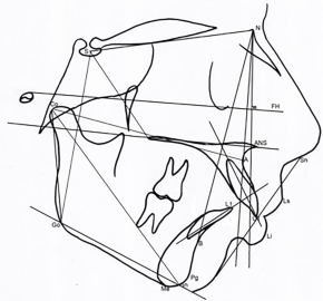
    Figure 1: The cephalometric points and measurements on lateral
cephalograms.
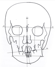
    Figure 2: The cephalometric points and measurements on postero-anterior
cephalogram.
Case Presentation
A 45-year-old woman was admitted to our department for the treatment of her facial asymmetry. Her only complaint was facial asymmetry without any discomfort, pain or a limitation in mouth opening. In her medical history, there was no trauma, hereditary or infection noted. She had noticed that her facial asymmetry had started after age of 17 years and progressed slowly. In her old pictures there was not a prominent asymmetry noticed. She reported that the increment of asymmetry had stopped during the age of thirties.
Clinically, a rotated facial appearance (a deviation to the left side) and the disproportion of the left and right mandible were very prominent (Figure 3). In her intraoral examination, occlusal plane was also tilted downward to the right side due to the over eruption of upper right molar and due to the HH (Figure 4). The patient had lost most of her posterior teeth for a long time. There was only one molar tooth in each buccal side in the maxilla and there were no posterior teeth after canines in the mandible. She had crown restorations with removable prosthesis in her mouth (Figure 4).
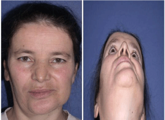
Figure 3: Preoperative extraoral views of the patient.
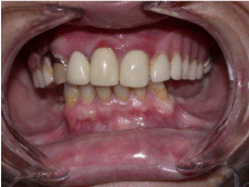
Figure 4: Preoperative intraoral views of the patient.
In the panoramic and the cephalometric radiographic examinations, the severe asymmetry of left and right condyle, ramal shapes and corpus heights were also revealed (Figure 5). Right condyle was enlarged together with elongation of the condylar neck, ascending mandibular ramus and body. In the posteroanterior cephalogram, right and left ramal heights were measured as 94.5 mm and 60 mm, respectively; which was deviated from the mean normal values of 60 mm on the right side (Table 1).
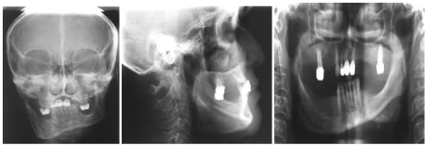
Figure 5: Preoperative radiographic views of the patient.
    Case


    Pre-
      Surgery
    Post-
      Surgery


    Skeletal    measurements


    SNA (°)
    84
    84


    SNB (°)
    87
    87


    ANB(°)
    -3
    -3


    SN/Go-Gn(°)
    19
    33


    N-A/FH (°)
    91
    91


    N-Pg/FH    (°)
    96
    93


    N-ANS    (mm)
    52
    52


    ANS-Me    (mm)
    62
    66


    N-Me    (mm)
    114
    118


    S-Go    right (mm)
    100.5
    67


    S-Go    left (mm)
    75
    71


    S-PNS    (mm)
    53
    53


    PP-SN    (°)
    4
    4


    Occlusal    Plane/SN (°)
    4
    6


    Co-Go    right (mm)
    80
    48


    Co-Go    left (mm)
    54
    54


    Co-A    right(mm)
    91
    94


    Co-A    left (mm)
    93.5
    93


    Co-Gn    right (mm)
    125
    128.5


    Co-Gn    left (mm)
    125.5
    129


    Go-Me    right (mm)
    79
    90


    Go-Me    left (mm)
    77
    79


    N?FH-A  (mm)
    -1
    -1


    N?FH-Pg  (mm)
    9.5
    4.5


    Dentoalveoler    measurements


    U1/NA(°)
    28
    28


    U1-NA    (mm)
    7
    7


    L1/NB(°)
    17
    25


    L1-NB    (mm)
    2
    4


    Overjet    (mm)
    1
    1


    Overbite    (mm)
    1
    1


    Soft    tissue measurements


    Nasolabial    angle (°)
    83
    83.5


    Ls^Sn-Pg’
    -1,5
    -1.5


    Li^Sn-Pg’
    0
    1.5


    Posteroanterior    measurements


    Co-Go    right (mm)
    94.5
    68


    Co-Go    left (mm)
    60
    60


    MSP-Me    (mm)
    11.5
    0


    Go    right-Go left(mm)
    102.5
    100


    Go-Me    right(mm)
    52.5
    43


    Go-Me    left (mm)
    50
    57


    U1-MSP    (mm)
    -2
    -2


    L1-MSP    (mm)
    -3.5
    -6


    U6    right-Z plane (mm)
    96
    96


    U6    left-Z plane (mm)
    84
    84


    Occlusal    plane tiltation
    12
    12
Table 1:  The pre and post-operative cephalometric measurements of the patient
(Cephalometric summary of the cases).
Surgical plan
- No maxillary surgery to correct the maxillary deviation was planned. Due to the lots of teeth missing, the overeruptedupper right molar and the deviated occlusal tilt were planned to correct with camouflage of with prosthetic restoration.
- Mandibular bilateral surgery preserving enlarged condylar head with inferior border ostectomy in right gonial area and in right inferior border of corpus were planned.
A 3D model obtained from the computerized tomographic data of the patient (Figure 6). All measurements were redone on this 3D model for the quantification of asymmetry. The treatment plan was applied on this model to see if the surgical plan was appropriate or not (Figure 7). Following the simulation of the surgery, it was observed that the vertical distance on the right posterior occlusion had not been enough (Figure 7). However, overerupted molar tooth with crown restoration could be used as with canal treatment or be extracted. The surgical guiding splint was prepared on the stone models according to this model surgery (Figure 8). The condylar head was preserved and no resection was performed on the condylar head and condylar neck during the surgery. Bilateral sagittal split osteotomy was done with the contouring of the right lower mandibular angular margin. Intermaxillary fixation was done between the screws placed into the maxillary and mandibular bones and remained for the next 4 weeks. Because of the surgery without condylectomy and of the position of inferior alveolar nerve, total symmetry was not possible but significant improvement and acceptable esthetic result were obtained (Figure 9). After 8 months follow-up period, the patient was referred to a prosthodontist. The postoperative results of the patient are presented including posteroanterior cephalometric, lateral cephalometric and panoramic radiograms (Figure 10) (Table 1). The results were stabile following three years (Figure 11).

Figure 6: Preoperative 3D model of the patient.
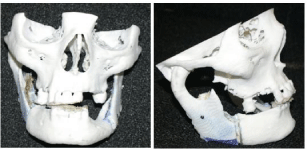
Figure 7: The simulation of surgery on this model.

Figure 8: The view of surgical splint during the operation.
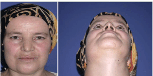
Figure 9: Postoperative views of the patient.
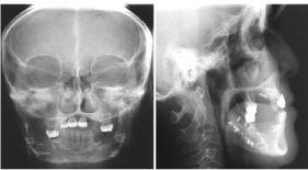
Figure 10: Preoperative radiographic views of the patient.
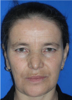
Figure 11: The extraoral view of patient following three years.
Discussion
In the present study, the case was diagnosed as facial asymmetry due to the ‘Hemimandibular Hyperplasia (HH)’ and mandibular surgery without condylar surgery with angular ostectomy was planned.
The general treatment plan in these types of anomalies to achieve symmetric facial appearance is bimaxillary surgery with or without condylar and lower mandibular border surgery. The treatment plan depends on the severity of cases, the location of inferior alveolar nerve, the severity of occlusal tilt, and condylar growth activity. The activity of the condylar overgrowth is the key in the treatment choice and in order to apply the correct surgical plan; active and inactive forms of HH must be distinguished which were first described by Norman and Painter [14]. The completetion of the condylar activity can be confirmed clinically by long-term serial follow-up, X-ray examination, bone scanning with Technetium-99 phosphate studies or by scintigraphy [15-20]. However, scintigraphy alone was reported as an unreliable technique, the anamnesis of the patient is also important as a reference [21]. Condylectomy is suggested in active condylar growth cases as high (the removal of upper 5 mm of the mandibular condyle to remove most active part condyle head growth) [16] and low (the removal of excess condyle to obtain symmetric ramal length) [22,21] type. In the early diagnosis of unilateral condylar hyperplasia; high condylectomy is recommended and orthodontic treatment is necessary after early condylar resection. Nevertheless, the studies showed that the condylectomy alone does not enough to correct all asymmetry and secondary orthognathic surgery is required [22]. And also, it is reported that condylectomy might cause lateral range limitation due to loss of the normal function of the lateral pterygoid muscle and might result in anterior open bite, deviation to the operated side when opening the mouth and loss of lateral excursion on the operated side due to the failure of reinsertion of the lateral pterygoid muscle [23-26].
However, some authors suggested that the hyperplastic condyle must be removed in every case [21,23,25,26], some authors recommend that the enlarged condyle should be left in-situ if the condylar activity was over and the movement of the condyle was acceptable [2,12,15,17]. Wolford et al. reported more stable and predictable outcomes in patients that diagnosed with active condylar hyperplasia and treated with high condylectomy, articular disc repositioning and orthognathic surgery when compared with the patients treated with orthognathic surgery only [12, 16]. Unilateral ramus osteotomy was favorably reported in cases with unilateral condylar hyperplasia of the mandible. However, bilateral ramus osteotomy was required in prognathic cases and in cases in which a unilateral procedure would cause excessive rotation of contralateral condyle [28]. In the current study, the patient stated that the facial asymmetry had not been increased for almost 17 years and the functional movements of the mandible were good. Clinically, no limitation of jaw movements was observed. The functional movement of the condyle might become worse postoperatively if the condylectomy would have been done [4]. The patient had also loss of posterior teeth and she was using a removable prosthetic restoration for the posterior area. Intraoral dental restoration was planned with prosthetic rehabilitation. For this reason, only mandibular surgery was performed following the model surgery simulation had done. In some HH cases, mandibular nerve is close to the inferior mandibular edge and positioning of inferior alveolar nerve [29]. In this case, as seen in the 3D model, maximum contouring osteotomy was performed at the rate allowed by the nerve location. 3D models of the patient guided the surgeon was very usefully about the considered surgical plan, and was also very useful to see clearly the thickness of the mandibular corpus, inferior alveolar nerve location, ramus and condylar shapes of the effected and noneffected sides before the surgery. The simulation of the operation on the 3D model showed that the surgical plan in the mandible only was effective to correct the facial asymmetry and eliminated the bimaxillary surgery need in the case.
Limitations
Intraoral dentition: The patience had not posterior buccal teeth except one molar with crown restoration in the both upper segments, so the vertical occlusal dimensions were disregarded to have a perfect prosthetic restoration. The socioeconomic condition of patient was not good enough to get the procedures like alveolar ridge reshaping or implant prosthetics done and the patient was referred for removable prosthesis.
Surgery without condylectomy: Even if preserving of the condyle is useful for TMJ functions, it limits the amounts of correction of asymmetry.
The position of inferior alveolar border: This is also important to determine the amount of surgical cut and it may limit amount of surgery if the alveolar nerve is close to the mandibular lower border.
Conclusion
HH cases have large condyles, increased ramal height and increased mandibular body height, so only condylar surgery does not enough to obtain symmetry. Bimaxillar surgery in addition to angular and lower mandibular border surgery in affected side are required in most cases.
In this case, condylar overgrowth had finished and multiple posterior teeth had missed; only Bilateral Mandibular Surgery (BSSO) with lower border ostectomy were selected and a good acceptable facial symmetry and profile was obtained.
References
- Adams R. Casehistory of Mary Keefe. Medical Section of the British Association, 1873. Bristol Meeting, September 1836.
- Marchetti C, Cocchi R, Gentile L, Bianchi A. Hemimandibular hyperplasia: treatment strategies. J Craniofac Surg. 2000; 11: 46-53.
- Obwegeser HL, Makek MS. Hemimandiular hyperplasia-hemimandibular elongation. J Maxillofac Surg. 1986; 14: 183-208.
- Xu M, Chan CC, Jin X, Lu J, Zhang C, Teng L. Hemimandibular hyperplasia: classification and treatment algorithm revisited. J Craniofac Surg. 2015; 25: 355-358.
- Khorsandian G, lapointe HJ, Armstrong JH, Wyosocki GP. Idiopatic noncondylar hemimandibular hyperplasia. Int J Paediatr Dent. 2001; 11: 298-303.
- Angiero F, Farronato G, Benedicenti S, Vinci R, Farronato D, Magistro S, et al. Mandibular condylar hyperplasia: clinical, histopathological, and treatment consideration. Cranio. 2009: 27: 24-32.
- Gray RJ, Sloan P, Quayle AA, Carter DH. Histopathological and scintigraph features of condylar hyperplasia. Int J Oral Maxillofac Surg. 1990; 19: 65-71.
- Walters M, Claes P, Kakulas E, Clement JG. Robust and regional 3D facial asymmetry assessment in hemimandibular hyperplasia and hemimandibular elongation anomalies. Int J oral Maxillofac Surg. 2013; 42: 36-42.
- Nolte JW, Verhoeven TJ, Schreurs R, Berge SJ, KarsemakersLHe, Becking AG, et al. 3-Dimensional CBCT analysis of mandibular asymemetry in unilateral hyperplasia. J Craniomaxillofac Surg. 2016; 44: 1970-1976.
- Ferguson JW. Definitive surgical correction of deformity resulting from hemimandibular hyperplasia. J Craniofac Surg. 2005; 33: 150-157.
- Ferreira S, Ferreira GR, Feverani LP, Francisconi GB, Sauza FA, Garcia IR. Unilateral condylar hyperplasia: A treatment strategy. J Craniofac Surg. 2014; 25: e256-258.
- Wolford LM, Moralles-Ryan CA, Garcia-Morales P, Perez D. Surgical management of mandibular condylar hyperplasia type 1. Proc (BaylUnivMedCent). 2009; 22: 321-329.
- Farina N, Olate S, Raposo A, Araya I, Alister JP, Uribe F. High condylectomy versus proportional condylectomy: is secondary orthognathic surgery necessary?. Int J Oral Maxillofac Surg. 2016; 45: 72-77.
- Norman JE, Painter DM. Hyperplasia of the mandibular condyle. A histological review of important early cases with a presentation and analysis of twelve patients. J Maxillofac Surg. 1980; 8: 161-175.
- Wolford LM, Mehra P, Reiche-Fischel O, Garcia-Morales P. Efficacy of high condylectomy for management of condylar hyperplasia. Am J Orthod Dentofac Orthop. 2002; 121: 136-151.
- Beirne OR, Leake DL. Technetium 99m pyrophosphate uptake in a case of unilateralcondylar hyperplasia. J Oral Surg. 1980; 38: 385-386.
- Matteson SR, Proffit WR, Terry BC, Staab EV, Burkes EJ Jr. Bone scanning with 99m technetium phosphate toassess condylar hyperplasia. Report of two cases. Oral Surg Oral Med Oral Pathol. 1985; 60: 356-367.
- Robinson PD, Harris K, Coghlan KC, Altman K. Bone scans and timing of treatment for condylarhyperplasia. Int J Oral Maxillofac Surg. 1990; 19: 243-246.
- Henderson MJ, Wastie ML, Bromige M, Selwyn P, Smith A. Technetium-99m bone scintigraphy and mandibular condylar hyperplasia. ClinRadiol. 1990; 41: 411-414.
- Sugawara Y, Hirabayashi S, Sussami T, Hyami S. The treatment of hemimandibular hyperplasia preserving enlarged condylar head. Cleft Palate Craniofac J. 2002; 39: 646-654.
- Farin˜a R, Pintor F, Pe´rez J, Pantoja R, Berner D. Low condylectomy as the sole treatment for activecondylar hyperplasia: facial, occlusal and skeletal changes. An observational study. Int J Oral MaxillofacSurg. 2015; 44: 217-225.
- Mehrotra D, Dhasmana S, Kamboj M, Gambhir G. Condylar hyperplasia and facial asymmetry: report of fivecases. J Maxillofac Oral Surg. 2011; 10: 50-56.
- Chen YR, Bendor-Samuel RL, Huang CS. Hemimandibular hyperplasia. Plast Reconstr Surg. 1996; 97: 730-737.
- Hampf G, Tasanen A, Nordling S. Surgery in mandibular condylar hyperplasia. J Maxillofac Surg. 1985; 13: 74-78.
- Raffaini M, Brevi B. Unaproposta di razionalizzazioneneltrattamenodelle asimmetrie facci alidell’adulto. Ortognatodonzia Ital.1994; 4: 513-530.
- Motamedi MH. Treatment of condylar hyperplasia of the mandible using unilateral ramus osteotomies. J Oral Maxillofac Surg. 1996; 54: 1161-1169.
- Bertolini F, Bianchi B, De Riu G, Di Blasio A, Sesenna E. Hemimandibular hyperplasia treated by early high condylectomy: a case report. Int J Adult Orthod Orthognath Surg. 2001; 16: 227-234.
- Epker BN, Stella JP, Fish LC. Dentofacial deformities: integrated orthodontic and surgical correction. St Louis: Mosby. 1996.
- Brasileiro BF, Sickels JEV. A modified sagittal split ramus osteotomy for hemimandibular hyperplasia and simultaneous inferior alveolar nerve repositioning. J Oral Maxillofac Surg. 2011; 69: 533-541.