Case Report
J Dent & Oral Disord. 2017; 3(3): 1061.
Maxillary Bilateral Canine-Premolar Transposition: A Rare Condition
Agarwala P¹, Agarwal V¹, Rekhade R¹ and Kulshrestha R²*
¹Department of Orthodontics and Dentofacial Orthopaedics, Haldia Institute of Dental Sciences & Research, India
²Consulting Orthodontist, Private Practice, Mumbai, India
*Corresponding author: Rohit Kulshrestha, Consulting Orthodontist, Private Practice, Mumbai, India
Received: March 28, 2017; Accepted: May 11, 2017; Published: May 18, 2017
Abstract
Tooth transposition is very rare developmental phenomena in which the adjacent teeth are switched or have their positions changed with one another and due to this aesthetical and functional problem may be present. The maxillary permanent canine is the tooth most frequently transposed, and it is often transposed with the first premolar followed by the lateral incisor and lastly the central incisor in very rare cases. Several etiologic factors may lead to transposition like genetics, developing tooth buds displacement, mechanical interferences, trauma and early loss of incisors. This paper reports a case of bilateral transposition in the maxillary arch involving the first premolar and canine.
Keywords: Ectopic eruption; Maxillary canine; Tooth transposition
Introduction
Tooth transposition is defined as the positional interchange in spatial area of two adjacent teeth including their roots, or eruption and development of a tooth in a position normally occupied by a non adjacent tooth. Tooth transposition can be a peculiar type of ectopic eruption in which the eruption order of teeth is changed [1]. It has been reported that transposition of maxillary teeth occurs approximately in one out of four hundred orthodontic patients. Transposition between the canine and first premolar (70%) is seen in maxillary dentition, followed by canine and lateral incisor (20%). Unilateral transpositions have been found more often than bilateral transpositions and it shows left side dominance [2]. In the literature, 6 types of transpositions have been clearly identified [3-5].
These are:
- Maxillary canine-first premolar (Mx.C.P1).
- Maxillary canine-lateral incisor (Mx.C.I2).
- Maxillary canine to first molar site (Mx.C to M1).
- Maxillary lateral incisor-central incisor (Mx.I2.I1).
- Maxillary canine to central incisor site (Mx.C to I1).
- Mandibular lateral incisor-canine (Mn.I2.C) transpositions.
Several etiologic factors like genetics, change in position of developing tooth buds, mechanical interferences, trauma and early loss of incisors have been seen to be related to tooth transposition [4,6- 9]. This article presents the rare occurrence of bilateral transposition of maxillary canine with maxillary first premolar.
Case Presentation
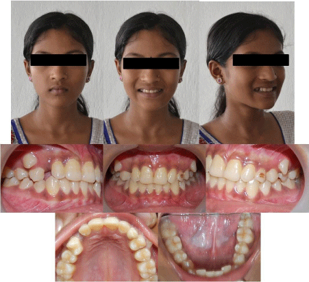
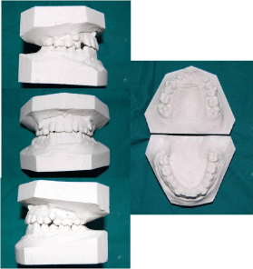
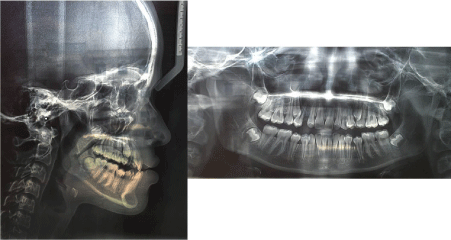
A female patient aged 13 years reported to the Department of Orthodontics and Dentofacial Orthopedics Haldia Institute of Dental Sciences Haldia with a chief complains of irregular teeth. A clinical examination revealed Angle Class I occlusion with 1.5 mm of overjet and 3 mm of overbite (Figure 1). Further examination revealed interchanged positions between maxillary first premolar and canine on both right and the left sides (Figure 2). Upper left deciduous canine was still present and it showed decalcification on its labial aspect. Remaining teeth in all the other quadrants were at normal location with normal morphology, overjet and overbite. Orthopantomogram (OPG) and Lateral Cephalogram showed no abnormalities (Figure 3). Medical and family histories were taken and no relevant finding was seen. The patient did not give any history of tongue thrusting, nail biting or lip biting habit. Treatment options included extraction of maxillary premolars to create space for the transposed canines or distalization of maxillary molars along with the premolars to create space for mesial movement of the canines.

Figure 1: Extra oral and intra oral photos.

Figure 2: Dental Casts.

Figure 3: Lateral Cephalogram and OPG.
Discussion
The maxillary canine is most frequently involved in transpositions. The term complete transposition is used when both crown and root structures of the involved tooth are seen in place of their transposed position. If the transposition is of the crown and not of the root apex is termed as incomplete transposition [10]. Retained deciduous canines (as seen in this case) have been suggested as a cause for extreme ectopic eruption of the maxillary canines into the incisor, premolar, or first molar areas.8 However, Peck et al. [3] stated that a retained deciduous tooth is a consequence of the anomaly, it does not to its causation. A genetic origin has been reported as the main etiologic factor. The main hypothesis of a genetic etiology for transposition is due to the disturbance in the order of development of the tooth buds. Genetic makeup plays an important role in laying the blueprints of the dentition. Expression of the homeobox genes which contain the transcription factors and pattern of development, have been assumed to provide the dental growth vector axis prior to odontogenesis. Dental initiation and manipulation of these genes due to mutation or any other factor can result in the transformation or transposition of the teeth. Mutations in several homeobox genes can cause selective tooth agenesis rather than transformation. This is certain because of the important genetic information these genes carry in them during the later stages of odontogenesis. Mx.C.P1 transposition has been currently considered as a tooth position anomaly caused by genetic factors, and it presents a multifactorial inheritance pattern. The maxillary canine tooth has the longest path of eruption among all the teeth and also it is the last tooth to erupt (apart from the third molars) in the oral cavity. Lack of space in the arch may also be a factor in ectopic positioning of the canine and theoretically due to this it is more susceptible to deflections during its long eruptive descent and hence it is frequently associated with transposition.
Conclusion
Early diagnosis of any transposition can help in correction with favorable prognosis and less chance of injuries to the periodontal tissues. This can be made possible with regular clinical and radiographic examination. Function and aesthetical restorations in patients with tooth transposition depends on the treatment plan and willingness/cooperation of the patient to achieve the best results. This case report suggests that abnormal eruptive migration of the canines, over-retained deciduous teeth, rather than a positional change causes this kind of transposition. Genes and early loss/over retained deciduous canines can lead to ectopic migration of the maxillary canine to premolar site. Therefore, the maxillary canine should be regularly monitored until it has completely erupted and reached its ideal location in the maxillary arch.
References
- Papadopoulosa MA, Chatzoudib M, Kaklamanosc EG. Prevalence of tooth transposition. A meta-analysis. Angle Orthod. 2010; 80: 275-285.
- Tripathi S, Singh RD, Singh SV, Arya D. Maxillary canine transposition – A literature review with case report. J Oral Biol Craniofac Res. 2014; 4: 155- 158.
- Peck S, Peck L. Classification of maxillary tooth transpositions. Am J Orthod Dentofacial Orthop. 1995; 107: 505-517.
- Peck S, Peck L, Kataja M. Mandibular lateral incisor-canine transposition, concomitant dental anomalies, and genetic control. Angle Orthod. 1998; 68: 455-466.
- Nishimura K, Nakao K, Aoki T, Fuyamada M, Saito K, Goto S. Orthodontic correction of a transposed maxillary canine and first premolar in the permanent dentition. Am J Orthod Dentofacial Orthop. 2012; 142; 4; 524-533.
- Peck L, Peck S, Attia Y. Maxillary canine-first premolar transposition, associated dental anomalies and genetic basis. Angle Orthod. 1993; 63: 99- 110.
- Teresa DM, Stefano M, Annalisa M, Enrico M, Vincenzo C, Giuseppe M. Orthodontic treatment of the transposition of a maxillary canine and a first premolar: a case report. J Med Case Rep. 2015; 9: 48.
- Gebert TJ, Palma VC, Borges AH, Volpato LER. Dental transposition of canine and lateral incisor and impacted central incisor treatment: A case report. Dental Press J Orthod. 2014; 19; 1: 106-112.
- Prasad V, Tandon P, Singh GP, Maurya RP. Management of maxillary lateral incisor: Canine transposition along with maxillary canine impaction on the contralateral side. J Orthod Res. 2015; 3: 61-64.
- Babacana H, Kilic B, Bicakc A. Maxillary canine-first premolar transposition in the permanent dentition. Angle Orthod. 2008; 78: 955-960.