
Research Article
J Dent & Oral Disord. 2017; 3(6): 1077.
Determining Facial Attractiveness for Orthodontic Treatment: A Study of Facial Characteristics of Female Caucasian Beauty Pageant Winners
Hylan-Cohen JA¹, Tsay TP² and Manasse RJ²*
¹Private Practice in Orthodontics in the Chicago, IL, USA
²Department of Orthodontics, College of Dentistry, University of Illinois at Chicago, USA
*Corresponding author: Robert J. Manasse, Department of Orthodontics, College of Dentistry, University of Illinois at Chicago, USA
Received: October 12, 2017; Accepted: November 13, 2017; Published: November 20, 2017
Abstract
Introduction: Soft tissue esthetics is a crucial component in orthodontic treatment planning. Previously, various norms or standards were derived as guidelines for evaluating facial esthetics and set as treatment goals. However, the validity of these standards was vague. This study described an attempt to obtain facial esthetic characteristics from publically judged attractive females using beauty pageant winners.
Methods: Twenty-seven current beauty pageant winners between the ages of 20 to 47 were photographed. Profile, frontal and smiling photos were obtained and composite images were generated. This data was also compared with the existing facial esthetic “norms”.
Results: Statistical tests indicated that 8 out of 13 variables in the study showed significant differences from published adult norms. Significantly the composite beauty pageant queens have a decreased lower facial height, retrusive upper lip, increased inter-labial gap, retrusive forehead and more nose projection.
Conclusion: These findings demonstrate subtle contributions of several facial components to an attractive face. Most importantly, this study presents facial esthetic guidelines to improve orthodontic and orthognathic surgical diagnosis and treatment planning.
Keywords: Facial attractiveness; Orthodontic treatment; Faces
Introduction
Obtaining facial attractiveness is an important consideration in orthodontic treatment. Patients seek treatment not to look average, but to look beautiful [1,2].
Historically, scientists and philosophers have attempted to define the parameters of attractive faces. Currently, Hollywood celebrities, beauty queens, and models represent ideal esthetic goals for many people. Medical professionals in various fields such as plastic surgery, oral surgery, orthodontics and prosthodontics have the ability to transform individuals’ facial tissues to reach these esthetic goals. The knowledge of what constitutes an attractive face is essential.
The importance of facial esthetics in orthodontic diagnosis and treatment planning has long been recognized. Edward Angle, the father of orthodontics, believed the most important outcome of treatment was a Class I occlusion. Angle suggested a properly aligned dentition would pave the way for acceptable soft tissue harmony [1]. Tweed, a student of Angle, began to emphasize facial proportions. Tweed believed premolar extractions were an important treatment protocol to promote dental stability. Unlike Angle’s belief that soft tissue balance would result from correcting the malocclusion, Tweed’s philosophy employed simultaneous consideration of soft tissue with occlusal stability [3].
In 1957, Riedel [4] proposed incorporating the public’s views of soft tissue attributes into orthodontic treatment norms or standards. In 1970, Peck and Peck [5] tried to decipher the difference between patients’ and health providers’ perceptions of attractive faces. They acknowledged the importance of the public’s opinion regarding facial attractiveness. The esthetic characteristics of beauty queens, professional models, and celebrities, known for their appearance, were studied using cephalometric analyses and photograph. This represented the departure from the traditional practice of imposing subjective opinion on treatment planning to incorporate public opinion in evaluating facial esthetics. This study concluded that the public preferred more protrusive dental and soft tissue characteristics than the established cephalometric standards.
In the 1980’s and the 90’s, Legan and Burstone [6], Powell and Humphreys [7], Arnett and Bergman [8], summarized attractive soft tissue norms suitable for adults based on models known for their beautiful facial attributes. This study was undertaken to determine whether the standards from 1980’s and 90’s are still currently valid by comparing existing norms of adult female facial characteristics to the facial esthetics of recent beauty pageant winners.
In 1992, Johnston et al. [9,10] developed a method called FacePrint that mimics evolution with a mathematical algorithm. This study using FacePrint had participants rank their most preferred male or female facial features over a series of generations. The participants used a computer keyboard to rank phenotypes’ attractiveness on a 10-point beauty scale. Ultimately, a beautiful composite face was produced and evaluated for attractiveness by Caucasian participants. The composite face was compared with anthropometric measurements of random female faces from their local population. Their study concluded that an attractive female face is not an average face and possesses unique facial proportions such as a decreased lower facial height and increased fullness of the lips. Their study found the highest ranked attractive face had an anterior lower facial height of an 11-year-old female according to the Farkas [11] growth curve.
Today, orthodontists use various norms for diagnosis and treatment planning. Since people seeking orthodontic treatment are largely motivated by a better dentition and cosmetic concerns [10], the treatment goals should aim at eliminating malocclusion while simultaneously improving facial attractiveness [11]. It is evident that an attractive facial norm or guideline is necessary for our profession.
This study describes an attempt to obtain facial esthetic characteristics from publically judged attractive females using beauty pageant winners.
Material and Methods
This research evaluated recent and current titled beauty pageant queens’ facial features. Their frontal and profile soft tissue attributes were compared with published norms (University of Illinois at Chicago Institutional Review Board Research Protocol Number: 2010-0927).
According to pageant documents and interviews with pageant judges, judging criteria for pageant queens are based on: beauty, poise, intelligence, personal interview, physical fitness, evening gown, on stage question and costume competition. Pageant judges stated that beauty and attraction were more than 80% of the judging criteria [12]. The pageant judges consisted of prior pageant queens, celebrities and medical professionals.
Pageant directors were contacted so that attendance to beauty pageants would be allowed for this study. On the interview day of the Mrs. Georgia America, Mrs. Missouri America, Mrs. Wisconsin America, and Mrs. Iowa America beauty pageants, the subjects were recruited with a “Verbal recruitment to participate in research” form. Before participation, subjects were asked to sign an informed consent. For participation in the study, each subject completed an eligibility questionnaire and a photographic questionnaire. Subjects were asked to state their age, previous pageant participation and titles, confirmation [13,14]. The inclusion criteria for participation in this study are as follows:
1. Caucasian female
2. Beauty pageant winner within the past five years.
The photographs were taken using a Sony Alpha Nex-5 digital SLR camera (Sony, New York, New York) mounted on a Velbon Victory 150 tripod with an 18-55 mm zoom lens, locked at 55mm. The images collected consisted of profile with lips at repose, frontal photographs with lips at repose, and Stage Two smiling frontal photographs [15]. Peck and Peck defined three stages in the genesis of a full smile. Stage Two is explained as the maximum movement of the upper lip and maximum appearance of the nasolabial fold using four facial muscles: levator labii superioris, zygomaticus major and superior fibers of the buccinators.
Participants were asked to sit in a chair and place their feet on a line in front of a black felt background five feet from the camera. To ensure natural head position, the participants were asked to look at a white sign behind the photographer as if looking off to the horizon. For the profile and frontal photograph at repose, participants were asked to lick their lips gently, and then relax [8]. For the Stage Two smiling frontal photograph, participants were asked to smile fully until their lips encountered resistance at the nasolabial fold [15]. With the camera in place, the participant was placed in a fixed position for the profile photo.
To ensure resolution standardization, the photographs were uploaded to Adobe Photoshop CS, version 7.0 and calibrated to 300 pixels per inch (Adobe, San Jose, CA). The profile photos were captured in Dolphin Solutions TM version 11.5 Premium (Dolphin, Chatsworth, CA) in the lateral x-ray view function. For standardization, the participants were oriented with the Frankfort horizontal plane parallel to the floor for later calibration using the digitizing landmark function in Dolphin for linear and angular measurements. The landmarks for the profile photographs are based on prior published definitions [5, 6,8,16] (Figure 1).
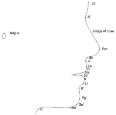
Figure 1: Profile composite of landmarks.
During the process of generating the average profile composite, the images were reduced from actual 2-D photographs to a single profile line. After landmark identification on the profile composite, angular and linear measurements were obtained from Dolphin and exported to Microsoft Excel (Redmond, WA) (Figure 2). The measurements were tested for intra and inter reliability using the Statistical Package for Social Sciences (SPSS), version 19, software for Windows (Chicago, IL).
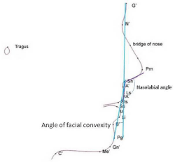
Figure 2: Legan angular soft tissue analysis.
After determination of high intra- and inter-reliability, the profile photographic sample was digitized. An average tracing composite of the twenty-seven profile photographs was completed in Dolphin’s superimposition screen (Chatsworth, CA). The traced images were registered at porion in the direction of soft tissue nasion.
Angular and linear measurements were calculated on the individual profile photographs and on the averaged composite image using Dolphin. The degree of variation between the soft tissue measurements of the study subjects were compared with the published “norms” [6-8]. (Figure 3-6).
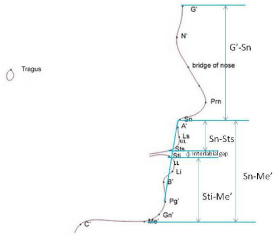
Figure 3: Legan linear and proportional soft tissue analysis.
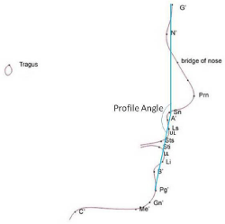
Figure 4: Arnett and Bergman soft tissue analysis.
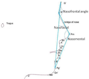
Figure 5: Powell and Humphreys soft tissue analysis.
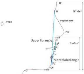
Figure 6: Additional soft tissue analysis.
For the frontal photographs, an average image at repose and smiling was generated using Jpsychomorph (Scotland, UK) [17] and saved as JPG (.jpg) files. One hundred and seventy-nine facial points were identified on each image. The landmarked images were then saved as TEM (.tem) which served as templates for averaging the photographs. Each .jpg file was paired with its respective .tem in a text file format (.txt) (Figure 7). The photographs were averaged in the Psychomorph window of the software and the “full Procrustes” method was used for shape normalization of each paired file to allow averaging of each point. From the twenty-seven photographs, an average frontal at repose and smiling face shape was generated. Finally, the color of 27 frontal repose and 27 smiling frontal faces were averaged into their respective composite face [18].
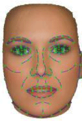
    Figure 7:  Text file (.txt): Frontal at repose photograph (.jpg) and template
(.tem).
After software processing to establish the frontal at repose and smiling frontal composite, the respective composite images were captured in Dolphin using the frontal x-ray view and printed at a 1:1 ratio. From the printed composite, fourteen points on two frontal images at repose and two smiling frontal images were hand traced 2 times 2 weeks apart.
The frontal at repose photographic landmarks are depicted in Figure 8. After the landmarks were digitized twice, the averages were taken. The measurements for comparison are displayed in Figure 9.
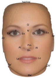
Figure 8: Frontal at repose photographic landmarks.
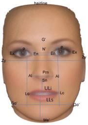
Figure 9: Frontal at repose photographic analysis.
For the smiling frontal photographic analysis, the landmarks were digitized twice and the averages were taken. The smiling frontal photographic analysis consisted of variables in Figure 9 compared to variables in Figure 10.
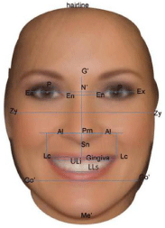
Figure 10: Smiling frontal photograph analysis.
After collection of the data from the profile photographs, the data were tabulated using Microsoft Excel and analyzed using the statistical software, SPSS (v.19.0), (Chicago, IL). A one-sample t-test was performed to evaluate significant mean differences between the beauty pageant queens’ soft tissue measurements and published norms.
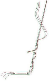
    Figure 11:  Profile tracing photographic composite in relation to subnasalesoft
tissue pogonion line: (-1) standard deviation (green), average composite
(black), (+1) standard deviation (red).
Results
Twenty-seven beauty pageant winners were evaluated in this study. The beauty pageant queens won the following pageants within the past five years: Mrs. Iowa, Mrs. Georgia, Mrs. Alabama, Miss North Central Georgia, Miss Georgia USA, Mrs. Savannah Georgia, Miss Illinois, 2 Mrs. Kansas, Mrs. Missouri, Miss Cupid, Miss Heartland, Mrs. America, Mrs. DC Galaxy, Mrs. Mid Missouri, Mrs. Marshfield, Mrs. Wisconsin, Mrs. Minnesota, Mrs. Elk River, Mrs. Des Moines Iowa, Mrs. Wisconsin, Mrs. Illinois America, 2 Mrs. Minnesota United States, 2 Mrs. Iowa America, and Mrs. Iowa America International. The ages ranged from 20-47 years old. The mean age was 34.96. Fifteen of the 27 women had orthodontic treatment in adolescence, and one woman had Invisalign as an adult. Three women reported having teeth extracted for orthodontic treatment. Per Institutional Review Board at the University, the participants must remain de-identified and it is not possible to list the years of the individual’s pageant queen titles.
The Kolmogorov-Smirnov and Shapiro Wilk test showed that all the variables in the study have approximately normal distribution.
After calibration and scaling, intra reliability was tested by tracings of 10 subjects’ photographs in Dolphin (Chatsworth, CA) with 19 different landmarks at 2 time periods, 2 weeks apart. A paired samples correlation showed a coefficient of correlation range between 0.982 and 1.00, indicating a high range of correlation thus providing good support for intra-operator reliability. To measure inter-operator reliability, the PI and a UIC orthodontic faculty member each traced the same 10 photographs with 19 different landmarks. A paired samples correlations showed coefficient of correlation range from 0.810 to 0.970, indicating good inter-operator reliability.
The profile photographic composite tracing within (+/-) one standard deviation was averaged in Dolphin and depicted in Figure 11.
Tables 1 and 2 show the descriptive statistics and the results of the one-sample t-test comparison of the sample and the published mean norms for profile facial measurements.
    Soft    Tissue Lateral Measurements
    N
    Mean    Norm
    Sample    Mean
    (±)    SD


    Legan Analysis


    Facial Convexity (intersection of G’-Sn and Sn-Pg’ )(°)
    27
    12
    11.64
    5.623


    Nasolabial Angle (Col-Sn-UL)(°)
    27
    102
    109.21
    10.341


    Interlabial Gap (Sts-Sti)(mm)
    27
    3.3
    4.044
    2.313


    Upper Lip Protrusion (UL-SnPg’)(mm)
    27
    3
    2.05
    2.062


    Lower Lip Protrusion (LL-SnPg’) (mm)
    27
    2
    1.44
    2.3113


    Lower facial thirds (Sn-Sts:Sti-Me’)(%)
    27
    33.3
    28.12
    2.543


    Arnett and Bergman Analysis


    Profile Angle (G’-Sn-Pg’)(°)
    27
    170
    168.77
    5.74


    Powell and Humphreys Analysis


    Nasofrontal angle (N’-nasal dorsum)(°)
    27
    122.5
    135.64
    7.076


    Nasomental angle (nasal dorsal line-nasomental line)(°)
    27
    126
    121.47
    5.976


    Nasofacial angle (G’-Pg’-dorsal plane of the nose)(°)
    27
    35
    37.5
    4.486


    Additional Analysis


    Upper lip angle (Pg’-UL:PG’-N’)(°)
    27
    10
    10.34
    3.541


    Mentolabial angle (LL-B’-Pg’)(°)
    27
    126
    128.19
    13.9


    Lower face percentage (Sn-Me’:G’-Me’)(%)
    27
    54
    51.17
    2.063
Table 1: Descriptive Statistics of the Beauty Pageant Queens.
    Profile    Soft Tissue Measurements
    N
    Mean  Difference
    t
    df
    p-value*
    95%    CI


    Legan


    Facial convexity(°)
    27
    -0.36
    -0.329
    26
    0.745
    -2.580 to 1.869


    Nasolabial angle (°)
    27
    7.21
    73.625
    26
    0.001*
    3.124 to 11.306


    Interlabial gap(mm)
    27
    0.74
    2.976
    26
    0.006*
    0.230 to 1.259


    Upper lip protrusion(mm)
    27
    -0.95
    -2.398
    26
    0.024*
    -1.768 to -0.135


    Lower lip protrusion (mm)
    27
    -0.56
    -1.265
    26
    0.217
    -1.478 to 0.352


    Lower facial thirds(%)
    27
    -5.18
    -10.581
    26
    0.000*
    -6.184 to  -4.172


    Arnett and Bergman


    Profile angle(°)
    27
    -1.23
    -1.117
    26
    0.274
    -3.504 to 1.037


    Powell and Humphreys


    Nasofrontal angle(°)
    27
    13.14
    9.652
    26
    0.000*
    10.345 to 15.944


    Nasomental angle(°)
    27
    -4.54
    -3.948
    26
    0.001*
    -6.905 to -2.177


    Nasofacial angle(°)
    27
    2.5
    2.896
    26
    0.008*
    0.725 to 4.275


    Additional


    Upper lip angle(°)
    27
    0.34
    0.505
    26
    0.618
    -1.056 to 1.745


    Mentolabial angle(°)
    27
    2.19
    0.82
    26
    0.42
    -3.306 to 7.691


    Lower face percentage(%)
    27
    -2.84
    -7.146
    26
    0.000*
    -3.653 to  -2.021
Table 2: One-Sample T-Test Results Of The Published Mean Norms Compared To Beauty Pageant Queens’ Mean Norms.
A one-sample t-test indicated that 8 variables out of 13 variables in the study showed statistically significant mean differences from the published norms with p-values ranging from 0.000 to 0.0024. For Arnett and Bergman’s [8] profile angle, Powell and Humphreys’ [7] nasomental, nasofacial, and nasofrontal angle, the midpoint of the published ranges was used as the mean for statistical testing in this study. Statistically significant differences were found in the following:
• Powell and Humphreys’ measurements: Nasolabial, Nasomental, Nasofacial, Nasofrontal angles
• Additional measurements: Lower face percentage.
The beauty pageant queens’ nasofrontal angle, nasofacial angle, nasolabial angle, upper lip protrusion, lower facial thirds and lower face percentage was significantly greater than the published norms. While the nasomental angle and interlabial gap was significantly less than the published norm.
The results show that 5 out of 13 variables in the study show no statistically significant differences. They are: the angle of facial convexity, profile angle, upper lip angle, mental labial angle, and lower lip protrusion.
The Jpsychomorph software does not allow extrapolation of the frontal at repose and smiling raw data; therefore, it was not possible to test their composite norms for statistical significance. However, measurements were made from both composite faces, which should represent usable norms for adult females frontal facial features at repose and when smiling (Table 3 and 4).
    Measurements
    N
    Published    Norm
    Beauty    pageant queens’ norm


    Powell and Humphrey Published Norm


    Inner canthal (En): Outer canthal (Ex) ratio (mm)
    27
    0.36
    0.38


    Basic facial shape

    Round, oval, square or diamond/pear
    Diamond/pear


    Eye variation
    27
    Round, small, ptotic


    Inter medial limbus: inter labial commissures
    27
    1
    Round


    Lip ratio- (Sn-ULi, LLS-Me’) (mm)
    27
    2.3
    0.925


    Upper facial height to lower facial height
    27
    1
    2.17


    (G’-Sn: Sn-Me’)
    27

    1.11


    Arnett’ and Bergman’s Published Norm


    Facial width- Zy-Zy: Go’-Go’ (mm)
    27
    1.36
    1.29
Table 3: Beauty Queens’ Frontal Soft Tissue analysis.
    Measurements
    N
    Beauty    pageant queens’ norm


    Gingiva display (mm)
    27
    2.25


    Inter labial commissure (Lc) at repose: Inter labial commissure (Lc) at smiling
    27
    0.77


    Nasion (N’) to subnasale (Sn) at repose: Nasion (N’) to    subnasale (Sn) at smiling
    27
    0.95


    Inter outer canthal (Ex) width at repose: Inter outer    canthal (Ex) width at smiling
    27
    1.06


    Nasion (N’) to upper lip inferiors (ULi) at repose: nasion    (N’) to upper lip inferiors (ULi) at smiling
    27
    1


    Inter inner canthal (En) at repose:Inter inner canthal    (En) at smiling


    Inter alare (Al) width at repose: Inter alare (Al) width at smiling
    27
    0.9


    Subnasale (Sn) to Upper lip inferiors: Alare to labial    commissure
    27
    0.77
Table 4: Beauty Pageant Queens’ Soft Tissue Frontal-Smiling analysis.
Discussion
The purpose of this study was to study facial features of publicly judged attractive adult females, assess and compare their esthetic characteristics with published norms, which were primarily Caucasian, and possibly provide orthodontists with data that could be used in diagnosis and treatment planning. This 2011 study sample was compared to norms for esthetics faces published at different time points: Legan norms published in 1980, Powell and Humphreys norms published in 1984 and Arnett and Bergman norms published in 1993. It can be inferred from the findings that the study sample in this research most closely resembles the models used by Arnett and Bergman, which was the most current published norm used for comparison.
Interestingly, the findings for the beauty pageant queens’ nasofrontal and nasofacial angle means were significantly greater than the published norm. An increased nasofrontal angle suggests a smaller nose or retrusive forehead; however a smaller nose in unlikely because our sample also had an increased nasofacial angle. Taking these findings together, the results suggests a flattened (retrusive) forehead.
The beauty pageant queens’ significantly decreased nasomental angle suggests a larger nose, a retrusive chin or a more deeply set soft tissue nasion. However, the significantly increased nasofacial angle, the non-significantly increased angle of facial convexity and profile angle suggest a more deeply set soft tissue nasion.
The beauty pageant queens’ significantly greater mean nasolabial angle suggests a retruded upper lip or tipped up nose. Their significantly decreased upper lip protrusion substantiates the retruded position of the upper lip. A retrusive upper lip may suggest a degree of maxillary hypoplasia, retruded upper incisors, or the effects of facial aging. It is not believed that this study sample has a vertical maxillary deficiency because of the normal gingival display. If the maxilla were vertically deficient there would be a tendency to show less of the maxillary incisors with the lips relaxed. It is possible that the beauty pageant queens have retruded incisors. Without cephalometric measurements, this could not be determined. Differentiating individuals of the sample depending on who did or did not have orthodontic treatment was not in the scope of this study.
One might expect a decreased interlabial gap and decreased incisor display when the lips are relaxed with aging; however, our sample displays an increased interlabial gap and displays more of the maxillary incisors at repose. This may suggest vertical maxillary excess and/or a short upper lip.
One of the most important findings in this study was the variation in facial heights. The study results showed significant differences between the sample norm in the lower facial thirds and lower face percentage. Our sample has a decreased stomion inferioris to soft tissue menton compared with the published norm [7]. These findings are consistent in both our profile and frontal facial analysis. This feature also contributes to a younger face.
Smiling facial esthetics was compared between the beauty pageant queens’ smile and the esthetic smile described by Kokich Jr et al. [19] who suggested that orthodontists, lay people and general dentists have differing opinions regarding smile esthetics and that orthodontists prefer 2 mm or less gingival exposure when smiling. It was interesting to see the beauty pageant queens’ smiling composite displayed more gingiva then some orthodontists may prefer.
This study was not in agreement with Riedel’s4 and Peck and Peck’s [5] findings for a more protrusive facial pattern. This sample is in agreement for an esthetic Class I soft tissue profile similar to Legan and Burstone [6], and Arnett and Bergman [8].
This study does support Johnston and Franklin’s [9] finding that a beautiful female face composite displays a decreased lower facial height. Additionally, this research supports Alley and Cunningham’s [20] beliefs that adult faces displaying unusually juvenile facial characteristics are considered more attractive. The slightly increased gingival display of the pageant queens’ also may contribute to their more youthful appearance.
This research supports the relationship between attraction, a youthful appearance and a decreased lower face. This implies that molar extrusion mechanics should be minimized or avoided in most orthodontic cases. To maintain the youthful appearance of the smile, treatment of adolescent individuals should avoid excessive reduction in gingival and incisor display at rest and smiling. The majority of adolescent individuals show that gingival and incisor display decreases with age. A person treated to “ideal” gingival and incisal display may appear prematurely old with aging.
Conclusion
This current study evaluated facial features of publically accepted attractive faces which were recent and current beauty pageant winners. The facial features evaluated were profile convexity, lip position, proportions of facial thirds and symmetry. This provided some evidence to support the idea that the perception of adult female facial attractiveness is associated with looking young. It was found that the obtuse nasolabial angle which is usually associated with ditched in profiles could be partially masked by an increased maxillary gingival and incisal display. Furthermore norms and ranges obtained from these attractive faces can act as a guide for orthodontists to help patients’ achieve their esthetic goals.
Acknowledgments
Special recognition is extended to Dr. Budi Kusnoto for his help and assistance with the technology used for this study and to Drs. Maria T. Sabater Galang, Pravin K. Patel and Carla A. Evans for their service on the Masters of Oral Sciences thesis committee in this project. Appreciation is extended to Mrs. Maria Grace Costa Viana for her help in the statistical analysis. Special thanks are extended to Carl and Faith Schway for their recruiting beauty pageant winners to be subjects for this study. Finally, with the help of the Jsphymorph software supplied by Bernie Tiddeman the photographic composite images were created.
References
- Profit, WR.: Contemporary Orthodontic Appliances. In: Contemporary Orthodontics. W.R. Proffit, H.W. Fields, and D.M. Sarver (edns.), Philadelphia: Mosby. 2007.
- Tuncer C, Canigur Bavbek N, Balos Tuncer B, Ayhan Bani A, Celik B: How do patients and parents decide for orthodontic treatment-Effects of malocclusion, personal expectations and media, J Clinical Pediatric Dentistry. 2015; 39: 392-399.
- Graber TM, Vanarsdall R, Vig K. Orthodontics Current Principles and Techniques. 4th edn. St. Louis: Mosby Inc. 2007.
- Riedel RA. An analysis of dentofacial relationships. Am J Orthod. 1957; 4: 103-119.
- Peck H, Peck S. A concept of facial esthetics. Angle Orthod. 1970; 284-318.
- Legan HL, Burstone CJ. Soft tissue cephalometric analysis for orthognathic surgery. J Oral Surg. 38; 744-751: 1980.
- Powell N, Humphreys B. Proportions of the Aesthetic Face. Thieme-Stratton Inc.1984.
- Arnett GW, Bergman RT. Facial keys to orthodontic diagnosis and treatment planning. Part I and II. Am J Orthod Dentofacial Orthop. 104; 299-312: 1993.
- Johnston VS, Franklin M. Is beauty in the eye of the beholder? Ethol Sociobiol. 14; 183-199: 1992.
- Johnston VS, Solomon CJ, Gibson SJ, Pallares-Bejarano A.Human facial beauty current theories and methodologies. Arch Facial Plast Surg. 5; 371- 377: 2003.
- Farkas LG, Munro I: Anthropometric Facial Proportions in Medicine. Springfield, Illinois, Charles C Thomas, 1987.
- Czarnecki ST, Nanda RS, Currier GF. Perceptions of a balanced facial profile. Am J Orthod Dentofacial Orthop. 104; 180-187: 1993.
- Sarver DM, Ackerman JL. Orthodontics about face: the re-emergence of the esthetic paradigm. Am J Orthod Dentofacial Orthop. 117; 575-576: 2000.
- Mrs. Minnesota-America. Judging. Retrieved on September 10, 2011.
- Peck H, Peck L, Kataja M. The gingival smile line. Angle Orthod. 62; 91-100: 1992.
- Arnett GW, Jelic JS, Kim J, Cummings DR, Beress A, Worley CM, et al. Soft tissue cephalometric analysis: diagnosis and treatment planning of dentalfacial deformity. Am J Orthod Dentofacial Orthop. 1999; 116: 239-253.
- Facereserach. (January 17, 2007). Protomethods. Retrieved on September 10, 2011.
- Tiddeman B, Stirrat, Perrett D. Towards realism in facial transformation: results of a wavelet MRF method. Computer Graphics Forum, Eurographics conference Issue. 2005; 24: 1-5.
- Kokich VO, Kiyak HA, Shapiro PA. Comparing the perception of dentists and lay people to altered dental esthetics. Journal of Esthet Dent. 1999; 11: 311- 324.
- Alley TR. Cunningham MR. Averaged faces are attractive, but very attractive faces are not average. Psychol Sci. 1991; 2: 123-125.