Clinical Image
J Dent & Oral Disord. 2018; 4(1): 1085.
Oral Papilloma Verruca Vulgaris
Stübinger S¹* and Berg BI1,2
¹Hightech Research Center of Cranio-Maxillofacial Surgery, University of Basel, Basel, Switzerland
²Department of Cranio-Maxillofacial Surgery, University Hospital Basel, Basel, Switzerland
*Corresponding author: Stefan Stübinger, Hightech Research Center of Cranio-Maxillofacial Surgery, University of Basel, Basel, Switzerland
Received: February 01, 2018; Accepted: February 07, 2018; Published: February 14, 2018
Clinical Image
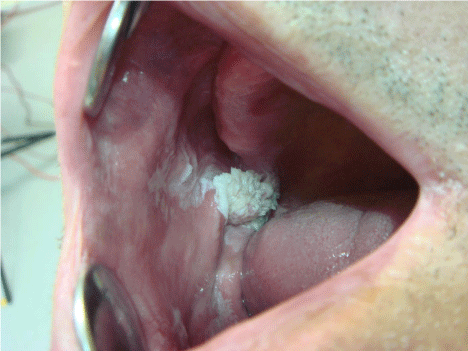
A 46-year-old man from Germany was referred for evaluation of an intra oral exophytic white tumor in the right distal part of his cheek. The patient was edentulous since 5 years and reported to smoke 20 cigarettes per day. According to his anamneses the neoplasm had initially grown rapidly and now showed up a steady state for more than 3 years. Physical examination revealed a neoplasm with a keratotic surface and a well-delineated border. There were no signs of bleeding or ulcerations. Overall, the lesion had a diameter of almost 2.0 mm. Additionally, scattered white plaques were seen on the surrounding mucosa. A small biopsy on the border was performed to exclude a malign tumor (Squamous-cell carcinoma). Histopathological examination revealed an intraoral Papilloma Verruca Vulgaris associated with Human Papilloma Virus (HPV) 2 and 4. The lesion was completely removed by surgical excision and the patient remained in a re-call for one year.
