
Research Article
A Case Report. J Dent & Oral Disord. 2018; 4(2): 1086.
Additional Mechanical Retention for Maryland Bridges - A Case Report
Smarandescu D*
Str. Banu Dumitrache 33, Bucharest, Romania
*Corresponding author: Dragos Smarandescu, Str. Banu Dumitrache 33, Bucharest, Romania
Received: January 03, 2018; Accepted: February 05, 2018; Published: February 22, 2018
Abstract
Statement of the Problem: There is often a need to obtain more mechanical retention of abutments that support a Maryland bridge.
Aim: To obtain more mechanical retention by zero degrees preparations but to be minimally invasive and as favorable as possible from the esthetic viewpoint.
Materials and Methods: The diagnostic cast was milled using a parallelometer. Four interlocks were created, connected by preparations on the lingual walls. After completing the milling of the cast, the margins were highlighted using a pencil. Then, after insulation of the plaster, resin templates were made, accurately adapted to the margins (occlusal line angle). The templates were then lightly bonded to the corresponding teeth. During intraoral preparation the bur was guided so as, at the end of the milling process, it would touch simultaneously the template margins and the finish line.
Results & Conclusion: The clinician was able to copy intraorally the preparation previously performed on the diagnostic cast. The four interlocks provide additional mechanical retention and the labial surfaces of the abutment teeth remain intact.
Keywords: Maryland bridge; Minimally invasive; Guided preparation; Mechanical retention
Introduction
Minimally invasive dentistry has led to quest for better preparations so as to reduce unnecessary loss of tooth structure. No doubt, adhesive dentistry has paved the way for minimally invasive preparations; still, mechanical retention, where possible, adds to the long term success of the restoration.
Maryland bridges have been in use for many years and have undergone various changes in design, as new materials became available. In a review article, Shimizu, Kawaguchi and Takahashi have pointed out that “Using functional monomers including VBATDT, MTU-6 or MDDT, gold alloy and Ag–Pd–Cu–Au alloy adhere directly to the resin materials. (…) A new era in this field began with the use of noble metal alloys including Ag–Pd–Cu–Au alloy, and the design of resin-bonded prostheses has been re-evaluated.”[1].
Back in 1986 Dummer and Gidden suggested “ A modification to the Maryland bridge design (…) which allows the restoration to be fitted in situations where one of the abutment teeth is heavily restored” [2]. The modification was intended to increase the mechanical retention.
As to the precission of intraoral preparations, Okada and Inoue showed in 2008 that “the ideal preparation is not easy even for skilled dentists, much less for students and beginners. If they use parallel measurements, they would save time and the accuracy gets higher” [3]. This statement refers to preparations for classical crown and bridges but is even more obvious for minimally invasive dentistry. Parallel measurements seem to date many years back. In 1943, Karlström presented a milling technique based on inserting paralleling pins into the teeth to be milled [4].
In the same review article mentioned above, published in 2013, the authors state the following: “Evaluation of the current status indicates that the design of posterior resin-bonded prostheses has almost become D-shaped. This design is almost complete with no clinically significant problems and will be used for the foreseeable future. On the other hand, there is no typical standard yet for the design of anterior resin-bonded prostheses. The available surfaces to be bonded are limited from the esthetic viewpoint to the lingual surface and a portion of the proximal surface in the anterior region. Hence, it is difficult to use a wrap-around design in this region. Consequently, there is a limitation to adding mechanical retention obtained by design although the improvement of the bond strength of the adhesive resinmaterial is definitely needed, particularly in this region” [1].
Materials and Methods
The clinical case presented in this article refers to a resin bonded Maryland bridge extending from canine to the first molar, therefore implying features of both posterior an anterior Maryland bridges. The intention was to obtain more mechanical retention by zero degrees preparations but to be minimally invasive and as favorable as possible from the esthetic viewpoint. The young female patient was unable to pay for a solution involving bone grafting and implants in the region of the two left upper premolars (Figure 1)
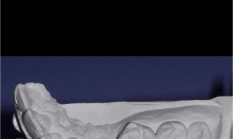
    Figure 1:  Diagnostic cast showing the state of the edentulous crest in the
region of the two left upper premolars.
Metal ceramic, resin bonded Maryland bridge immediately became an option. To increase mechanical retention the diagnostic cast was milled using a parallelometer and the preparation was then copied intraorally. Four interlocks were created, connected by preparations on the lingual walls (Figure 2).
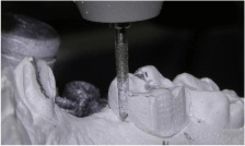
    Figure 2:  Diagnostic cast milled using torpedo turbine bur, same size as the
one that was to be used intraorally. Zero degrees interlocks and lingual wall
preparation.
After completing the milling of the cast, the margins were highlighted using a pencil (Figure 3).
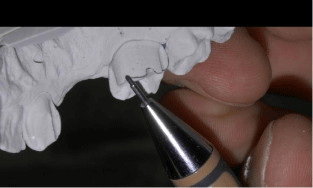
Figure 3: Marking the margins that resulted after the preparation of the cast.
Then, after insulation of the plaster, resin templates were made, accurately adapted to the margins (Figure 4).
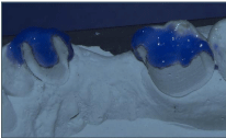
Figure 4: Resin templates made on the modified diagnostic cast.
The templates were then lightly bonded to the corresponding teeth (Figure 5). The bur used intraorally was identical to the one used to prepare the diagnostic cast. It was guided so as, at the end of the milling process, it would touch simultaneously the template margins and the finish line (Figure 6).
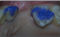
Figure 5: Templates bonded to corresponding teeth.
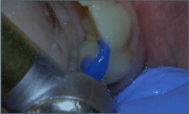
    Figure 6:  Bur guided so as, at the end of the milling process it would touch
simultaneously both the margins of the template and the finish line.
Results & Conclusion
The result was that the clinician was able to copy intraorally the preparation previously performed on the diagnostic cast. The author of this article had been using this technique for many years and in numerous cases, but in the case presented here the intraoral preparation was done under supervision by a dentist with very limited clinical experience. The outcome can be seen in Figure 7. The winglets of the bridge were designed with retentive holes. Core material resin was used to bond the restoration. (Figure 8). The esthetic outcome is shown in Figure 9. After two years the bridge is perfectly functional.
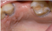
Figure 7: Preparation completed.
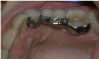
    Figure 8:  Occlusal aspect of the cemented bridge. Winglets with retentive
holes.
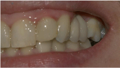
    Figure 9:  Final esthetic outcome. Intact labial surface of the abutment teeth
(canine and first molar).
Template guided preparation, developed by the author of this article in 2008, seems to be an efficient clinical solution and has been used by many dentists throughout Romania, where the technique is being taught since 2009. Other extraoral paralleling methods and devices have been developed and patented in the last 7 years, including laser projectors and virtual model manipulation.
References
- Shimizu H, Kawaguchi T, Takahashi Y. The current status of the design of resin-bonded fixed partial dentures, splints and overcastings. Japanese Dental Science Review. 2014; 50: 23-28.
- Dummer PM, Gidden J. The Maryland bridge: a useful modification. J Dent. 1986;14: 42-43.
- Okada M, Inoue H. Introduction of parallel measurement and supplementary apparatus newly developed. Prosthodont Res Practice. 2008; 7: 243-245.
- Karlström KAS. Preparation of dental bridge. U.S. Patent 2,318,402. 1943.