
Research Article
A Case Report. J Dent & Oral Disord. 2018; 4(2): 1087.
Simple Templates for Predictable, Minimally Invasive Axial Walls Preparation
Smarandescu D*
Str. Banu Dumitrache 33, Bucharest, Romania
*Corresponding author: Dragos Smarandescu, Str. Banu Dumitrache 33, Bucharest, Romania
Received: January 04, 2018; Accepted: February 07, 2018; Published: February 22, 2018
Abstract
Statement of the Problem: There is often a problem to make parallel preparation of multiple abutments
Aim: To reproduce intra-orally the preparation that has been previously carried out on a diagnostic cast.
Materials and Methods: After initial impression in silicones, the diagnostic cast is being developed. The abutments are milled individually, the tip of the bur (torpedo) accurately following the finish line. On the remaining occlusal/ incisal surface of each tooth block-out resin is applied. Resin occlusal templates are obtained, which are placed intraorally on respective teeth and fixed with bonding, without etching the tooth surface. During preparation the bur (same dimension and shape as the one used while milling the cast) follows a vector that stretches from the margins of the template to the finish line. Parallel axial surfaces are prepared. Further vestibular and occlusal/incisal reduction is then carried on.
Results & Conclusion: The abutments will be ideally parallel, with minimum loss of tooth substance.
Keywords: Minimally invasive; Predictable preparation; Guided preparation
Introduction
Preparing parallel abutments may be a difficult task, especially when:
- Teeth are malpositioned, rotated, etc.,
- Multiple teeth must be milled,
- There is a large distance between the abutments, and/or
- Molars must be milled, especially on the distal surface.
Geometric concepts have been presented and thought to help improve the quality of preparations [1-3]. They are currently the most widely used method of teaching students in dental universities worldwide. However, Gold [4]. has shown that “casts from a number of preparations by dentists with a variety of fixed prosthodontic experience levels were surveyed, and the preparations were commonly found to contain undercuts, over tapered surfaces, and lack of a parallel path of insertion.” He therefore suggested the use of an intra-oral paralleling device.
The option for such a device for intra-oral surveys has stirred the imagination of inventors [5-7], and many articles in prestigious journals point to the importance of their use [8-13].
Dental inspection mirrors with paralleling guiding lines have also been patented [13,14].
In 1989, Möllersten showed that “compared with results from a previously performed model study carried out during ideal preparatory conditions, the current investigation showed that the paralleling precision of guiding instruments used clinically in fact decreases considerably, remaining, however, at an acceptable level.” [15]. In the same year [16], he compared guided and freehand preparations and concluded that “both instrument preparation and freehand preparation were influenced by the dentist’s dexterity and technical capability”, showing that paralleling devices do not seem a reliable solution thus far, though they might be of help. Other options for parallel abutment preparation include pins, jigs, and templates, placed intra-orally on the teeth to be milled.
Resin jigs have been designed as aids to parallel guiding plane preparation for removable partial dentures [17,18].
Resin templates have also been designed for easing the task of abutment preparation [19,20]; a preparation technique of this type is described in this article.
Aim
The method described below allows the clinician to reproduce accurately in the oral cavity the preparation of the patient’s teeth that was previously carried out on a diagnostic cast. Ideally, the cast is to be milled with the use of a parallelometer. What has been achieved on the diagnostic cast can then be easily reproduced intra-orally.
Methods and Materials
For the experiments on casts and on extracted teeth, impressions were taken with condensation-type silicones, and casts were developed with class III plaster. For the clinical cases, initial impressions were taken with C-type silicones, and the final impressions were taken with A-type silicones. For the casts of the clinical cases, we used class IV plaster. The technique described below also implies the use of self- or light-curing pattern resins.
Description of the method
The example is of two molars and the corresponding diagnostic casts.
1. One lower right second molar is placed in a plaster base (class III plaster; color: pink) that reaches the dentin-enamel junction. An impression is taken, and the diagnostic cast is developed (class IV plaster; color: ivory) (Figure 1). Then, a parallelometer and a torpedo diamond bur are used for the preparation of the cast (Figure 2). A pencil is used to mark the occlusal line angle resulting after the preparation of the axial walls (Figure 3). For accurate reproduction, on the real tooth, of the preparation that was carried out on the diagnostic cast, the occlusal line angle is first transferred from the cast to the tooth. This is easily done by making a resin template on the cast after preparation and insulation with water base gel (Figure 4), then fixing the template with regular bonding to the occlusal surface of the tooth to be milled (Figure 5). The tooth preparation is then carried out, with the same type of bur as that used to mill the cast. As the tip of the bur follows the finish line marked by the retraction cord (blue), the turbine is guided to bring the bur into contact with the resin template (Figure 6). In this way, the “vector” of the bur is guided by the template, which is why we call this the “template-guided vector” (TGV) technique.
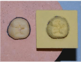
Figure 1: One lower molar and its corresponding cast.
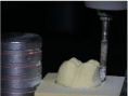
Figure 2: jdod-v4-id1087-g002
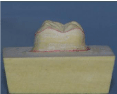
Figure 3: Occlusal line angle and finish line, after preparation.
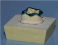
Figure 4: Template on cast.
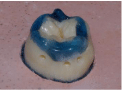
Figure 5: Template on tooth.

Figure 6: Guided preparation.
2. The same procedure can be followed if the cast is milled freehand. (Please see the Figures below for the 3rd left lower molar.) Once the cervical and occlusal lines of the freehand-milled diagnostic cast are marked the remaining occlusal surface is insulated with water-based gel and the resin template is prepared over it (Figure 7 and 8). The template is then fixed with bonding to the tooth (Figure 9), and the tooth is milled to bring the bur into contact with the two lines: the finish line and the margin of the template (Figure 10).
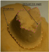
Figure 7: Prepared axial walls.
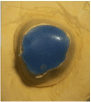
Figure 8: rResin template on cast, occlusal view.
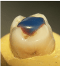
Figure 9: Resin template placed on tooth, distal view.
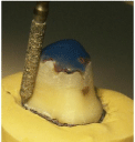
Figure 10: Bur guided by the template.
Clinical case
The author developed the technique in February 2008, so much experience has been accumulated; many dentists in Romania attended the hands-on courses that were held in those 8 years. The case described below was one of the most difficult because of the malposition of the teeth.
Although strongly exhorted, the 23 year old female patient refused an orthodontic treatment and asked for a prosthodontic approach. (Figure 11). Initial impression was taken and the cast was placed on the parallelometer table, after the individual dies were separated (Figure 12). Dies were milled one by one, using a 016 torpedo bur (Figure 13). Then templates were made on the remaining incisal and palatal surfaces. The templates were fixed intraorally (Figure 14) and the preparation of the teeth was carried on, using a 016 torpedo bur (Figure 15). The final outcome, after peroidontal plastic surgery and consecutive reshaping of the labial finish line, is shown in Figure 16
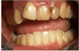
Figure 11: Before treatment. The patient refused any orthodontic intervention.
Figure 12: Separated dies ready to be milled.
Figure 13: Parallelometer milling of individual dies.

Figure 14: Templates bonded to corresponding teeth.

Figure 15: The four abutments after preparation.
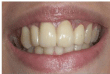
Figure 16: Final aspect.
Discussion
Sequential preparation: The method described offers the possibility to prepare the teeth sequentially even for a bridge, applying the temporary crown immediately. The patient will have the abutments prepared one by one, the length of each appointment being reduced. Because of the templates, there is no need to remove the temporary crown from one abutment when preparing another tooth, because the template gives the guarantee of parallel preparation. When all abutments have been prepared, the temporary crowns are removed and impression is taken.
Flat occlusal surface: Experience showed that flat occlusal surface makes uncertain the positioning of the templates intra-orally. This problem is solved by digging locating grooves on these surfaces before initial impression.
Step by step: With the experience gained in eight years, on both model experiments and clinical cases the following steps are recommended to be taken in order to benefit most from this preparation technique:
Clinical
- Preparation of locating grooves on selected teeth
- Retraction cord packed in the sulcus
- Precise impression in silicones
- Impression of the opposing teeth and bite registration
- Development of the cast and sectioning of individual dies. Model placed in articulator.
- Parallelometer preparation of the diagnostic cast using the same type of bur as the one that will be used intraorally
- Resin templates made on the milled dies
- Resin templates laid aside; occlusal reduction carried on in relation to the antagonists, then making the height rings
- Depth marking of the buccal/labial surface
- Resin templates fixed intra-orally
- Zero degrees preparation of abutments, followed by occlusal
- Reduction according to the height rings.
- Slight inclination of proximal surfaces according to clinical judgment;
- Further buccal/labial reduction according to the depth markings.
- Preparation of locating grooves on selected teeth
- Retraction cord packed in the sulcus
- Precise impression in silicones
- Development of the cast and sectioning of individual dies.
- Freehand preparation of the diagnostic cast using the same type of buras the one that will be used intra-orally
- Resin templates made on the milled dies
- Resin templates fixed intra-orally
- Preparation of the abutments, followed by occlusal reduction according to clinical judgment; further labial/buccal reduction.
Lab
Clinical
The simplified technique would be as follows:
Clinical
TECHNICAL (Steps 4 to 6 can be taken by dentists, without involving the lab)
Conclusion
The great disadvantage of this technique is that it takes extra time to produce the templates; that is the reason why some of our colleagues have decided to use the method in the simplified way described above, so they:
Do a freehand cast milling themselves (it is much easier and more precise than the preparation done intra-orally and in case of mistake it can be repeated by developing another cast in the same impression)
Do the templates themselves there are at least four advantages when using this technique.
- It is especially useful in difficult cases, as those mentioned in the introduction (where teeth are malpositioned, rotated etc.; multiple teeth must be prepared; there is a big distance between the abutments; or when milling molars, especially on the distal surface).
- It may be a good solution for young, inexperienced doctors; students are especially happy as they can preview the result before even touching the teeth.
- It increases the precision of the preparation, easing the task of the dental technician and ensuring better fixation of the restoration.
- It decreases the length of the appointments, because:
- There is no need to prepare all abutments in the same appointment
- The amount of time needed for the milling of an abutment is far shorter, as there is no need to check for the geometry of the preparation, provided that the bur vector is respected.
References
- Johnson JF. Preparation of mouths for fixed and removable partial dentures. J Prosthet Dent. 1961; 11: 456-462.
- Tjan AHL, Miller GD. Biogeometric guide to groove placement on threequarter crown preparations. J Prosthet Dent. 1979; 42: 405-410.
- Rudd RW, Bange AA, Rudd KD, Montalvo R. Preparing teeth to receive a removable partial denture. J Prosthet Dent. 1999; 82: 536-549.
- Gold HO. Instrumentation for solving abutment parallelism problems in fixed prosthodontics. J Prosthet Dent. 1985; 53: 172-179.
- Walton CB. Operative and prosthetic paralleling instrument. Application December 30, 1957, Serial No. 705,984. Patented Nov. 3, 1959, US2910773.
- Stefanoff BT, inventor. Parallel wall indicator. 33/1 N; 33/174 D; 33/349.
- Fridge DS. Dental paralleling handpiece. US Patent 3346959. 1967.
- Brown H. Alignment of abutments for fixed partial dentures. J Prosthet Dent. 1962; 12: 940-946.
- Bacelar de Rezenda A. A new parallelometer. J Prosthet Dent. 1969; 21: 79-85.
- Vitsentzos SI. A new device to directly examine parallelism of abutment teeth. J Prosthet Dent. 1989; 61: 531-534.
- Todorov GR. Preparation of abutment teeth with a specialized clinical parallelometer. Folia Med (Plovdiv) 2001; 43: 69-72.
- Nishida M, Sohmura T, Takahashi J. Training in tooth preparation utilizing a support system. J Oral Rehabil. 2004; 31: 149-154.
- Loft M. Dental inspection mirror. US Patent App. 10/944,490, 2004.
- Robert W, inventor. Dental mirror and method of using same. Int. a.5A61B 1/24, U.S. Cl 433/30, U.S. Patent Oct. 1, 1991, sheet 1 of 2, 5,052,925 sheet 2 of 2, 5,052,925.
- Möllersten L. Clinical precision of paralleling instruments. Eur J Oral Sci. 1989; 97: 66-75.
- Möllersten L. Comparison between guided and freehand preparation. J Prosthet Dent. 1989; 62: 130-139.
- Waghorn S, Kuzmanovic DV. Technique for preparation of parallel guiding planes for removable partial dentures. J Prosthet Dent. 2004; 92: 200-201.
- Canning T, O’Sullivan M. Acrylic resin jigs as an aid to parallel guiding plane preparation. J Prosthet Dent. 2011; 99: 162–164.
- Smarandescu D. Template Guided Vector: a high-precision abutment preparation technique, Poster presentation at FDI Congress 2008, Stockholm.
- Papaspyridakos P, Lal K. Use of vacuum-formed templates to guide tooth preparation and insertion of interim restorations. J Prosthodont. 2010; 19: 303-306.