Clinical Image
J Dent & Oral Disord.2018; 4(2): 1090.
Oral Tuberculosis Mimicking Denture Sore
Lee ST, Jang SB and Choi SY*
Department of Oral & Maxillofacial Surgery, School of Dentistry, Kyungpook National University, Daegu, South Korea
*Corresponding author: So-Young Choi, Department of Oral and Maxillofacial Surgery, School of Dentistry, Kyungpook National University, Republic of Korea
Received: January 11, 2018; Accepted: February 15, 2018; Published: February 23, 2018
Clinical Image
A 66 -year -old male patient visited our department due to unhealed ulceration in the upper lip mucosal area. The patient was wearing a full denture in the maxilla for 10 years and the maxillary anterior vestibule area was inflamed 5 months before. The patient had no fever and difficulty in swallowing. And, he was taking cholesterol drugs and gastrointestinal drugs. The patient was admitted to the department of otorhinolaryngology, and prescribed medication for inflammation, but the symptoms did not improve and he visited the department of oral and maxillofacial surgery for accurate diagnosis.
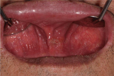
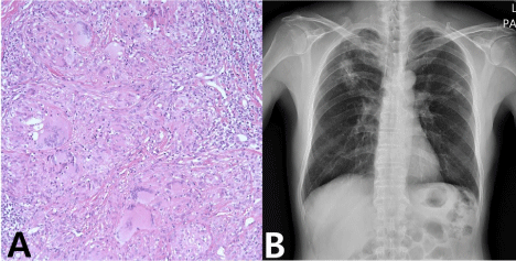
On the right side of the patient’s maxillary anterior vestibule, an ulcerative lesion with a well-defined border of 1 cm in diameter was observed (Figure 1). For accurate diagnosis, incisional biopsy was performed. Histopathological examination revealed granulomatous inflammation of tuberculosis in the lesion, the granuloma formation with multinucleate giant cell, epithelioid histiocytes and lymphocytes. (hematoxylin and eosin staining, original magnification ×200) Based on the results of clinical and histopathological findings, chest x-rays were obtained (Figure 2). Chest x-ray examination revealed consolidation and cavitation of the upper lobes, consistent with the pulmonary tuberculosis. The patient was admitted to the respiratory medicine clinic and confirmed for tuberculosis.

Figure 1: Well-defined ulacerative lesion in oral cavity.
