
Case Report
J Dent & Oral Disord. 2018; 4(3): 1092.
A Dermoid Cystic Lesion on the Forehead- A Case Report
Surabhi V*, Shetty N, Vachhani D, Bangera D, Athiramol CK and Boricha V
Department of Oral & Maxillo-Facial Surgery, A.J. Institute of Dental Sciences, Kuntikana, Mangaluru, Karnataka, India
*Corresponding author: Surabhi V, Department of Oral & Maxillo-Facial Surgery, AJ Institute of Dental Sciences Kuntikana, Karnataka, India
Received: February 19, 2018; Accepted: March 14, 2018; Published: March 21, 2018
Abstract
The epidermoid cyst is a development cyst, filled with keratin and entrapped by stratified squamous epithelium. Epidermoid cyst is histological a benign lesion that usually affects the scalp regions, face, neck and back. They are usually asymptomatic and its etiology is mostly related to trauma, as well as the entrapment of epithelial rests during embryonic fusion. This study aims to present a case report of a 13-year-old patient, with an epidermal cyst in the anterior fontanelle region.
Keywords: Epidermoid cyst; Fore head; Ultrasonography
Introduction
Epidermoid cysts are non-odontogenic cysts which is benign in nature. These rare lesions are derived from germinal epithelium and can be seen throughout the body, particularly in sites where embryonic elements fuse together. Mostly occurring in the ovaries and testicles, 7% occurring in the oro-facial region and 1.6% in the oral cavity, and represents 0.01% of all cysts in the oral cavity. Male to female ratio is 3:1 [1,2]. It is commonly asymptomatic, however, it may become symptomatic due to secondary infection. They contain a central core of keratin proteins, desquamating cells and cholesterol, and lined with stratified squamous epithelium. Epidermoid cysts commonly spread along pathways of least resistance such as natural cleavage planes and anatomic canals, extend into more than one cranial fossa and envelop the neural and vascular structures [3].
Case Presentation
A medically fit, 13-year-old patient came to our department seeking help for an asymptomatic mass over the fore head, which was present since childhood, and had increased in size over the years. Patient complained of unaesthetic appearance of the face because of the lump. There was no family history either. On detailed examination, the lesion was single, well-defined seen in the forehead at the midline, measuring about 2x2 cm in its greatest diameter. Skin over the swelling was smooth and of the normal color without any secondary changes. It was firm in consistency, slightly compressible, non-tender without local rise in temperature. No dental abnormality was detected. Based on the clinical examination a provisional diagnosis of dermoid cyst was given. Epidermoid cyst, Lipoma, and sebaceous cyst were considered under differential diagnosis of this lesion. Ultra sound of the lesion was carried out which revealed well defined heterogeneously hyperechoic lesion measuring approximately 25x8x27 mm in the subcutaneous plane of the forehead which was seen extending intracranially through defect in adjacent frontal bone measuring 4mm. Features were suggestive of epidermoid cyst.
Therefore, based on the clinical information and laboratory test, it was decided to perform an excisional biopsy of the lesion. The excision of the lesion was performed under local anesthesia. Ring block local infiltration was given around the lesion, patient was painted and drapped aseptically. Horizontal incision was given over the lesion, blunt dissection was done and cyst was completely excised and was sent for histopathological examination which revealed it to be an epidermoid cyst. There were no surgical complications and the patient recovered without complaints and was very happy with aesthetical results.
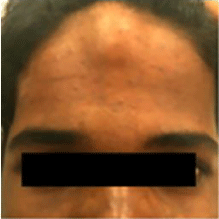
Figure 1: Showing Cystic lesion over the forehead.
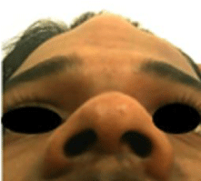
Figure 2: Showing Cystic lesion over the forehead.
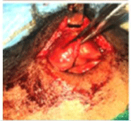
Figure 3: Intra operative excision of the lesion.
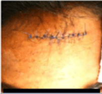
Figure 4: Post operative lesion after suturing.
Discussion
Roser, in 1859 first described epidermoid cyst [3]. They are the developmental lesions that result from embryologic displacement of ectoderm into the meninges, ventricles, or rarely into the parenchyma of the brain [2]. Epidermoid cysts are usually reported in the face, trunk, neck, extremities and scalp, intra-orally- floor of the mouth is the most common site. Buccal mucosa, tongue, lips, uvula and intraosseous location within the mandible and maxilla are also reported. Based on the pathogenesis, epidermoid cysts are classified into congenital and acquired. Congenital cysts are dysembryogenic lesions which arise from ectodermal elements trapped in between, at the time of midline fusion, of the first and second branchial arches in the third and fourth week of the intrauterine life. They may also arise from tuberculumimpar of His. Acquired cyst are derived from traumatic or iatrogenic inclusion of epithelial cells or from occlusion of sebaceous gland duct. It was first noticed by Werhner in 1855 and was referred to as “Implantation cyst” by Sutton in the year 1895 [4-6]. The two theories for epidermoid cyst formation are firstly, occurrence of the cyst when two epidermal surfaces fuse together during early intrauterine life and ectodermal implant is retained deep inside the surface. Secondly, traumatic entrapment of surface epithelium in the connective tissue; later these cells may differentiate to form cyst [1,6]. Fine Needle Aspiration Cytology (FNAC), Ultrasound, CT and MRI provides required information on the cyst location that allows optimal treatment planning [7]. US is the first choice of specialized investigation because of its feasibility, low cost and efficacy. And this modality was used in our case. Some authors consider MRI superior to other methods, as it shows the exact position, extension and demarcation of cysts [8]. Epidermoid cysts are insensitive to radiation or chemotherapy the treatment is surgical excision, which depends on the location, size, nearby structures and symptoms associated. Aim is to completely remove the lesion without damage to adjacent neurovascular bundles. Few doctors advocate radical excision of the lesion to prevent recurrence, and others advocate conservative approach to minimize morbidity and mortality [3]. Care should be taken not to rupture the cyst, as cystic contents will act as irritants to fibrovascular tissues, causing postoperative inflammation [7]. The epidermoid cyst rarely discloses malignancy [9]. The occurrence of Basal cell carcinoma, and Squamous cell carcinoma has been reported in the literature that had evolved from epidermoid cyst [1].
Conclusion
Thorough understanding about this slow growing swelling is very important not only because of the aesthethic concern but also because of its malignant potential.
References
- Rastogi V, Puri N, Kaur G, Yadav L, Sharma R. Unusual Cases of Epidermoid cyst: Case Series. International Journal of Scientific Study. 2013; 1: 72-76.
- Cambruzzi E, Presa K, Silveira LC, Perondi GE. Epidermoid cyst of the posterior fossa: a case report. J Bras Patol Med Lab. 2011; 47: 79-82.
- Giustino FD, Pecci R, Giannoni B, Vannucchi P. Cerebellopontine Angle Epidermoid Cyst: Case Report. International Journal of Otolaryngology and Head & Neck Surgery. 2013; 2: 5-7.
- Walstad WR, Solomon JM, Schow SR, Ochs MW. Midline cystic lesions of the floor of the mouth. J Oral MaxillofacSurg. 1998; 56: 70-74.
- De Ponte FS, Brunelli A, Marchetti E, Bottini DJ. Sublingual epidermoid cyst. J CraniofacSurg. 2002; 13: 308-310.
- Noffke CE. Implantation-type epidermoid cyst of the mandible. Dentomaxillofac Radiol. 1999; 28: 383-385.
- Patil K, Mahima VG, Malleshi SN. Sublingual epidermoid cyst: a case report. Cases J. 2009; 2: 8848.
- Elias LS, Oton-Leite AF, Silva CM, Ribeiro-Rotta RF, Batista AC, Mendonça EF. Epidermoid cyst: highlights on diagnosis and magnetic resonance imaging features. Rev odontociênc. 2010; 25: 204-207.
- Ozan F, Polat HB, Ay S, Goze F. Epidermoid cyst of the buccal mucosa: A case report. J Contemp, Dent Pract. 2007; 8: 90-96.