
Case Report
J Dent & Oral Disord. 2018; 4(4): 1096.
Non-Syndromic Bilateral Impacted Supernumerary Premolar Patient with Associated Oresidual Cyst: A Case Report and Review of Literature
Bharti HV¹, Bharti C²*, Ratre RK³ and Singh B4
¹Private Practitioner, Dr. Bharti’s Multispecialty Clinic, Bhopal, India
²Department of Orthodontics, People’s College of Dental Sciences & Research Center, Bhopal, India
³Department of Orthodontics, Government Dental College, Indore, India
4Private Practitioner, The Dentist Ayodhya Bypass, Bhopal, India
*Corresponding author: Chandni Bharti, Department of Orthodontics, People’s College of Dental Sciences & Research Center, Bhopal, India
Received: March 15, 2018; Accepted: April 05, 2018; Published: April 12, 2018
Abstract
A radicular cysts are inflammatory cysts at the apices of teeth with necrotic pulp. A rare case report is presented of an individual with a radicular cyst involving treated mandibular molar along with impacted bilateral supernumerary premolars.
Keywords: Radicular cyst; Impaction; Supernumerary premolars
Introduction
Radicular cyst being a true cyst is a lesion found in association with non-vital or deep carious lesion or restoration consisting of pathologic fluid filled epithelium lined cavity. Supernumerary teeth are those teeth in addition to the normal series of deciduous or permanent dentition, occurring anywhere in the mouth, having a prevalence of 0.1% and 3.8% [1], occurring 3 times more in males than in females indicating a possible sex linked inheritance [2].The prevalence of supernumerary premolars is between 0.075-0.26%, [3] and may occur single or multiple, mostly remaining impacted, unerupted, and usually asymptomatic. This article reports the concomitant presence of bilateral superrnumery premolar in the lower arch along with radicular cyst with the endodontically treated.
Case Presentation
A 24 year old male patient reported to a private clinic with the chief complaint of irregularly placed upper and lower front teeth. Family, medical was non-contributory. Patient has a history of undergoing root canal treatment 1 year back. Extra oral examination shows no gross asymmetry, straight profile, competent lips, and straight divergence (Figure 1). Intra-oral examination reveals crowding in the upper and lower arches with Class II (end-on) molar and class I incisor relationship (Figure 2).
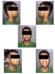
Figure 1: Pre-treatment extra-oral photographs.
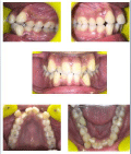
Figure 2: Pre-treatment intra-oral photographs.
As the patient was willing for orthodontic treatment, so diagnostic records of the patient was obtained like lateral cephalogram and orthopantomogram. On exta-oral examination no abnormality was observed. Patient showed no gross asymmetry of the face, straight profile with average growth pattern, competent lips.
Intra-oral examination, upper and lower anterior crowding with Class I molar relationship, restoration wrt 36 5 years back.
Cone beam examination, revealed that radiolucent lesion extended from distal root of the lower left first molar along with resorption of root. The radiolucent lesion was surrounded by welldefined radiopaque margin running down to involve the pericoronal radiolucency of the left supernumerary premolar. On the right side, supernumerary premolars were placed near the apex of the first molar and second premolar with its follicle almost in approximation with the root apices of the first molar (Figure 3).
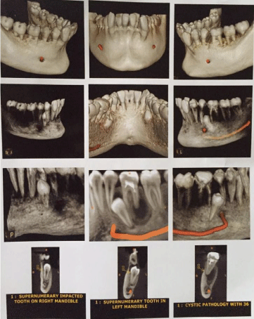
Figure 3: CB CT images.
Discussion
Radicular and residual cysts are the most common cystic lesions of the jaws. The frequency of radicular cysts in permanent dentition is about 7-54 %, while in primary dentition is approximately 0.5-3.3 % of the total number of radicular cysts in both the primary and permanent dentition. [4], radicular cysts originate from epithelial remnants of the periodontal ligament as a result of inflammation and associated infiltration of inflammatory cells which is generally a consequence of pulp necerosis [5]. These cysts commonly involve the apex of the affected tooth. The radicular cyst is generally cyst which arises from epithelial residues (cell rests of Malassez) in the periodontal ligament as a result of inflammation, usually following the death of the dental pulp found at the apices of the involved teeth. The radicular cyst arises from epithelial remnants stimulated to proliferate by an inflammatory process originating from pulpal necrosis of a non-vital tooth. Radio graphically; the classical description of the lesion is a round or oval, well circumscribed radiolucent image involving the apex of the tooth. Over the years, the cyst may regress, remain static or grow in size. The treatment of the cysts can be either non-surgical management or surgical management being either marsupialization or enucleation. The choice of the treatment may be determined by factors such as the extension of the lesion, relation with noble structures, origin, and clinical characteristics of the lesion, and co-operation and systemic condition of the patient [6].
Cases involving one or two supernumerary teeth are most commonly involving the anterior maxilla, followed by mandibular premolar region. When multiple supernumerary teeth are present (>5), the most common site is mandibular premolar region [7]. Cases having bilateral supplemental premolar teeth developing later than their counter parts have been reported in literature, having the highest frequency of occurrence in the mandibular arch(7%) [8,9]. Supernumerary premolars account between 8 % and 9.1 % of all supernumerary teeth. The various possible mechanism of development theories include phylogenetic theory of atavism, dichotomy theory, heredity, hyperactivity of dental lamina, and aberrations during embryologic formation, progress zone; usually associated with Gardener’s syndrome, Cleidocranial dysostosis, cleft lip and palate [10]. Although mandibular supernumerary premolars are common, their bilateral occurrence and its association with clinical complications are uncommon.
Because large percentage of supernumerary premolar remain impacted, unerupted, and are usually asymptomatic and most cases are diagnosed during inspection of radiograph prior to commencement of orthodontic treatment [11].
Dentigerous cyst and root resorption have been cited as frequent complication associated with supernumerary premolar.
In planning the treatment alternatives for the impacted supernumerary premolars, the potential risk of leaving them insitu and hazards of surgical removal of these teeth especially around the lower premolar region, where the teeth are close proximity to the inferior alveolar nerve and blood vessels should be assessed judiciously [3]. The timing of surgical removal of supernumerary premolars is as much debated among clinicians as are the treatment methods. Whenever these teeth are associated with any pathological formation or when hinder eruption of or give rise to malpositioning of permanent teeth, they should be removed as soon as possible [3,12]. Periodic follow up of such patients is extremely important.
Conclusion
An unusual case of a radicular cyst was noted with impacted supernumeri premolars were noted bilaterally. The impacted supernumerii premolars having an unknown etiology while chronic lesion lead to radicular cyst development. Bilateral occurrence of mandibular supernumerary premolar is unusual. Impacted supernumerary premolars should be carefully removed to avoid complications of damage to the mental nerve and vessels with appropriate follow up.
References
- Parolina A, Kundabala M, Dahal M, Mohan M, Thomas MS. Management of supernumerary teeth. J Conserv Dent. 2011; 14: 221-224.
- Saini T, Keene JJ Jr, Whetten J. Radiographic diagnosis of supernumerary premolars: Case Reviews. J Dentistry for Childrem. 2002: 69: 184-190
- Kaya GS, Yapici G, Omezli MM, Dayi E. Non-syndromic supernumerary premolars. Med Oral Pathol Oral Cir Bucal. 2011; 16: e522-525.
- Mass E, Kaplan I, Hirshberg HIrshberg A. A clinical and histopathological study of radicular cysts associated with preimary molars. J Oral Pathol Med. 1995; 24; 458-461.
- Ramakrishna Y, Verma D. Radicular cyst associated with a deciduous molar: A casereport with unusual clinical presentation. J Indian Soc Pedod Prev Dent. 2006 ; 24: 158-160.
- Caroline RA, Valois Edson DC. Periapical cyst repair after non-surgical endodontic therapy: Case report. Branz Dent J. 2005; 16: 254-258.
- Scheiner MA, Sampson WJ. Supernumerary teeth: A review of the literature and four case reports. Aust Dent J. 1997; 42: 160-165.
- Bradwaj VK, Kaundal JM,Chug A,Vaid S, Soni A, Chandel M. Rare occurrence of bilaterally impacted mandibular supernumerary teeth. Dent Hypothesis. 2012; 3: 83-85.
- Gibson N. A late developing mandibular premolars supernumerary tooth. Aust Dent J. 2001; 46: 51-52.
- Yusof Wz. Non-syndrome multiple supernumerary teeth: Literature review. J Can Dent Assoc. 1990; 56: 147-149.
- Solarez R, Romero MI. Supernumerary premolars : A literature review. Pediatr Dent. 2004; 26: 450-458.
- Kasat VO, Saluja H, Kalburge JV, Kini Y, Nikam A, Laddha R. Multiple bilateral supernumerary premolar in non-syndromic patient with associated orthokeratininsed odontogenic cyst: A case report and review of literature. Contemp Clin Dent. 2012; 3: 248-52.