Special Article - Pediatric Dentistry
J Dent & Oral Disord. 2018; 4(4): 1099.
Ameloblastic Fibro-Odontoma: A Diagnostic Dilemma
Dangore Khasbage S*
Department of Oral Medicine and Radiology, Datta Meghe Institute of Medical Sciences, Maharashtra, India
*Corresponding author: S. Dangore Khasbage, Department of Oral Medicine and Radiology, Datta Meghe Institute of Medical Sciences, Maharashtra, India
Received: May 28, 2018; Accepted: June 07, 2018; Published: June 15, 2018
Clinical image
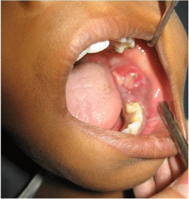
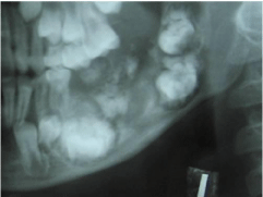
A 11 year-old boy presented with 5 months history of a slowly enlarging, painless swelling on the left side of mandible. His medical, family, and social history were unremarkable and so were the results of the physical examination. There was no history of trauma. Oral examination revealed a large lobulated lesion extending from deciduous first molar to the retromolar region. Second deciduous molar and the first permanent molar were clinically missing. The swelling measured about 4× 2 cm; there was obliteration of the labial and buccal vestibule. The lesion was reddish, non-tender, and soft to firm in consistency. Ameloblastic fibroma was considered as the provisional diagnosis and odontoameloblastoma, and ameloblastic fibroodontoma in differential diagnosis (Figure 1). Radiographic examination was performed as a first option in investigation (Figure 2).

Figure 1: Ameloblastic fibroma.

Figure 2: Radiographic examination.
A panoramic radiograph revealed a large mixed lesion, with the displacement of teeth towards the periphery of the lesion. The lesion was well circumscribed except along the posterior aspect, where the margin was irregular and ill defined. The differential diagnosis included ameloblastic fibroodontoma and immature complex odontoma, The presence of radioopacities in the lesion ruled out ameloblastic fibroma. On the other hand, the presence of radiooapcities characterizes another entity, ameloblastic fibroodontoma. Odontoameloblastoma usually is an unencapsulated lesion, whose behavior resembles the ameloblastoma.
In the present case, the histopathological examination with hematoxylin and eosin staining reported as a cellular lesion composed of immature mesodermal component along with islands of odontogenic epithelium and areas of dentin. No evidence of malignancy, such as nuclear pleomorphism, was found. The case was finally confirmed as ameloblastic fibro-odontoma. The lesion was excised intraorally under general anesthesia.
References
- Hegde V, Hemavathy S. A massive ameloblastic fibro-odontoma of the maxilla. Indian J Dent Res 2008; 19: 162-164
- Sivapathasundharam B, Manikandan R, Sivakumar G, Goerge T. Ameloblastic fibro-odontoma. Indian J Dent Res 2005; 16: 19-21.
- Silva GC, Jham BC, Silva EC, Horta MC, Godinho SH, Gomez RS. Ameloblastic fibro-odontoma. Oral Oncol Extra 2006; 42: 217-220.