
Case Report
J Dent & Oral Disord. 2018; 4(5): 1102.
Surgical Orthodontic Treatment of Acquired Openbite Attributed to Bilateral Idiopathic Condylar Resorption of the Temporomandibular Joints
Kajii TS¹*, Takata S¹ and Izumi K²
¹Department of Oral Growth & Development, Fukuoka Dental College, Japan
²Department of Oral & Maxillofacial Surgery, Fukuoka Dental College, Japan
*Corresponding author: Takashi S. Kajii, Section of Orthodontics, Department of Oral Growth & Development, Division of Clinical Dentistry, Fukuoka Dental College, Japan
Received: July 30, 2018; Accepted: August 29, 2018; Published: September 05, 2018
Abstract
Purpose: To prevent significant relapse after orthognathic surgery for acquired openbite with bilaterally Idiopathic Condylar Resorption (ICR) of the temporomandibular joint, it is postulated that the gonial region of the distal segment of the mandible should not be positioned downward by orthognathic surgery.
Clinical Presentation: The 27-year-old Japanese female presented with acquired openbite attributed to bilateral ICR. Orthodontic treatment combined with two-jaw surgery was planned for the patient.
Intervention: Le Fort I osteotomy with upward position of posterior aspect of the maxilla and Bilateral Sagittal Split Osteotomy (BSSO) without downward position of the gonial region of the mandibular distal segment were performed. The positions of both segments of the mandible were maintained at 1 year after orthognathic surgery.
Conclusion: For acquired openbite patients, the present report suggests that BSSO positioning the gonial region of the distal segment of the mandible upward may be a stable procedure to prevent significant relapse.
Keywords: Orthodontic treatment combined with surgery; Orthognathic surgery; Temporomandibular joint; Idiopathic condylar resorption; Backward rotation of the mandibular ramus; Digastric and mylohyoid muscles
Abbreviations
DC/TMD: Diagnostic Criteria/Temporomandibular Disorders; ICR: Idiopathic Condylar Resorption; TMJ: Temporomandibular Joint; AICR: Adolescent Internal Condylar Resorption; BSSO: Bilateral Sagittal Split Osteotomy
Introduction
In the diagnostic criteria for temporomandibular disorders (DC/TMD), idiopathic condylar resorption (ICR) [1-3] and degenerative joint disease (osteoarthrosis and osteoarthritis) are included among the joint diseases of TMD [4]. Wolford, et al. [5- 8] reported that mandibular condyle resorption is likely caused by temporomandibular joint (TMJ) pathologies such as adolescent internal condylar eesorption (AICR, formerly called ICR), reactive arthritis, and connective tissue/autoimmune diseases. In AICR [6], it is postulated that the articular disc becomes displaced anteriorly, and the condyle then is surrounded by the hyperplastic synovial tissue that continues to release chemical substrates [1,2,9] which penetrate the condylar head, causing condylar resorption.
Kajii, et al. [10] reported that Angle Class II patients with bilateral ICR show shorter condylar height attributable to resorption or osseous changes of the TMJ condyle. The shorter condylar height may affect subsequent backward (clockwise) rotation of the mandibular ramus, because the muscles attached to the ramus and other soft tissues might retract the ramus upward [5] and forward [11] and the digastric and mylohyoid muscles could retract the mandibular body backward and downward [5,8,12] in the patients with short ramus height. The subsequent backward rotation of the ramus causes “acquired openbite” [13]. Not only orthodontic treatment but also orthognathic surgery is frequently necessary for treating the acquired openbite (Figure 1).
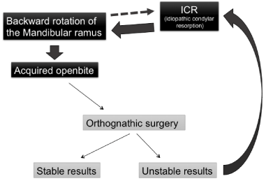
Figure 1: Cascade of events causing acquired openbite.
After orthognathic surgery such as sagittal splitting ramus osteotomy for acquired openbite, Aymach and Kawamura [11] suggested that the sphenomandibular and stylomandibular ligaments attached to the proximal (condylar) segment of the mandible produce an upward directed force on the proximal segment. Hwang, et al. [12] suggested that the digastric and mylohyoid muscles produce a backward and downward directed force on the distal segment of the mandible. In consequence, the force on the distal segment may cause the proximal segment of the mandible to be pushed upward, and the superior-anterior surface of the condyle may be compressed into the glenoid fossa, and significant relapse (this is also acquired openbite due to ICR) sometimes occurs [12,14,15]. It is therefore postulated that the gonial region of the distal segment of the mandible should not be positioned downward by orthognathic surgery such as sagittal split ramus osteotomy for acquired openbite with bilateral ICR [10].
Based on the postulation, here we show orthodontic treatment combined with two-jaw surgery for a patient with acquired openbite attributed to bilateral ICR of the TMJ.
Materials and Methods
History and diagnosis
The 27-year-9-month-old Japanese female presented with retruded mandible and openbite. Her chief complaint was an esthetic problem due to maxillary protrusion, disturbance of mastication and lip closure, and masticatory muscle pain. Before 20 years of age, she had no openbite and no pain around TMJ although she had clicking in the TMJs. However, after 20 years of age, she had been conscious of her pain of right TMJ and subsequent openbite. During the past several years, the openbite and bilateral TMJ pain were slightly increased and she had been conscious of her pain of bilateral masticatory muscle. She had no rheumatism or any other significant medical history.
She had a symmetrical face and a convex profile with retrognathic mandible (Figure 2A). Overjet and overbite were 9.5 mm and -1.5 mm, respectively. The molar relationship was Angle class II on both sides. Significant facets were found on the occlusal surface of anterior teeth and premolars although only molars contacted at centric occlusion. The maxillary and mandibular dental midlines were almost coincident with the facial midline. Mild crowding of the maxillary and mandibular incisors was accompanied (Figure 3A). Maximum mouth opening was 38 mm.
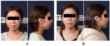
    Figure 2:  A: Pretreatment (27 years 9 months), B: posttreatment (30 years 2
months) facial photographs.
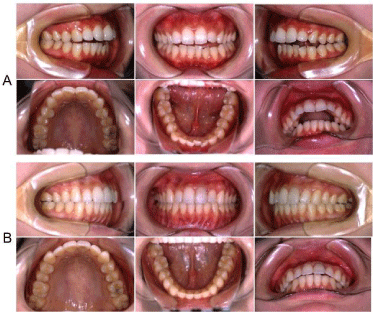
    Figure 3:  A: Pretreatment (27 years 9 months), B: posttreatment (30 years 2
months) intraoral photographs.
Panoramic radiograph and lateral TMJ panoramic radiograph revealed severe resorption and deformation in both condyles of the TMJ with slightly limited mouth opening in both TMJs (Figures 4A,5A), so that magnetic resonance imaging was taken. The image confirmed deformation in both condyles and revealed anterior disc displacement without reduction with slightly limited opening (Figure 6). Lateral cephalometric analysis showed a skeletal Class II type with extremely backward rotation of the mandibular ramus, extremely high angle type with short ramus, and lingual inclination of the maxillary incisor (Table 1).
    Pre treatment
    Post treatment
    Norm value


    Age
    27 yrs 9 mos
    30 yrs 2 mos


    SNA (°)
    76.5
    78
    82.1


    SNB (°)
    66.5
    70
    78.5


    ANB (°)
    10
    8
    3.6


    Y-axis (°)
    77.5
    73
    65.2


    Ramus height(mm)
    38
    37
    50.1


    GZN (°)
    108
    98.5
    93


    Gonial angle (°)
    129.5
    139
    121.2


    FMA (°)
    50
    49
    28.6


    FMIA (°)
    34.5
    34
    55.2


    IMPA (°)
    95.5
    97
    96.2


    Occlusal to SN (°)
    37
    32.5
    16.8


    U1 to SN (°)
    93
    89
    103.8


    Interincisol angle (°)
    112
    117
    125.6
Table 1: Cephalometric measurements.
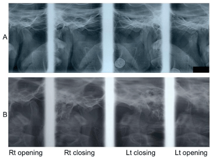
    Figure 5:  A: Pretreatment (27 years 9 months), B: posttreatment (30 years 2
months) lateral TMJ panoramic radiographs. Rt: right; Lt: left; closing: mouth
closing; opening: mouth opening.
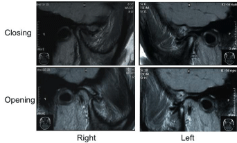
    Figure 6:  Pretreatment (27 years 9 months) magnetic resonance imaging of
the TMJ. Closing: mouth closing; Opening: mouth opening.
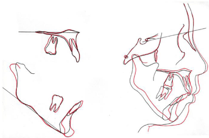
    Figure 7:  Cephalometric superimposition. Black: pretreatment; red:
posttreatment.
This patient was diagnosed as Angle Class II, skeletal Class II and acquired openbite with backward rotation of the mandibular ramus attributed to bilateral ICR of the TMJ.
Treatment plan
The treatment plan for this patient was orthodontic treatment combined with surgery including pre-surgical orthodontic treatment after her pain of bilateral masticatory muscle improved, orthognathic surgery, and post-surgical orthodontic treatment.
The pre-surgical orthodontic treatment plan was a fixed orthodontic treatment without tooth extraction.
The orthognathic surgery plan was two-jaw surgery: (1) Le Fort I osteotomy to move the posterior aspect of the maxilla upward, induce subsequent mandibular auto-rotation, and increase the occlusal plane angle of the maxilla, and (2) bilateral sagittal split osteotomy (BSSO) with advancement of 8.0 mm at pogonion and without downward position of the gonial region of the mandibular distal segment by increasing the occlusal plane angle of the maxilla. Genioplasty for augmenting the prominence of the chin was also included in the orthognathic surgery plan.
Treatment progress
Before starting the pre-surgical orthodontic treatment, the patient took non-steroidal anti-inflammatory drug for 2 months and her pain of bilateral masticatory muscle improved. Then .018 x .025 inch slots are adjusted edge wise braces were bonded on both maxillary and mandibular dentitions at 28 years 3 months of age.
After alignment of teeth and coordination of maxillary and mandibular dental arches were achieved, two-jaw surgery was performed at 29 years 3 months of age. Genioplasty for augmenting the prominence of the chin was also performed at the same time. Then the post-surgical orthodontic treatment started. During surgical orthodontic treatment, her pain of muscle had eased off. An ideal occlusion was obtained without any pain of masticatory muscle and TMJs at 30 years 2 months of age (active treatment time: 1 year 11 months), and all of the appliances were removed (Figures 2B,3B,4B,5B,7). Wrap-around [16] and lingual fixed [16] retainers were set on the maxillary and mandibular arches, respectively.
Results
Results show that treatment based on pretreatment planning was successful. The facial profile of patient was improved (Figure 2B). The acquired openbite was corrected. Solid intercuspation of the teeth was achieved, and a Class I molar relationship was achieved (Figure 3B). Panoramic radiograph and lateral TMJ panoramic radiograph after treatment showed no resorption image at the condyle of the TMJs (Figure 4B). The positions of proximal and distal segments of the mandible were maintained at 1 year after orthognathic surgery (Figure 4B,6).
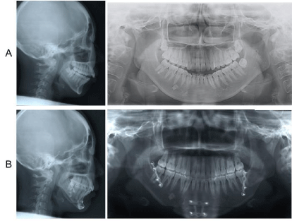
    Figure 4:  A: Pretreatment (27 years 9 months), B: posttreatment (30 years 2
months) cephalograms and panoramic radiographs.
Discussion
In the present acquired openbite case, two reasons why a relapse of acquired openbite did not occur at 1 year after orthognathic surgery were considered to be as follows; the positions of distal and proximal segments of the mandible. We avoided positioning the gonial region of the distal segment of the mandible downward at two-jaw surgery. In cases with excessively counterclockwise rotated mandible by twojaw surgery, a backward and downward directed force produced by the digastric and mylohyoid muscles attached to the distal segment may cause the proximal segment to be pushed upward, and if the gonial region of the distal segment is positioned downward by two-jaw surgery compared with that of pretreatment, the superioranterior surface of the condyle may be compressed into the glenoid fossa, and significant relapse sometimes occurs [12,14,15].
Another reason was considered to be the position of proximal segment of the mandible. We avoided excessively rotating bilateral proximal segments of the mandible counter clock wisely at two-jaw surgery. In cases of maxillary intrusion and mandibular advancement by two-jaw surgery, Hwang, et al. [12] reported that a step is created at the inferior border of the mandibular buccal site because the inferior border of the proximal segment is lower than that of the distal segment. To avoid creating an osseous step at the site, the proximal segment, namely the condylar head, is rotated further in counterclockwise direction. However, this further rotation of the condyle may lead to exposure of the less dense, previously unloaded anterior-superior condylar surface, and the condylar surface may be exposed to compressive loading. Hwang, et al. [12] proposed that the exposure of the less dense anterior-superior condylar surface leads to postoperative condylar resorption. Therefore, a reason for no relapse of acquired openbite of the present case was probably that excessively rotating bilateral proximal segments of the mandible counter clock wisely was avoided.
Wolford and Gonçalves, [7] proposed that maxillo-mandibular surgical advancement by two-jaw surgery, accompanied by counterclockwise rotation of the occlusal plane and by downward potion of the gonion of the distal segment of the mandible, is a stable procedure for patients with healthy TMJs and for patients undergoing simultaneous TMJ articular disc repositioning with the Mitek minianchor. The rotation of the mandible by two-jaw surgery is the opposite direction described above. The present acquired openbite case suggested that the gonion of the distal segment of the mandible should not be positioned downward by BSSO for acquired openbite patients without articular disc repositioning.
Conclusion
For two-jaw surgery of acquired openbite patients with bilateral ICR, the present case report suggests that BSSO positioning the gonial region of the distal segment of the mandible upward may be a stable procedure to prevent significant relapse.
Acknowledgment
The authors would like to thank Dr. Hiroyuki Ishikawa (Fukuoka Gakuen), Dr. Sachio Tamaoki (Section of Orthodontics, Fukuoka Dental College), and Dr. Tetsuro Ikebe (Section of Oral Surgery, Fukuoka Dental College) for their contributions to this case report.
References
- Arnett GW, Milam SB, Gottesman L. Progressive mandibular retrusion - idiopathic condylar resorption. Part I. Am J Orthod Dentofacial Orthop. 1996; 110: 8-15.
- Arnett GW, Milam SB, Gottesman L. Progressive mandibular retrusion - idiopathic condylar resorption. Part II. Am J Orthod Dentofacial Orthop. 1996; 110: 117-127.
- Wolford LM, Cardenas L. Idiopathic condylar resorption: Diagnosis, treatment protocol, and outcomes. Am J Orthod Dentofacial Orthop. 1999; 116: 667- 677.
- Schiffman E, Ohrbach R, Truelove E, Look J, Anderson G, Goulet JP, et al. Diagnostic criteria for Temporo Mandibular Disorders (DC/TMD) for clinical and research applications: Recommendations of the international RDC/TMD consortium network and orofacial pain special interest group. J Oral Facial Pain Headache. 2014; 28: 6-27.
- Gonçalves JR, Cassano DS, Wolford LM, Santos-pinto A, Marquez IM. Postsurgical stability of counterclockwise maxillomandibular advancement surgery: Affect of articular disc repositioning. J Oral Maxillofac Surg. 2008; 66: 724-728.
- Wolford LM, Cassano DS, Gonçalves JR. Common TMJ disorders: Orthodontic and surgical management. In: McNamara JA, Kapila SD, editors. Temporomandibular disorders and orofacial pain: Separating controversy from consensus. Volume 46, Craniofacial growth series. Ann Arbor (MI): The University of Michigan; 2009. p. 159-198.
- Wolford LM, Gonçalves JR. Condylar resorption of the temporomandibular joint: How do we treat it? Oral Maxillofac Surg Clin North Am. 2015; 27: 47-67.
- Al-Moraissi EA, Wolford, LM. Does temporomandibular joint pathology with or without surgical management affect the stability of counterclockwise rotation of the maxillomandibular complex in orthognathic surgery? A systematic review and meta-analysis. J Oral Maxillofac Surg. 2017; 75: 805-821.
- Kajii TS, Okamoto T, Yura S, Mabuchi A, Iida J. Elevated levels of ß-endorphin in temporomandibular joint synovial lavage fluid of patients with closed lock. J Orofac Pain. 2005; 19: 41-46.
- Kajii TS, Fujita T, Sakaguchi Y, Shimada K. Osseous changes of the mandibular condyle affect backward-rotation of the mandibular ramus in Angle Class II orthodontic patients with idiopathic condylar resorption of the temporomandibular joint. Cranio. 2018.
- Aymach Z, Kawamura H. Facilitating ramus lengthening following mandibulardependent surgical closing of a skeletal open bite with short ramus: a new modified technique. J Craniomaxillofac Surg. 2012; 40: 169-172.
- Hwang SJ, Haers PE, Sailer HF. The role of a posteriorly inclined condylar neck in condylar resorption after orthognathic surgery. J Craniomaxillofac Surg. 2000; 28: 85-90.
- Tanaka E, Yamano E, Inubushi T, Kuroda S. Management of acquired open bite associated with temporomandibular joint osteoarthritis using miniscrew anchorage. Korean J Orthod. 2012; 42: 144-154.
- Kobayashi T, Izumi N, Kojima T, Sakagami S, Saito I, Saito C. Progressive condylar resorption after mandibular advancement. Br J Oral Maxillofac Surg. 2012; 50: 176-180.
- Nogami S, Yamauchi K, Satomi N, Yamaguchi Y, Yokota S, Abe Y, et al. Risk factors related to aggressive condylar resorption after orthognathic surgery for females: retrospective study. Cranio. 2017; 35: 250-258.
- Daskalogiannakis J. Glossary of Orthodontic Terms. Berlin: Quintessence Publishing. 2000.