
Research Article
J Dent & Oral Disord. 2020; 6(1): 1121.
A Call for Maxillofacial Prosthodontist-Rehabilitation of Bilateral Total Maxillectomy Defect with Limited Mouth Opening: A Case Report
Jain V and Gupta C*
Department of Prosthodontics, Center for Dental Education & Research, All India Institute of Medical Sciences, India
*Corresponding author: Gupta C, Department of Prosthodontics, Center for Dental Education & Research, All India Institute of Medical Sciences, India
Received: December 09, 2019; Accepted: January 03, 2020; Published: January 10, 2020
Abstract
Maxillofacial defects are highly individualized and require the maxillofacial prosthodontist to think differently to fabricate a good functional prosthesis within economical constraint along with the basic principle of prosthesis making. In the case of bilateral total maxillectomy defects fabrication of well retentive functional prosthesis itself a challenging task for a prosthodontist. Limited mouth opening further makes it complicated. This case report describes a simple technique of fabrication of two-piece hollow obturator prosthesis joined by magnets for rehabilitation of bilateral total maxillectomy patient with limited mouth opening.
Keywords: Bilateral total maxillectomy; Two-piece obturator; Magnets
Introduction
Obturator rehabilitation remains a viable treatment option for most maxillary defects due to low cost, limited morbidity, decreased dental visit, and the ease of prosthesis modification [1]. In bilateral total maxillectomy defects due to large defect and lack of anatomic structures to support and retain the prosthesis, it’s quite difficult to fabricate well retentive & functional obturator prosthesis. Along with this other oral morbidity such as delicate soft tissues, xerostomia, and trismus also hamper the prosthetic outcome [2]. For placement and function of one-piece hollow obturator prosthesis requires an adequate mouth opening, lack of lip contracture, sufficient anterior & posterior soft tissue undercut and good neuromuscular coordination. Bilateral total maxillectomy patients generally lack in one or more factors and need innovative and economical solution for problems [3]. In this case report, we discuss the rehabilitation of a bilateral total maxillectomy patient with restricted mouth opening by making two-piece hollow bulb definitive obturator to ease the placement and assist in constant retention and stability of the prosthesis.
Case Presentation
A 23-year-old male patient reported to the department of maxillofacial prosthodontics with chief complaints of unaesthetic appearance and difficulty in chewing. His past medical history revealed that he had undergone bilateral total maxillectomy due to sinonasal hemangiopericytoma of maxillary sinus in December 2017. The inferior portion of the nasal septum was also removed. No post-surgical radiotherapy was prescribed. After the surgery, interim obturator plate was given to him to avoid nasal regurgitation.
Extraoral examination shows collapsed midface, drooping lip, prognathic appearance, depressed nasal bridge, restricted mouth opening, and disturbing appearance of the face (Figure 1). Intraoral examination shows well-healed large bilateral total maxillectomy defect with sufficient anterior and posterior undercut, remaining portion of nasal septum, inferior nasal conchae, and the openings of both maxillary sinuses (Figure 2). Soft palate was anatomically & functionally normal. In the lower arch there was a full complement of teeth with mild crowding in lower anterior.
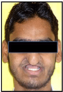
Figure 1: Extra-oral profile.
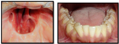
Figure 2: Intraoral image.
Now the treatment objectives were to separate the nasal and oral cavities, restore the mid-facial contour, and improve masticatory functions by providing a full complement of maxillary teeth with keeping in mind the restricted mouth opening. To accomplish these objectives patient had given different treatment options (osteocutaneous fibular free flap reconstruction with implant retained/supported obturator prosthesis or conventional obturator prosthesis) and patient choose conventional obturator prosthesis due to economical constraint and fear of the further surgical procedure. Two-piece magnet retained hollow obturator definitive prosthesis was designed and fabricated for the rehabilitation of this patient. Informed consent was taken before starting the procedure.
Fabrication of hollow obturator part
A preliminary impression of maxillary defect was made with a highviscosity irreversible hydrocolloid (Algiplast, DPI, Mumbai, India), covering all the margins of the defect and blocking all unfavorable undercuts with wet gauge piece. The cast was obtained by pouring it with type III gypsum material (Kalstone, Kalabhai Karlson Pvt. Ltd, India) and trimmed carefully to avoid loss of deliberate posterior and lateral extension. A custom impression tray was fabricated on the obtained cast. The tray was border molded with low fusing impression compound (DPI-Pinnacle; Dental Products of India Ltd) and a physiologic definitive impression was made of the defect using a medium viscosity polyvinyl siloxane impression material (Reprosil; Dentsply, Germany). Hollow bulb definitive obturator plate (using lost salt technique) [4] was made over obtained cast. Obturator was finished & polished and delivered to the patient (Figure 3). The patient was instructed about wearing of the prosthesis as first seat the prosthesis in a posterior undercut, then rotate the prosthesis slightly superiorly in defect area and engage the anterior defect area by pulling the upper lip outward. During removal firstly disengage obturator from anterior defect area then remove the prosthesis in an inferior and downward direction. The patient was also instructed regarding hygiene maintenance of defect and prosthesis both. Patient was instructed to take out the prosthesis before sleeping and storage in water while not in use. Patient was recalled after 24 hours following placement of the definitive obturator plate to assess its function and to examine the defect area for the early signs of irritation. Necessary adjustments were made and patient was scheduled for the recall visits.
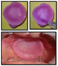
Figure 3: Hollow bulb definitive obturator plate placed in the mouth.
Fabrication of hollow denture part
After 2 weeks when patient had no complaints regarding comfort or tissue soreness with obturator plate, vertical and centric relation records were made. The cast was mounted on Hanau Wide-Vue II articulator and teeth were arranged in centric occlusion. Try-in was done for evaluation of aesthetics and verification of centric and vertical relation. At the same time, palatal contour customized to improve the intelligibility of speech (Figure 4) [5].
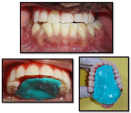
Figure 4: Recorded palatal contour.
To make two-piece obturator prosthesis teeth part of the obturator was separated from definitive obturator plate. To maintain the patency of hollowness of denture part laboratory condensation silicone (Zetalabor Lab Putty C Silicone) placed in hollowed denture part and routine procedure of dewaxing, packing, curing was performed on teeth part of obturator (Figure 5). After curing processed teeth part of the obturator attached with an obturator plate with the help of two magnets, one anteriorly and another posteriorly (Figure 6). The magnets were embedded in the respective contacting surfaces at a depth of 0.5 mm. At last, obturator transferred to previously mounted articulator and necessary adjustments were done in occlusion that occurs due to a processing error.
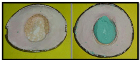
Figure 5: Hollowing of denture part of definitive obturator.
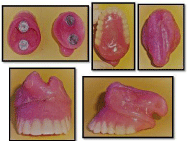
Figure 6: Final prosthesis.
Obturator was finished & polished and delivered to the patient (Figure 6). The patient was again instructed regarding the care of prosthesis and hygiene maintenance as formerly. Patient was also instructed to wear the obturator plate first and once this was comfortably seated, denture part was to be inserted. During removal, the patient was instructed to first remove the denture part gently to disengage magnetic attraction after that obturator part as formerly instructed. Patient was recalled after 24 hours for a follow up. A total of six months of periodic follow up was reported. The patient was quite satisfied with the aesthetic and function of the prosthesis.
Discussion
Two-piece of prosthesis provides several advantages as ease of placement in restricted mouth opening, more efficient use of available soft tissue retentive areas, constant retention and stability to prevent movement of the prosthesis, avoid undue stress on the underlying soft tissues of the defect [6].
A closed field, permanent, rare earth (Nd-Fe-B) commercially available magnet was used for joining of two-piece prosthesis in this case. Each magnet was 1cm in diameter and 1.5mm in thickness. Triple plating of Ni-Cu-Ni present over it which makes it smooth, shiny, non-scratching surface, that is corrosion resistant. (Figure 7). Due to such a small size of the magnet, the obturator was possible to made retentive with reduced weight and due to an automatic reseating property it was easy for the patient to insert two-piece prosthesis in a proper position [7].

Figure 7: Magnet.
There are some disadvantages also of this approach such as additional laboratory steps, need of replacement of magnets due to lack of long-term durability in oral conditions, frequent follow-up appointments.
Conclusion
Maxillofacial defects are highly individual and require the clinician to call upon all his knowledge and experience to fabricate a functional prosthesis. Two-piece obturator joined with magnets is a good treatment alternative to restore bilateral total maxillectomy defects with restricted mouth opening and economical constraint, aesthetically and functionally.
References
- Chigurupati R, Aloor N, Salas R, Schmidt BL. Quality of life after maxillectomy and prosthetic obturator rehabilitation. J Oral Maxillofac Surg Off. 2013; 71: 1471-1478.
- Minsley GE, Warren DW, Hinton V. Physiologic responses to maxillary resection and subsequent obturation. J Prosthet Dent. 1987; 57: 338-344.
- Brown KE. Peripheral consideration in improving obturator retention. J Prosthet Dent. 1968; 20: 176-181.
- Nanda A, Jain V, Nafria A. Light is right-various techniques to fabricate hollow obturators. Cleft Palate-Craniofacial J Off Publ Am Cleft Palate-Craniofacial Assoc. 2013; 50: 237-241.
- Gopi A, Dhiman RK, Kumar D. Customizing the palatal contour of a complete denture using a Palatogram in a case of partial glossectomy. Med J Armed Forces India. 2015; 71: S452-455.
- Wang RR. Sectional prosthesis for total maxillectomy patients: a clinical report. J Prosthet Dent. 1997; 78: 241-244.
- Riley MA, Walmsley AD, Harris IR. Magnets in prosthetic dentistry. J Prosthet Dent. 2001; 86: 137-142.