
Case Report
J Dent & Oral Disord. 2020; 6(4): 1137.
Endodontic Management of Mandibular Canine Having Two Roots: A Rare Presentation
Sapna Y1, Garg M1, Shreya1 and Chauhan R2*
¹Post-Graduate Student, Department of Conservative Dentistry and Endodontics, Saraswati Dental College, Lucknow, Uttar Pradesh, India
²Professor, Department of Conservative Dentistry and Endodontics, Saraswati Dental College, Lucknow, Uttar Pradesh, India
*Corresponding author: Raju Chauhan, Department of Conservative Dentistry and Endodontics, Saraswati Dental College, Lucknow, Uttar Pradesh, India
Received: May 01, 2020; Accepted: May 21, 2020; Published: May 28, 2020
Abstract
As the knowledge of development and natural anatomy of the teeth are applicable to clinical practice, especially during root canal therapy, the consideration of anatomical variations of teeth are equally important to avoid endodontic failure. Mandibular canines are recognized as usually monoradicular i.e. single rooted, in most of the cases although approximately 15% may have two canals. Further the incidences of two rooted canine with two canals is as low as 1.7%. Therefore our case is a rare presentation, which was successfully managed with non-surgical endodontic treatment.
Keywords: Mandibular canine; Anatomical variations; Two roots; Two canals; Endodontics
Introduction
Deep knowledge of mechanical and chemical debridement of the entire root canal, followed by a 3-dimensional obturation and a final coronal restoration to prevent access to microorganisms is the key for successful root canal therapy.
The mandibular canine has its long and stable root, useful for prosthetic support due to its proprioceptive properties that regulate or guide masticatory function, combined with its role in occlusal guidance during the eccentric movements and posterior disocclusion [1]. A proper knowledge of the root canal anatomy is a basic prerequisite for successful endodontic treatment. A careful evaluation of radiographs and the pulp chamber floor can help to detect unusual root canal system morphology and prevent failure of root canal therapy [2].
Generally, mandibular canines contain a single root and canal. The occurrence of two roots and two canals is a rare entity ranging from 1 to 5% [3].
Although the prevalence of two roots and two canals in a mandibular canine is very low, the clinician should be mindful of variations in the number of roots and canals for proper case management. Unpredictable findings in the root canal morphology of mandibular canines have a great impact in endodontic treatment.
Case Presentation
A 45 year-old women with a non-significant medical history reported to the Department of Conservative Dentistry and Endodontics, with the chief complaint of pain in lower right front tooth. Patient gave history of getting root canal treatment done at a private clinic 2-3 years back. Clinical examination revealed generalized severe attrition of her teeth with tenderness to percussion on the mandibular right canine. Intraoral periapical radiographs in different horizontal angulations were taken which revealed improper root canal treatment in tooth #42 & 43 and periapical thickening in tooth #44 (Figure 1). A closer examination of the radiograph revealed the presence of two separate roots in tooth #43, with only one root obturated and a broken instrument in tooth #42. A clinical diagnosis of symptomatic apical periodontitis was made and nonsurgical retreatment was planned in tooth # 42 & 43, while root canal treatment in tooth #44.
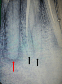
    Figure 1: Pre-operative radiograph showing poorly obturated tooth #43 with
presence of two separate roots (black arrows), broken instrument in tooth #42
(white arrow) and periapical thickening in tooth #44 (red arrow).
Treatment planning discussed with the patient and an informed consent was taken. After administration of local anaesthesia, tooth #43 was accessed under rubber dam isolation. Obturation material was removed from the distal canal and the missed mesial canal was located under dental operating microscope (Carl Zeiss, USA) (Figure 2(a)). Root can patency was achieved with #10 k-file. Root canal length was determined with an electronic Apex Locator (Root ZX, J Morita, Japan) and confirmed radiographically (Figure 2(b)).
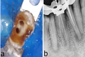
    Figure 2: (a) Access opening in tooth #43 showing two distinct orifice, (b)
Working length radiograph of tooth #43.
Biomechanical preparation was done up to ProTaper file size F2 using 17% EDTA lubrication (Glyde, Dentsply Switzerland) under constant irrigation of 3% NaOCl. Similar protocol was also followed for endodontic treatment of tooth #42 and #44. After taking master cone radiograph (Figure 3(a)) obturation was done using F2 ProTaper GP cones using AH Plus Sealer (Dentsply Maillefer, Switzerland) (Figure 3(b)). After 1 week, patient reported asymptomatically and final restoration was performed using composite resin.
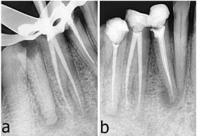
    Figure 3: (a) Master cone radiograph of tooth #43, (b) Post-operative
radiograph with obturation of tooth #42, 43 and 44.
Discussion
This case reported the atypical finding of biradicular canine. This case report stands out from the normal routine case because in 95% cases single root and single canal reported by many of the literature. The vast majority of the problem that occurs during the root canal treatment is due to the insufficient knowledge of the anatomy of the endodontic space [4]. Extra root or root canal if not detected is a major reason for failure of the treatment. Incomplete removal of all the irritants from the pulp space may increase the possibility of treatment failure.
The initial diagnostic radiograph is extremely important because it allows for the identification or suspicion of root and root canal anatomical variations [5]. An ideal scout radiograph provides information about the mesio- distal root canal orientation and a subsequent IOPA radiograph with mesial or distal angulations will provide information about the faciolingual root canal orientation [6]. Also, pre- operative radiographs should be observed with careful attention. A sudden change in the radiographic density of the pulp space usually indicates an additional canal, whereas a sudden narrowing of or even disappearance of the root canal space indicates a bi or tri furcation of root. In case of post- operative radiograph, if obturating material is not centered within the root, it may be a sign of a missing canal [7]. Successful endodontic management of the involved tooth starts with good preoperative radiographs, a proper access cavity preparation, and optimum obturation of all the root canal system. An adequate armamentarium is required, namely recent diagnostic techniques like RVG, dental operating microscope, CBCT which help complete diagnosis and plan appropriate treatment.
Conclusion
Although the literature indicates that the occurrence of mandibular canines with two roots and two root canals is not common, these anatomical variations are associated with technical difficulties during endodontic treatment. A thorough knowledge of the tooth and root canal morphology, clinical exploration, and detailed radiographic interpretation as well as use of advanced radiographic technique may be helpful in detecting root canal aberrations and to achieve success.
References
- J. Abduo, M. Tennant M, McGeachie J. Lateral occlusion schemes in natural and minimally restored permanent dentition: a systematic review. J Oral Rehabil. 2013; 40: 788–802.
- Chauhan R, Singh S. Management of a 3-canal mandibular premolar in a patient with unusual root canal anatomy in all mandibular premolars. Gen Dent. 2013; 61: 16-18.
- Wang L, Zhang R, Peng B. Clinical features and treatment of mandibular canines with two root canals: two case reports. Chin J Dent Res. 2009; 12: 61-62.
- Al Qudah AA, Awawdeh LA. Root canal morphology of mandibular incisors in a Jordanian population. Int Endod J. 2006; 39: 873-877.
- Victorino FR, Baldi JV, Moraes IG, Bernardineli N, Garcia RB, Bramante CM. Bilateral mandibular canines with two roots and two separate canals- case repot. Braz Dent J. 2009; 20: 84-86.
- Jadhav GR. Endodontic management of a two rooted, three canaled mandibular canine with a fractured instrument. J Conserv Dent. 2014; 17: 192-195.
- Cantatore G, Berutti E, Castellucci A. Missed anatomy, frequency and clinical impact. Endo Topics. 2009; 15: 3-31.