
Mini Review
J Dent & Oral Disord. 2020; 6(4): 1138.
Physioterapeutic Intervertion on Facial Palsy
Ribeiro MVMR1*, Da Silva AAS2, Alves ALCS2, Ferreira CVO2, Jucá JVO2, Praxedes BSL2, Barbosa FT3 and Ribeiro EAN4
1Master in Health Sciences from the Federal University of Alagoas, Professor at the Centro Universitário Tiradentes Maceió-Alagoas, Specialist in Ophthalmology; Maceió, Alagoas, Brazil
2Medical student at the Centro Universitário Tiradentes Maceió-Alagoas, Maceió, Alagoas, Brazil
3PhD in Health Sciences from the Federal University of Alagoas, Professor at Tiradentes University Maceió-Alagoas; Maceió, Alagoas, Brazil
4PhD in Pharmacology from the Federal University of Paraíba, Professor of the Postgraduate Program in Health Sciences at the Federal University of Alagoas; Maceió, Alagoas, Brazil
*Corresponding author: Marina Moura Rezende, Master in Health Sciences from the Federal University of Alagoas, Professor at the Centro Universitário Tiradentes Maceió-Alagoas, Specialist in Ophthalmology, Maceió, Alagoas, Brazil
Received: May 11, 2020; Accepted: June 01, 2020; Published: June 08, 2020
Abstract
Peripheral facial palsy is a peripheral facial nerve lesion that affects 8-55 people per 100,000 and is associated with a deficit in the patient’s psychic, physical, and social health. Its main etiology is idiopathic which corresponds to 50-60% of the cases. There are several types of treatment; however, the physiotherapeutic is the most used. This should be associated with other therapies such as pharmacology and surgery. Nevertheless, 10-40% of cases do not progress to full recovery and remain with chronic facial paralysis or paresis.
Keywords: Facial palsy; Rehabilitation; Review; Therapeutic approaches
Introduction
Since Avicenna (979-1037 DC.) [1], peripheral facial paralysis has been described as a peripheral neural lesion of the facial nerve, and may be at any level of its path, from the nerve nucleus to the neuromuscular junction [2,3]. The involvement of this nerve results in complete or partial paralysis of facial muscles and may be associated with disorders of taste, salivation and lacrimation, hyperacusis and hypoesthesia in the external ear canal [4].
The first major incidence is idiopathic peripheral facial palsy, or Bell’s Palsy, and the second is traumatic, with other associated conditions such as hypertension, diabetes mellitus, virosis, pregnancy and puerperium, herpesviruses, vascular damage and neoplasms [3- 5]. Therefore, Bell’s facial paralysis is defined as primary, and the others, as secondary.
The peripheral facial paralysis’s incidence is of 8-55 cases per 100,000 inhabitants [6-8]. Among these Bell’s palsy has the highest incidence, accounting for 50-60% [6,9].
Maximal deficiency occurs in the first 48-72 hours, and the severity of paralysis is related to duration of facial dysfunction, the extent of recovery time, and deterioration in quality of life [10].
The effects of facial nerve paralysis are debilitating and often depress emotional conditions, with a variety of possible functional and aesthetic problems [11]. Although 60-90% of cases can recover completely, 10-40% will remain with chronic facial paralysis or paresis [6,7,9,12]. It is believed that spontaneous recovery of the facial nerve occurs in up to nine months [13]. The psychological impact of facial disfigurement can result in fear of public places and undermine the socialization [11,14].
Surgical techniques and drug treatment may minimize physical deficits, but rarely improve facial function with respect to selective muscular control and symmetry of expression [14].
The purpose of this narrative review is to list the literature on the concept, diagnosis, and treatment of this pathology, since it is a relevant clinical condition and it causes several aesthetic, psychological, functional and social impairment to the patient.
Methods
A review of the PubMed (National Institutes of Health), Scielo and Medline databases was conducted in the Portuguese, English, Spanish and French languages, with the descriptors “facial paralysis”, “physiotherapy”, Bell’s palsy “and” rehabilitation. “We found 50 articles and selected 34 for this review, considering the relevance of the meaning by the authors.
Clinical manifestations
In Bell’s palsy the characteristic findings are acute manifestations of facial motor neuron palsy that affect the upper and lower face muscles with a peak manifestation in 48 to 72 hours [10]. These findings are often accompanied by symptoms of neck, mastoid and ear pain, dysgeusia, hyperacusis or altered facial sensation [10]. Full recovery of the lesion is often impaired by synkinesia [4].
In some cases of chronic evolution of peripheral facial palsy, the most common complication is corneal dryness, which occurs due to the inability to completely close the eyelid, the reduced production of tears and the loss of the eye blinking reflex [4,10]. Lagoftalm may also occur, characterized by depression of the lower eyelid and consequent tearing, a phenomenon called epiphora [6]. Long-term aesthetic or functional sequelae of facial asymmetry include oral dysfunction, contractures, nasal obstruction, difficulty drinking, tasting, salivation and speech, psychosocial problems8, disuse atrophy, lip commissure deviation, tissue adhesion, muscle stretching, hypoesthesia, dysesthesias and hemifacial spasm [4,5].
Classification
There are approximately two dozens clinical types of facial paralysis/paresis, being subdivided into group I (perip heral facial paralysis):
1. Peripheral facial palsy (Bell’s palsy)
2. Alternate facial palsy
3. Bilateral facial palsy
4. Facial palsy in multiple cranial neuropathy
5. Recurrent facial palsy
6. Transient facial paralysis
7. Familial facial paralysis, in group II (central facial paralysis):
8. Central facial palsy
9. Emotional facial palsy
10. Paralysis of the face with paradoxical hypermimia and in group III (Others types of facial palsy):
11. Congenital facial palsy in children
12. Ramuscular facial palsy
13. Bilateral volitional facial palsy with preserved emotional motility (Foix-Chavany-Marie syndrome)
14. Facial paralysis of the upper floor - unilateral
15. Paralysis of the upper floor - bilateral
16. Muscular facial palsy and neuromuscular junction
17. Psychogenic facial palsy, these p atrophies are associated with the involvement of one or both of the neural systems, the facial nerve, the myonural plaque, or the facial muscles [15].
Staging system
The best known systems for assess facial palsy are the House- Brackman scale and the Sunnybrook system [10].
The House-Brackman classification consists of six degrees: I - Normal; II - Mild dysfunction; III - Moderate dysfunction; IV - Moderately severe dysfunction (Figure 1); V - Severe dysfunction; VI - Total paralysis, with description of changes that can be observed in each degree, addressing the gross evaluation of the face, static (symmetry and tonus) and in movement - forehead, eye and mouth [16]. It is the most used scale in the international literature [10,15,16], but it fails to provide satisfactory information about regional facial function [6]. For a more detailed analysis, the Sunnybrook [9,10,18] system is used, by evaluating static symmetry, voluntary movement and synkinesia [13,19].
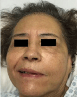
    Figure 1: Left facial paralysis, House-Brackman IV. Fonte: Paiva ALC, Araujo
JLV, Ferraz VR, Veiga JCE. Facial paralysis due to Ramsay Hunt syndrome
- A rare condition. Rev. Assoc. Med. Bras. vol.63 no.4 São Paulo Apr. 2017.
For the calculation of the Sunnybrook scale, “voluntary movement x4 - static symmetry x5 – synkinesia = total” [19]. The value for a normal face, with all movements, is 100, and a total paralysis is classified as 0 [18].
However, according to Coulson et al., The Sunnybrook scale, despite having good accuracy for voluntary movements, does not do so well for synkinesia [17].
In addition to these scales, others may be used for the evaluation and classification of facial palsy and its functional consequences, such as Facial Disability Index Yanagihara, Babak and Kimberly (BK) and Sydney Scale [1,9,11,16-21].
The Facial Disability Index is a small and self-administered questionnaire that assesses the physical and psychosocial aspects associated with facial neuromuscular function [22].
The Yanagihara scale evaluates ten facial movements, each of which can reach four points; the facial function is evaluated by adding to these scores. A total of 40 points indicates normal facial movement, while 0 indicates no movement [23].
The Babak and Kimberly (BK) classification evaluates facial function in five categories. Going from A to E, A would be the normal facial function, B partial palsy with mild synkinesia, C partial palsy with moderate to severe synkinesia, D partial palsy without synkinesia, and finally, E would be complete facial paralysis [19].
Finally, the Sydney scale is based on the anatomical relevance of each branch of the facial nerve, relating them to their respective movements, such as brow wrinkle, ocular closure, lip opening, smiles, whistles and movement of the plastism, as well as synkinesis in general [17].
Pharmacological/surgical treatments
Pharmacological and surgical treatments target a more incisive treatment of facial palsy. They are usually done in association with physiotherapeutic treatments [24].
Pharmacological treatment is related to the use of corticosteroids, starting at the earliest possible to reduce edema and inflammation [10,24]. The use of antiviral drugs is also widely used, since viral infection may be one of the causes - mainly herpes virus type
[1.7,10,12,24]. However, the combined use is controversial in the literature, and would have been more appropriately according to the clinical condition, degree of paralysis and with the confirmation of viral infection [7,10]. In addition, the use of botulinum toxin also applies in cases of synkinesia, improving quality of life and social interaction, but is associated with sequelae such as ptosis and dry eye [19,25,26]. There can also be used eye drops to avoid ocular dryness and ulcerations in cases of palpebral closure deficits [7,10,12].
Also aiming at recovery with nerve regeneration and reinnervation of the musculature, may be used: pentoxifylline vasodilator, at a dose of 400 mg 12/12 h orally, complex B Vitamins, prednisone as soon as possible at a dose of 1 mg/kg body weight, at 8:00 h, for five days [27].
Surgical treatment is indicated only in cases of correction of synkinesia, relief of facial nerve compression and neoplasias [7,12,28] such as temporal muscle elongation myoplasty and hypoglossal-facial anastomosis [29].
Physiotherapeutic treatment
The physiotherapeutic treatment consists of electrostimulation, massage, thermotherapy, cryotherapy and kinesiotherapy, associated or not to biofeedback [3,4,6,10,13,14,30-32]. The treatment give the patient a more satisfactory quality of life, improving social interaction through the functionality and spontaneity of facial movements and emotional expressions [14].
Biofeedback: Using resources such as verbal commands, a mirror (visual biofeedback) or surface electromyography, these resources generate a new flow of information about the activity performed, improving voluntary control [5,13,14]. Patients usually do visual biofeedback in association with facial mimic; it is based on neural plasticity - however, since the proprioceptive sensitivity of the facial muscles is low, the patient needs to use methods that help him visualize the extent of movement, being active during the process and having control over the results [14].
The use of a mirror, which is inexpensive and accessible, allows the patient to perform the movements daily, giving continuity to treatment outside the clinic setting [14,25].
A variation is the use of video recordings, called self-modeling, where the patient can follow his own evolution through videos of his best expressions, enabling improvement in the follow-up of the treatment [13].
Surface electromyography, usually used in association with biofeedback, provides information about muscle electrical activity, captured through surface electrodes. Here, the signals captured by the electrodes are amplified, filtered and converted by a computer into graphs that represent muscle activity [14]. The use of biofeedback by surface electromyography in patients with peripheral facial paralysis has shown improvement in facial symmetry and synkinesis [4]. The patient receives visual and hearing stimulation, and that is important because it provides immediate feedback to the patient, as well as an objective way of measuring movements and results [5,14].
The use of facial mimic or isolated mime therapy in patients with peripheral facial paralysis has shown an improvement in facial symmetry, as well as a lower incidence of the following symptoms: pain, stiffness, involuntary movements, difficulties to, drink and speak, and also an improvement in quality of life [9].
Treatment with biofeedback allows positive results to patient, such as improvements in facial function, symmetry, and decrease of synkinesis [5]. In addition, it is effective in preventing synkinesis during the eye closure by performing friction of the lips [25]. With the use of self-modeling it was possible to verify not only the symmetry of the smiles but also an improvement in the general facial appearance [13].
Thermotherapy: Through warm compresses, it is possible to maintain vascularization and cell exchanges, increasing metabolism and decreasing local tension - heat promotes muscle relaxation and prepares muscle stretching [3,30]. Thermotherapy can be used previously to exercise and massage; however, it should not be used in the region of the mastoid apophysis because they may aggravate the lesion of the inflamed facial nerve and cause injury to the parotid gland [3].
Cryotherapy: It is used ice cubes to promote the depolarization of the nerve fibers, obtaining muscle contraction in the flaccid phase [3,30].
Massage: It is made in cases of post-traumatic edema or if there are tetanizations of the facial muscles in the phases of anarchic regeneration of the facial nerve [3]. Rapid massages and facial mimic exercises should be stimulated to improve facial symmetry and synkinesis.
Electrostimulation: Non-surgical or low-intensity lasers for the treatment of Bell’s facial paralysis may increase the range of action potentials (stimulated nerve function) and the capacity to accelerate the regeneration of nerve structures [33].
There are controversies over its use, which should be done in muscle areas distant from nerve bundles, avoiding the risk of hypertonia and synkinesis [3,21]. In addition, it should be expected about eighteen months to begin treatment, which should be done only if sagging persists [3].
According to some studies, the only muscles that may eventually have a beneficial result through electrostimulation are the orbicularis oris muscles [3].
Kinesiotherapeutic and mechanical resources: Physiotherapeutic resources such as Proprioceptive Neuromuscular Facilitation (Kabat Method or PNF) are used - diagonal patterns and spirals of passive and active movements are used, since most of the muscles act in a spiral direction, using facilitation in the stretch reflex, against resistance. The resistance in the facial muscles should be performed on the healthy side and the paralysis is maintained or facilitated to perform the desired movement. The therapist uses verbal command to reinforce learning and stimulate the correct execution of the requested exercise [34].
Another physiotherapeutic resource is sensory stimulation (ROOD Method), which aims to increase the sensitivity of stretch receptors and voluntary muscle contraction. It is performed by rapid brushing (it must be performed in the opposite direction to the inclination of the hairs in order to reinforce the activity of the motoneurons alpha and gamma neurons), slow massage and cold application (applied in rapid stimuli on the muscle belly towards of its contraction, producing a localized facilitation effect), tapping (vigorous and rapid tactile stimuli in the target musculature), placing (passive maintenance of a corporeal segment in space, requiring active patient support), and rapid stretching [34], in addition to exercises as contraction and relaxation, aiming from the motor stimulation and the muscular strengthening [30].
Conclusion
Considering the high incidence of peripheral facial paralysis in the population and its effects on the physical, psychic and social health of the patient, it is observed how important is the study of this pathology. It is also worth mentioning the need for clinical trials and cross-sectional studies on this clinical condition to establish a pattern in diagnosis and treatment.
References
- Souza IF de, Dias ANM, Fontes FP, Melo LP de. Métodos fisioterapêuticos utilizados no tratamento da Paralisia Facial Periférica: uma revisão. Rev Bras Ciências da Saúde. 2016; 19: 315-320.
- Chevalier A. Rééducation des paralysies faciales centrales et périphériques. Encycl Méd Chir (Editions Sci Médicales Elsevier SAS, Paris, tous droits réservés), Kinésithérapie-Médecine Phys. 2003; 1: 15.
- Matos C. Paralisia Facial Periférica: o papel da medicina física e de reabilitação. Acta Med Port. 2011; 24: 907-914.
- Garanhani MR, Cardoso JR. Capelli ADMG, Ribeiro MC. Fisioterapia na paralisia facial periférica. Estudo retrospectivo. Braz J Otorhinolaryngol. 2007.
- Cronin GW, Steenerson RL. The effectiveness of neuromuscular facial retraining combined with electromyography in facial paralysis rehabilitation. Otolaryngol - Head Neck Surg. 2003.
- Nicastri M, Mancini P, De Seta D, Bertoli GA, Prosperini L, Toni D, et al. Efficacy of Early Physical Therapy in Severe Bell’s Palsy. Neurorehabil Neural Repair. 2013; 27: 542-551.
- de Almeida JR, Guyatt GH, Sud S, Dorion J, Hill MD, Kolber MR, et al. Management of Bell palsy: clinical practice guideline. CMAJ. 2014; 186: 917- 922.
- Hg Beurskens C, Al Burgers-Bots I, Kroon DW, Oostendorp RA. Literature Review of Evidence Based Physiotherapy in Patients with Facial Nerve Paresis. J Jpn Phys Ther Assoc. 2004; 7: 35-39.
- Beurskens CHG, Heymans PG. Physiotherapy in patients with facial nerve paresis: Description of outcomes. Am J Otolaryngol - Head Neck Med Surg. 2004.
- Eviston TJ, Croxson GR, Kennedy PGE, Hadlock T, Krishnan A V, Krishnan A. Bell’s palsy: aetiology, clinical features and multidisciplinary care. Neurol Neurosurg Psychiatry. 2015; 0: 1-6.
- Sassi F, Toledo P, Mangilli L, Alonso N, Andrade CRF de. Correlação entre eletromiografia e índice de inabilidade facial em pacientes com paralisia facial de longa duração: implicações para o resultado de tratamentos. Rev Bras Cir Plást. 2011; 26: 596-601.
- Hohman MH, Hadlock TA. Etiology, diagnosis, and management of facial palsy: 2000 patients at a facial nerve center. In: Laryngoscope. ; 2014.
- Beurskens CH, Heymans PG. Mime therapy improves facial symmetry in people with long-term facial nerve paresis: A randomised controlled trial. Aust J Physiother. 2006; 52: 177-183.
- Goulart F, Vasconcelos KS de S, Souza MRV de, Pontes PB. A Utilização Do Biofeedback No Tratamento Fisioterápico Da Paralisia Facial Periférica. Acta Fisiatr. 2000.
- Maranhão-Filho P, Maranhão ET, Aguiar T, Nogueira, R. Paralisia facial: quantos tipos clínicos você conhece? Parte I. Rev Bras Neurol. 2013; 124: 177-183.
- Santos RMM, Guedes ZCF. Estudo Da Qualidade De Vida Em Indivíduos Com Paralisia Facial Periférica Crônica Adquirida. Rev CEFAC. 2011.
- Coulson SE, Croxson GR, Adams RD, O’Dwyer NJ. Reliability of the “Sydney,” “Sunnybrook,” and “House Brackmann” facial grading systems to assess voluntary movement and synkinesis after facial nerve paralysis. Otolaryngol - Head Neck Surg. 2005.
- Hultcrantz M. Rehabilitation of Bells’ palsy from a multi-team perspective. Acta Otolaryngol. 2016; 136: 363-367.
- Maria CM, Kim J. Individualized management of facial synkinesis based on facial function. Acta Otolaryngol. 2017; 137: 1010-1015.
- Prakash V, Hariohm K, Vijayakumar P, Thangjam Bindiya D. Functional Training in the Management of Chronic Facial Paralysis. Phys Ther. 2012.
- Tuncay F, Borman P, Taser B, Ünlü I, Samim E. Role of Electrical Stimulation Added to Conventional Therapy in Patients with Idiopathic Facial (Bell) Palsy. Am J Phys Med Rehabil. 2015.
- Gonzalez-Cardero E, Infante-Cossio P, Cayuela A, Acosta-Feria M, Gutierrez- Perez JL. Facial disability index (FDI): Adaptation to Spanish, reliability and validity. Med Oral Patol Oral Cir Bucal. 2012; 17: 1006-1012.
- Hato N, Fujiwara T, Gyo K, Yanagihara N. Yanagihara facial nerve grading system as a prognostic tool in Bell’s palsy. Otol Neurotol. 2014; 35: 1669- 1672.
- Ferreira M, Marques JA, Santos PC. Physical Therapy with Drug Treatment in Bell Palsy. Am J Phys Med Rehabil. 2015; 94.
- Nakamura K, Toda N, Sakamaki K, Kashima K, Takeda N. Biofeedback rehabilitation for prevention of synkinesis after facial palsy. Otolaryngol Head EE, uarte Neck Surg. 2003 Apr; 128: 539-43.
- Monini S, De Carlo A, Biagini M, Buffoni A, Volpini L, Lazzarino AI, et al. Combined protocol for treatment of secondary effects from facial nerve palsy. Acta Otolaryngol. 2011; 131: 882-886.
- Valença MM, Valença LPA de A, Lima MCM. Paralisia facial periférica idiopática de Bell: a propósito de 180 pacientes. Arq Neuropsiquiatr. 2001; 59: 733-739.
- Hadlock TA, Greenfield LJ, Wernick-Robinson M, Cheney ML. Multimodality Approach to Management of the Paralyzed Face. Laryngoscope. 2006.
- Martin F. Rééducation des paralysies faciales. Ann Chir Plast Esthet. 2015; 60: 448-453.
- Granado Lage L, Antonio Sérgio Fidel Júnior -Rivail, Elias -Roberto, Fadel -Fernando, Guitmann -Jayme, Resumo C. Paralisia Facial e Parestesia: condutas terapêuticas. 2003. Acervo CISPRE.
- Watson GJ, Glover S, Allen S, Irving RM. Outcome of facial physiotherapy in patients with prolonged idiopathic facial palsy. J Laryngol Otol. 2015; 129: 348-352.
- Tansini S, Gaspodini K, Pimentel GL. Intervenção fisioterapêutica na paralisia facial: uma revisão de literatura. EFDeportes.com, Rev Digit. 2015; 209.
- Snyder SK, Byrnes KR, Borke RC, Sanchez A AJ. Quantitation of calcitonin generelated peptide mRNA and neuronal cell death in facial motor nuclei following axotomy and 633 nm low power laser treatment. Lasers Surg Med. 2002; 31: 216-222.
- Gonçalves M, Bérzin F. Estudo Eletromiográfico comparativo de movimentos de Facilitação Neuromuscular Proproceptiva com os realizados nos planos sagital. Rev Bras Fisioter. 2000; 4: 55-64.