Case Report
J Dent & Oral Disord. 2020; 6(5): 1142.
Multiple Dental Impactions and Agenesis: A Case Report
Penido FO1, Barbosa FI2, Oliveira BJ2, Aguiar TR3 and Consani RLX4*
1Post-graduate Student, University of Itauna, Brazil
2Professor, University of Itauna, Brazil
3Specialist in Buccomaxillofacial Surgery and Traumatology, University of Vale do Rio Verde, Brazil
4Professor, State University of Campinas, Piracicaba Dental School, Department of Prosthodontics and Periodontology, Brazil
*Corresponding author: Consani RLX, Piracicaba Dental School, Department of Prosthodontics and Periodontology, Brazil
Received: June 05, 2020; Accepted: July 01, 2020; Published: July 08, 2020
Abstract
Like the agenesis of the first molars, simultaneous impaction of second and third molars are uncommon events. This case report aims to expose and discuss the surgical treatment of a second and third molars impacted bilaterally and simultaneously associated with agenesis of all first molars involving with bone cement dysplasia.
Keywords: Tooth impacted; Anodontia; Oral surgical procedures; Dentistry; Maxillofacial abnormalities
Introduction
The tooth to be classified as retained should not have been erupted in the oral cavity according to the normal chronology of eruption [1]. Beyond that it must be evident that the spontaneous eruption will not occur [2]. This condition represents a predisposition for the advent of several pathologies such as dental caries, periodontal problems, root resorption of the adjacent tooth and even the development of cysts and tumors [3].
Among the dental groups, third molars are the ones with the highest prevalence of retention and impaction. This is due, among other factors, as ectopic position, lack of space, late maturity and angulation variation [3]. However, the occurrence of impacted permanent second molars is a rare phenomenon, recently became more prevalent occurring mainly in the mandibular region (0,2%) [4].
Under these circumstances, the treatment includes four possible maneuvers: extraction, intervention, transfer and observation. Extraction refers to removal the dental organ, while observation is the follow-up of the case without application of any therapy. Another option would be the intervention, an orthodontic or surgical attempt to remove the factor that prevents the eruption, while the transfer tries to reposition the tooth in the árcade [5].
The choice of treatment will depend on the specific characteristics of each case, making the exact location of the tooth crucial, with a critical analysis of the anatomical structures nearby and the tooth position. Classical radiology does not provide the necessary precision for the event while the tomography would be a better option [6].
The congenital absence of teeth accompanies the evolution of the human species. In the posterior segment of the arch, agenesis of the third molars is normal. It is normal the agenesis of the third molars in the posterior segment of the arch, which can lead to several occlusal disturbances and dental migrations [7].
The present article aims to present an uncommon case of agenesis of the first four molars associated with simultaneous impaction of maxillary and mandibular in both sides of the second and third molars with concomitant presence of bone cement dysplasia. Besides, to expose the treatment proposed.
Case Presentations
Female, melanoderma 29 years old, sought dental care owing to non-eruption of the second and third permanent molars. Dental history revealed the ectopic eruption of a maxillary central incisor tooth on the anterior third of the palate, requiring orthodontic treatment. Was also reported the mutual impaction of the second and third molars in maxilla and mandible in both sides, associated with the congenital absence of the first molars. The others teeth erected conventionally.
The patiente did not report having any systeic disease and family history did not reveal significant changes in relation to this dental problem.
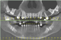
In the pre-surgical procedure, radiographic and tomographic examinations were performed (Figure 1), revealing the horizontal position of the impacted teeth, which presented complete rhizogenesis. A hyodense bone mass and an anatomical alteration in the mandibular region close to the dental elements 37 and 47 were also observed.

Figure 1: Tomographic image showing agenesias and impacted teeth in the maxilla and mandible region.
Hematological examinations (blood count, coagulogram, fasting blood glucose, dosage of urea and creatinine) did not reveal any changes that contraindicate the surgical procedure.
The treatment protocol incluided the extration of the left and right second and third molars from arches. The surgery was performed under general anestesia in hospital ambient due to its complexity.
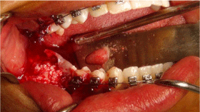
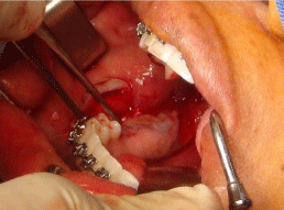
After removal of the teeth, an inorganic bovine graft (Geistlich Bio-Oss- Geistlich Pharma AG, Wolhusen, Switzerland) and fibrin rich in platelets and leukocytes were used for the reconstruction of the lost bone tissue (Figures 2 and Figures 3).

Figure 2: Use of inorganic bovine graft (Geistlich Bio-Oss - Geistlich Pharma AG, Wolhusen, Switzerland).

Figure 3: Placement of fibrin rich in platelets and leukocytes.
Due to the presence of a rarefied bone in the region of the teeth 37 and 47, part of the bone tissue was removed from the mandible, stored in 10% formaldehyde solution, and submitted to histopathological analysis. The result confirmed the presence of cement-bone dysplasia.
In the postoperative period, the patient remained under hospital follow-up for two days, using cephalothin (1 g IV 6/6 h), ketoprofen (100 mg IV 12/12 h) and tramadol hydrochloride (50 mg IV 6/6 h). After hospital discharge, the patient was advised to remain at rest and to use trometamol cetarolac for 5 days (10 mg SL 6/6 h).
After fifteen days, the first return occurred, the suture was removed, and normal healing was observed. Thirty days after surgery, the patient returned, presenting excellent healing.
Discussion
Usually, the simultaneous dental inclusion of second and third molars is associated with other morphological anomalies occurred in the dentition [8]. As reported, the patient presented atypical positioning of the permanent central incisor and congenital absence of the first molar.
For treatment of the case, the literature suggests some clinical alternatives [9]:
• Surgical extraction of the third molars and orthodontic repositioning of the second molars;
• Molars;
• Surgical extraction of the second molars with orthodontic traction of the third molars;
• Surgical extraction of both teeth.
Due to the degree of impaction and with the patient’s agreement, the treatment chosen was to remove the second and third molars and the affected alveolar bone.
An essential moment of the surgical planning was the preoperative evaluation, which allowed to analyze all obstacles described in the literature and that could influence the extraction procedure of the teeth [10]. Thus, it was evaluated the formation and angulation of the root, position and depth of the retained teeth and proximity of the mandibular canal and maxillary sinus.
This analysis resulted in the decision to section all the teeth involved in the surgical area, thus avoiding the possibility of injuring the inferior alveolar nerve, exposing the maxillary sinus and removing large amounts of bone that could result in the reduction of mechanical resistance of the mandible and possible fracture.
Final Considerations
In view of the above, it can be concluded that the treatment of generalized inclusion cases leading to success is based on good planning associated with the appropriate surgical technique and patient agreement.
References
- Hupp JR, Ellis E, Tucker MR. Contemporary Oral, and Maxillofacial Surgery. 7th edition. Elsevier; 2018.
- Varpio M, Wellfel B. Disturbed eruption of the lower second molar: clinical appearance, prevalence, and etiology. ASDC J Dent Child. 1988; 55: 114-118.
- Akarslan ZZ, Kocabay C. Assessment of the associated symptoms, pathologies, positions and angulations of bilateral occurring mandibular third molars: is there any similarity? Oral Surg Oral Med Oral Pathol Oral Radiol Endod. 2009; 108: 26-32.
- Bondemark L, Tsiopa J. Prevalence of ectopic eruption, impaction, retention and agenesis of the permanent second molar. Angle Orthod. 2007; 77: 773-778.
- Frank CA. Treatment options for impacted teeth. J Am Dent Assoc. 2000; 131: 623-632.
- Larmour CJ, Mossey PA, Thind BS, Forgie AH, Stirrups DR. Hypodontia-A retrospective review of prevalence and etiology. Part I. Quintessence Int. 2005; 36: 263-270.
- Ziegler CM, Woertche R, Brief J, Hassfeld S. Clinical indications for digital volume tomography in oral and maxillofacial surgery. Dentomaxillofac Radiol. 2002; 31: 126-130.
- Vedtofte H, Andreasen JO, Kjaer I. Arrested eruption of the permanent lower second molar. Eur J Orthod. 1999; 21: 31-40.
- Magnusson C, Kjellberg H. Impaction and retention of second molars. Angle Orthod. 2009; 79: 422-427.
- Gbotolorun OM, Arotiba GT, Ladeinde AL. Assessment of factors associated with surgical difficulty in impacted mandibular third molar extraction. J Oral Maxillofac Surg. 2007; 65: 1977-1983.