
Case Report
J Dent & Oral Disord. 2021; 7(1): 1155.
Lipomatous Neurofibroma of the Tongue: Report of an Exceptionally Rare Case
Hiroshi Harada1*, Takafumi Ogura2, Mari Namikawa3, Shin-ichi Nakatsuka4 and Akira Kurose1
1Department of Anatomic Pathology, Hirosaki University, Japan
2Department of Oral and Maxillofacial Surgery, Sakai City Medical Center, Japan
3First Department of Oral and Maxillofacial Surgery, Osaka University, Japan
4Department of Diagnostic Pathology, Sakai City Medical Center, Japan
*Corresponding author: Hiroshi Harada, Department of Anatomic Pathology, Hirosaki University Graduate School of Medicine, 5 Zaifu, Hirosaki 036-8562, Japan
Received: February 18, 2021; Accepted: March 12, 2021; Published: March 19, 2021
Abstract
Lipomatous neurofibroma is a special subtype of neurofibroma, which includes abundant mature adipoid tissue, and usually presents subcutaneous location. We describe herein an exceptionally rare case of lipomatous neurofibroma of the tongue. The patient was a 46-year-old Japanese female, who had a well demarcated mass lesion of the tongue. The tumor was composed of mature adipoid tissue and haphazardly arranged spindle cells, which lacked cellular atypia and mitotic activity. Also lipoblast-mimicking cells with intracytoplasmic vacuoles were intermingled within the tumor. Immunohistochemical study revealed strong positive reaction for S100 protein in the tumor. While spindle cells were negative for CD34, alpha-smooth muscle actin, neurofilament, Leu-7, glial fibrillary acidic protein, p63, epithelial membrane antigen and cytokeratin AE1/AE3, type IV collagen highlighted delicate fibrillary cytoplasmic process. Although tumor cells were negative for SOX10 as well, positive cells were sporadically identified within the fibrous capsule, which could suggest intraneural occurrence of the tumor.
Keywords: Lipomatous neurofibroma; Peripheral nerve sheath tumor; Tongue; S100 protein; SOX10; Immunohistochemistry
Abbreviation
LpNF: Lipomatous Neurofibroma
Introduction
Adipose tissue is ubiquitously distributed throughout the body. There are many histological types of neoplastic lesion originated from adipose tissue or comprising adipoid tissue among so-called soft tissue tumors and their frequency of occurrence is relatively high. On the other hand, adipoid tissue may appear in tumors of various histological types in large quantities through the process of metaplasia and other secondary changes, and its predominance in the lesion sometimes results in misdiagnosis in practical histopathology. Lipomatous Neurofibroma (LpNF) is a special subtype of neurofibroma, which includes abundant mature adipoid tissue, and usually presents subcutaneous location [1,2].
We describe herein an exceptionally rare case of LpNF of the tongue.
Case Presentation
The patient was a 46-year-old Japanese female and was otherwise healthy with no particular history. She had been aware of a tumor on the right underside of her tongue for several years, but left it untreated as she had no other symptoms. However, her acquaintance recommended her surgical removal and she visited a regional general hospital. In the initial examination, a well-defined submucosal mass was found and measured 15x12x10 mm in size with no adhesions to the surrounding tissue. Under the diagnosis of a benign tongue tumor, she underwent enucleation of the tumor. There were no symptoms of recurrence or distant metastasis for a year until the follow-up course was completed.
Pathological findings
The tumor was covered with a thin capsule-like connective tissue, and a small amount of skeletal muscle was attached to the outside of the connective tissue. It comprised mature adipoid tissue and haphazardly arranged spindle cells, which lacked cellular atypia and mitotic activity (Figure 1). Spindle cells were often sparsely arranged at the boundary between the two regions, and exhibited a myxoid appearance. In this part, some cells had large or small intracytoplasmic vacuoles, which exhibited a lipoblast-like morphology by compressing and deforming the nucleus (Figure 2).
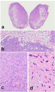
    Figure 1: Light-microscopy. The mass is covered with a thin fibrous capsule
with a small amount of skeletal muscle fibers attached to the outside of the
capsule (a). Within the tumor, there are two components, a mature adipose
tissue and a dense region of spindle cells, which are irregularly mixed and
migrated to each other (b). Spindle cells have small, round to spindle-shaped
nuclei with scant atypia (c,d).
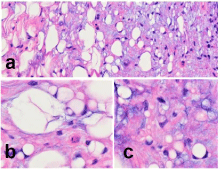
    Figure 2: Light-microscopy. Spindle cells often sparsely arranged at the
interface between the two regions, and exhibit a myxoid appearance (a).
In the same part, some cells have large or small cytoplasmic vacuoles and
exhibit lipoblast-like morphology (b,c).
Immunohistochemical study revealed strong positive reaction for S100 protein in the tumor. Spindle cells were negative for CD34, alpha-smooth muscle actin, neurofilament, Leu-7 and glial fibrillary acidic protein in addition to p63, Epithelial Membrane Antigen (EMA) and cytokeratin AE1/AE3. Type IV collagen visualized delicate fibrillary cytoplasmic processes similarly to S100 protein, suggesting the presence of delicate basement membrane structures around the processes. Although tumor cells were negative for SOX10 as well, positive cells were sporadically identified within the fibrous capsule (Figure 3). Labeling rate with Ki-67 (MIB1) was low overall, averaging less than 1%.
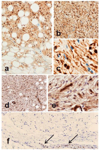
    Figure 3: Immunohistochemistry. Adipoid tissue and fascicle of spindle cells
are strongly positive for S100 protein (a,b), and some spindle cells have a
fibrillary processes with a characteristic waving (c). Also in type IV collagen,
fine fibrillary processes are highlighted (d,e). Tumor cells were negative
for SOX10 (f). However, positive cells are sporadically identified within the
fibrous capsule (arrows).
Discussion
LpNF is a concept proposed by Val-Bernal et al. [1,2]. According to their theory, the adipoid tissue contained in a LpNF is produced by the metaplastic changes in the pluripotent cells of the neuroectoderm, and more than 20 cases have been reported to date, including cases added later. The proportion of LpNF among all neurofibromas occurring under the skin was less than 7% [1-4]. Val-Bernal et al. stated that the proportion of head and neck cases was higher than that of ordinary neurofibromas, and that the CD34 negative rate was significantly higher [2]. The present case is consistent with the characteristics of LpNF, and this case is considered to be a counterpart of this subtype that occurs under the mucosa. To our knowledge, no distinct example of LpNF that occurs beneath the oral mucosa has been previously described in the literature.
Oral neurofibromas may occur either associated or unassociated with neurofibromatosis type I [5]. Broly et al. described a solitary tumor arising in the floor of the mouth, and reviewed total 26 cases of solitary neurofibromas of the oral cavity with various locations such as tongue, gingiva, oral floor, palate, lip and cheek [6]. Neurofibromas usually spread diffusely, but occasionally grow in a nodular fashion. Similarly to our present case, the case reported by Broly et al. presented a solitary encapsulated mass, which was considered to have arisen within the lingual nerve because of EMA-positive residual perineurial sheath at the periphery [6]. On basis of thin fibrous capsule, intraneural occurrence was considered as to our present case as well, but EMA-positive perineurial sheath was not confirmed. Antoni A region of schwannoma with nuclear palisading was not observed. No evidence related to neurofibromatosis type I was noted in general examination of the present patient.
Because a large amount of adipoid tissue is clearly recognized histologically and is extremely eye-catching, a benign lipomatous tumor is mentioned as a differential diagnosis in the present case, and in fact, spindle cell lipoma or lipoblastoma were to be considered. However, the present tumor differs from spindle cell lipoma in that spindle cells are CD34-negative, and differs from lipoblastoma in that it lacks the characteristic lobulated structure. In addition, both of these tumor types are found predominantly in men, and lipoblastoma is epidemiologically incompatible because it is a tumor usually arising in adolescences or young adults.
In most cases, the S100 protein shows a clear positive reaction in peripheral nerve sheath tumors, but it has poor specificity. Coupled with lack of expression of other more specific neural markers, the present case was not easy to diagnose. However, type IV collagen visualized fine fibrillary cytoplasmic processes, suggesting the existence of delicate basement membrane structures around the processes, and thus confirming its characteristics as a peripheral nerve sheath tumor. Furthermore, SOX10 expression could support differentiation toward peripheral nerve, and actually Bajpai et al. stated that combination panel of CD34 and SOX10 was useful for differentiation between spindle cell lipoma and neurofibroma of the oral cavity [7]. Although tumor cells were negative for SOX10, intraneural occurrence of the present tumor is possible despite that EMA-positive perineurial sheath was not confirmed, because SOX10-positive cells were sporadically identified instead within the fibrous capsule. The original characteristics of neurofibroma may have partially lost through metaplastic process similarly to loss of CD34 expression noted above and, in addition, SOX10 originally labels subpopulations of tumor cells ranging only from 20 to 40% of neurofibromas [8].
On the contrary, if the immunohistochemical characteristics are not sufficiently examined, it is assumed that even spindle cells could not recognized to be positive for S100 protein. As also lipoblastmimicking cells with cytoplasmic vacuoles were intermingled within the tumor, it could be assumed that this unique subtype, LpNF was misidentified as a well differentiated liposarcoma of the tongue, especially in some cases previously reported with favorable final outcome in the literature.
From the submucosal condition, it is necessary to distinguish LpNF from lipomatous pleomorphic adenoma (lipomatous mixed tumor), which also has a prominent adipoid tissue [9,10], but no typical characteristics of pleomorphic adenoma is seen in any part of the present tumor, and AE1/AE3 and p63 were completely negative, which could detect epithelial and myoepithelial cells included in pleomorphic adenoma. SOX10 itself expresses not only in schwannian and melanocytic neoplasms but also myoepithelial cell tumors [8], however there is no convincing evidence that the present tumor is an epithelial neoplasm as noted above.
In any case, it should be fully recognized from the pathologist’s point of view that in some cases what stands out at first glance is not necessarily the identity and essence of the lesion.
Conclusion
We described an exceptionally rare case of LpNF arising in the tongue. Almost all of the conventional reported cases occurred subcutaneously, but the counterpart that occurred beneath the oral mucosa has not yet been described on basis of sufficient morphological and immunohistochemical examination, and thus this article could be the first report of the oral example of LpNF.
References
- Val-Bernal JF, de sa Dehesa J, Garijo MF, Val D. Cutaneous lipomatous neurofibroma. Am J Dermatopathol. 2002; 24: 246-250.
- Val-Bernal JF, Gonzalez-Vela MC. Cutaneous lipomatous neurofibroma: characterization and frequency. J Cutan Pathol. 2005; 32: 274-279.
- Yoshida Y, Yamamoto O. Cutaneous lipomatous neurofibroma. J Dermatol. 2009; 36: 674-675.
- Canelas M, Reis JP, Cordeiro M, Figueiredo A. Lipomatous neurofibroma associated with segmental neurofibromatosis. J Cutan Pathol. 2010; 37: 705- 706.
- Thompson LDR, Koh SS, Lau SK. Sporadic neurofibroma of the tongue unassociated with neurofibromatosis type I: a clinicopathologic study of ten cases. Head Neck Pathol. 2020; 14: 374-380.
- Broly E, Lefevre B, Zachar D, Hafian H. Solitary neurofibroma of the floor of the mouth: rare localization at lingual nerve with intraoral excision. BMC Oral Health. 2019; 19: 197.
- Bajpai M, Pardhe N, Kumar M. Immunohistochemical differentiation between spindle cell lipoma and neurofibroma of oral cavity using CD34 and SOX10. Indian J Pathol Microbiol. 2018; 61: 561-563.
- Miettinen M, McCue PA, Sarlomo-Rikala M, Biernat W, Czapiewski P, Kopczynski J, et al. Sox10-a marker for not only schwannian and melanocytic neoplasms but also myoepithelial cell tumors of soft tissue: a systematic analysis of 5134 tumors. Am J Surg Pathol. 2015; 39: 826-835.
- Seifert G, Donath K, Schafer R. Lipomatous pleomorphic adenoma of the parotid gland. Classification of lipomatous tissue in salivary glands. Pathol Res Pract. 1999; 195: 247-252.
- Shah SS, Moustafa TZ. An unusual variant of a common palatal salivary gland tumor: case report of a pleomorphic adenoma with significant lipomatous metaplasia. Case Rep Dent. 2018; 2018: 2052347.