Case Report
J Dent & Oral Disord. 2023; 9(1): 1176.
Simultaneous Bilateral Dentigerous Cysts at the Mandibular Notch with Ectopic Third Molars: A Case Report
Sugauchi A1, Uchihashi T1,2*, Yokota Y1, Kitaoka Y1, Inubushi T3, Ogaya Y4 and Tanaka S1
1First Department of Oral and Maxillofacial Surgery, Graduate School of Dentistry, Osaka University, Japan
2Unit of Dentistry, Osaka University Hospital, Japan
3Department of Orthodontics and Dentofacial Orthopedics, Graduate School of Dentistry, Osaka University, Japan
4Department of Pediatric Dentistry, Graduate School of Dentistry, Osaka University, Japan
*Corresponding author: Toshihiro Uchihashi First Department of Oral and Maxillofacial Surgery, Graduate School of Dentistry, Osaka University, 1-8, Yamadaoka, Suita, Osaka, Japan
Received: December 17, 2022; Accepted: January 18, 2023; Published: January 25, 2023
Abstract
Dentigerous cysts usually develop in the 2nd to 4th decades of life and contain the crown of an unerupted or impacted tooth. It is commonly observed in the mandibular third molar region. Typically, this kind oflesion presents withno symptoms, is often discovered during routine radiographic examination, and presents unilaterally. We report a case of a 59-year-old woman with bilateral radiolucent lesions located at the incisula mandibulae, containing the third molars. Histology of the specimens extracted from both sides showed findings of infected dentigerous cysts. It is an unusual case with regard to the site of the cysts as well as their simultaneity and bilaterality.
Keywords: Dentigerous cyst; Incisula mandibulae; Impacted teeth; Tooth retention
Introduction
Dentigerous cysts account for approximately 20% of the cysts developing in the jawbone and the second most common odontogenic cyst after radicular cyst [1-3]. They are common in the mandibular third molar and maxillary canine regions andcontain the crown of the impacted tooth [4]. There are a few reports on the unilateral development of this cyst in the mandibular notch; however, its bilateral occurrence at the mandibular notches has never been reported [5,6]. Dentigerous cysts, in general, present withno symptoms and are found accidentally on dental radiographic examination. However, as this lesion grows, it displaces adjacent teeth and becomes palpable. An infected lesion can cause an abscess or a phlegmon. Here, we present a rare case of bilateral dentigerous cysts in the vicinity of the mandibular notches with impacted third molars unexpectedly discovered following infection.
Case Report
A 59-year-old woman presenting with pain in her mandibular right molar and difficulty with opening her mouth was referred to our hospital by a private dentist. She was hypertensive and was taking antihypertensive medication (bisoprolol fumarate and perindopril erbumine).
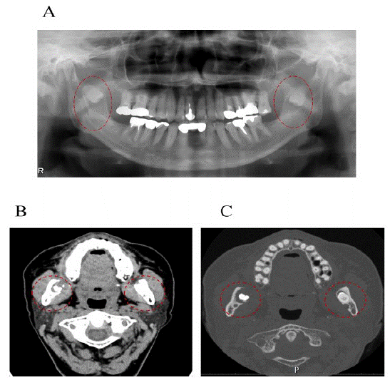
At the first visit, her facial findings were normal, and there was neither swelling in the oral cavity nor natural drainage; however, drainage of pus was confirmed distal to the bilateral second molars on application of pressure. Bacterial test results for pus detected only indigenous bacteria of the oral cavity. A panoramic radiograph showed two unilocular lesions containing impacted teeth located in the vicinity of the bilateral mandibular notches, with root apexes of the buried teeth reaching the mandibular notches (Figure 1A). Computed tomography showed bilateral circular lesions continuing to the crowns of the impacted teeth in the mandible. The boundary of the lesions was clear, with slight buccolingual bulging and no effect on the surrounding soft tissue(Figure 1B&C). Despite the atypical positions of the impacted teeth, dentigerous cysts with infection were suspected based on radiographic findings.

    Figure 1: Evaluation of the positions of the impacted third molars

and cysts on dental radiography (A) and computed tomography

(CT) (B; soft tissue window and C; bone window) before treatment.

Radiography and CT showing bone resorption from the mesial edge

of the incisula mandibulae to the impacted third molar teeth on

both sides. The dotted red circles indicate the lesion.
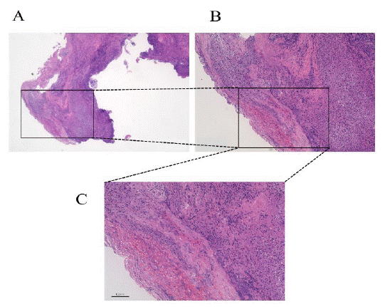
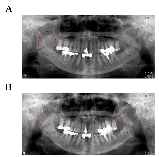
We suggested two treatment options to the patient: 1) biopsy followed by surgical management under general anesthesia based on the histopathologic findings and 2) direct surgical management followed by determination of the need for additional treatment. The patient preferred the latter. Following antibiotic therapy for a few days, we performed bilateral maxillary cystectomy and bilateral tooth extraction with an intraoral approach under general anesthesia. Histopathological examination mainly revealed inflammatory granulation tissue and findings suggestive of cystic epithelium and tooth cysts with infection or inflammation (Figure 2). A panoramic photograph taken on the first postoperative day showed that the impacted teeth were completely removed (Figure 3A). Judging from the histological result, we did not perform an additional operation and followed up on the patient. There was no recurrence, and normal bone healing was observed 6 months postoperatively (Figure 3B).

    Figure 2: Histpathological image of a sample stained with hematoxylin

and eosin showing cystic epithelium mainly composed of

inflammatory granulation tissue (magnification, A: ×40, B: ×100

and C: ×200.).

    Figure 3: Panoramic radiographs taken 1 day (A) and 6 months (B)

postoperatively.

The teeth and cysts completely removed (A), and normal bone regeneration

observed with no recurrence of the lesion (B).
Discussion
In the present case, the impacted third molars with dentigerous cysts were present bilaterally at the mandibular notches. Simultaneous impaction of both teeth at an atypical site, such as the mandibular notch, is unusual.
Ectopic impacted teeth are located away from the normal path of eruption because of abnormal positioning of the tooth germ or movement of impacted teeth. The mandibular third molars are the most frequently impacted teeth, and the impaction may occur in any part of the mandible [7]. However, impaction at the mandibular process or mandibular notch is rare. On reviewing the literature, only 30 cases, including our cases, of impacted wisdom teeth in the ascending ramus of the mandible were found. Of these, only 2 cases were recognized as occurring bilaterally [8-10]. There are various hypotheses regarding their etiology, such as aberrant position of the tooth germ, trauma, and displacement due to pathological lesions [11]. However, in most cases, dentigerous cysts were implicated. Of the 30 cases, 20 were accompanied by dentigerous cysts on the impacted wisdom teeth. Therefore, in relation to the third molars, ectopic tooth germs are rare, and it is speculated that pathologic lesions may push and displace the superior teeth to an ectopic position. In addition, it is also reported that inflammation of the pathological lesions contributes to further displacement of the teeth [9]. Dentigerous cysts usually occur in the mandibular third molar, mandibular premolar,and maxillary canine regions. Previous reports indicate bilateral mandibular odontogenic cysts are often correlated with syndromes, such as basal cell nevus syndrome, mucopolysaccharidosis, cleidobranchia dysplasia, and and drugs, such aslong-term use of cyclosporine and calcium channel blockers [12,13]. Including our own case, 12 cases of cysts at both sides of the mandible body or ramus of mandible region without prior history of syndromes have been reported [14].
In this case, we found impacted wisdom teeth bilaterally in the mandibular notch region. It is unlikely that the tooth germs on both sides existed originally in the mandibular notch region andthat the cyst had caused resorption of the cortical bone from incisula mandibulae without enlargement in the bone marrow and had reached the oral mucosa. Instead, it is more likely that cystic internal pressure caused the teeth to move apically, simultaneously. In addition, it is surmised that the intraluminal pressure of the cyst further increases because of infection, displacing teeth more than usual to the mandibular notch. Although rare, the presentation of the bilateral cysts in our case may be attributed to their simultaneous infection.
Acknowledgement
We would like to thank Editage (www.editage.com) for English language editing.
Conflicts of Interest
The authors declare no conflicts of interest.
References
- Tsukasa K, Shintaro S, Sawako O, Satoko N, Midori A, et al. Endoscope- assisted enucleation of mandibular dentigerous cysts. J Oral Maxillofac Surg Med Pathol. 2021; 33: 126-130.
- Atul K, Astha C, Pulin S, Munish K, Monika V. Non-syndromic bilateral dentigerous cysts of maxillary and mandibular canines: A case series and review of literature. J Oral Maxillofac Surg Med Pathol. 2015; 27: 562-566.
- Kenji H, Shuhei T, Sumitaka H, Masahito F, Akira S, Hideharu H. A dentigerous cyst associated with a supernumerary tooth (fourth molar) in the mandibular ramus: A case report. J Oral Maxillofac Surg Med Pathol. 2019; 31: 98-102.
- Pant B, Carvalho K, Dhupar A, Spadigam A. Bilateral nonsyndromic dentigerous cyst in a 10-year-old child: A case report and literature review. Int J Appl Basic Med Res. 2019; 9: 58–61.
- Chen CY, Chen YK, Wang WC, Hsu HJ. Ectopic third molar associated with a cyst in the sigmoid notch. J Dent Sci. 2018; 13: 172–4.
- Castillo JLD, DeMaria G, Chamorro M. Ectopic third molar in sigmoid notch: Report of a case. Adv dent& Oral Health. 2018; 8: 555731.
- Nodine AM. Impacted and aberrant teeth: Their history causes and treatment. Dent Items Interest. 1947; 69: 30.
- Findik Y, Baykul T. Ectopic third molar in the mandibular sigmoid notch: Report of a case and literature review. J Clin Exp Dent. 2015; 7: e133–7.
- Hanisch M, Fröhlich LF, Kleinheinz J. Ectopic third molars in the sigmoid notch: Etiology, diagnostic imaging and treatment options. Head Face Med. 2016; 12: 36.
- Wang CC, Kok SH, Hou LT, Yang PJ, Lee JJ, Cheng SJ, et al. Ectopic mandibular third molar in the ramus region: Report of a case and literature review. Oral Surg Oral Med Oral Pathol Oral Radiol Endod. 2008; 105: 155–61.
- Salmerón JI, del Amo A, Plasencia J, Pujol R, Vila CN. Ectopic third molar in condylar region. Int J Oral Maxillofac Surg. 2008; 37: 398–400.
- Devi P, Bhovi TV, Mehrotra V, Agarwal M. Multiple dentigerous cysts. J Maxillofac Oral Surg. 2014; 13: 63–6.
- Ustuner E, Fitoz S, Atasoy C, Erden I, Akyar S. Bilateral maxillary dentigerous cysts: A case report. Oral Surg Oral Med Oral Pathol Oral Radiol. 2003; 95: 632–5.
- Vasiapphan H, Christopher PJ, Kengasubbiah S, Shenoy V, Kumar S, et al. Bilateral dentigerous cyst in impacted mandibular third molars: A case report. Cureus. 2018; 10: e3691.