Case Report
Austin J Dent. 2014;1(1): 1005.
Sequelae of Intrusive Trauma to the Primary Predecessors: Odontoma Like Malformation and Enamel Hypoplasia
Rajni Nagpal1 and Naveen Manuja2*
1Department of Conservative Dentistry, Kothiwal Dental College, India
2Department of Pediatric Dentistry, Kothiwal Dental College, India
*Corresponding author: Naveen Manuja, Department of Paediatric Dentistry, Kothiwal Dental College, Moradabad, Uttar Pradesh, India
Received: May 22, 2014; Accepted: June 30, 2014; Published: July 01, 2014
Abstract
Traumatic injury to the primary teeth can cause significant alteration of the underlying permanent dentition because of the close anatomic relationship between the developing permanent teeth and the apices of the overlying primary incisors. These developmental disturbances may range from white or yellow brown enamel discoloration with or without enamel hypoplasia, crown- root dilaceration, odontoma like malformation, root duplication or angulation, arrest of root development, germ sequestration and eruption disturbances. This case report describes a rare complication of odontoma-like malformation along with hypoplasia of the permanent teeth resulting from intrusive injury to primary predecessors at a very young age. The management along with esthetic considerations is discussed to achieve the physio-psychological rehabilitation of such patient.
Keywords: Odontoma like malformation; Penamel hypoplasia; Intrusive injury; Sequelae of trauma; Permanent maxillary incisor; Primary predecessors
Introduction
The major concern with primary tooth trauma is the potential for damage to the underlying permanent successor [1]. This may occur directly from the injury or from the residual infection associated with the traumatized primary tooth [2]. The topographic relationship of the primary teeth to the permanent tooth germs explains the potential for possible developmental disturbances. Several developmental anomalies have been reported in the permanent dentition as sequelae of injuries to their primary predecessors [2-4]. These developmental defects may be simple or complex, extensive or local, affecting the crown, root, or the entire germ. In the coronal region, structural alterations may occur such as enamel hypoplasia, crown dilaceration and white, yellow or brown discoloration. Disturbances affecting the root region include root duplication, root dilaceration and partial or complete arrest of root formation. When the entire permanent tooth germ is affected, the following may be noted: alterations to the process of eruption of the permanent tooth, retention of the permanent tooth or malformation of the permanent tooth germ giving the appearance of an odontoma. Do Espírito Santo Jácomo DR et al. [5] in their longitudinal study of eight years reported that the most common developmental disturbances were discoloration of enamel and/or enamel hypoplasia (46.08%) and eruption disturbances (17.97%) due to the traumatic injury in their predecessors.
The extent of the disturbance of the developing tooth germ is related to the stage of germ development (the child's age at the time of injury), the spatial relationship of the involved teeth, the type of trauma, the severity, and the direction of impact [6-9]. Intrusion injuries which constitute 4.4-22% of traumatic injuries in the primary dentition have been reported to be associated with the highest likelihood of damage to the underlying permanent teeth [1,3,10-15]. An intrusive injury is the consequence of impact by a force in an axial direction that results in displacement of the tooth within the socket. Displacement of the primary tooth root may affect development of the permanent tooth by altering the secretary phase of the ameloblasts resulting in enamel hypoplasia or, in subsequent stages, changing the root formation process [16]. Sennhenn-Kirchner S et al.[13] reported that intrusion injuries are the most frequent cause of developmental disturbances in permanent teeth (47%). In half of the cases, a comparatively mild form of lesion like enamel discoloration was observed. Remaining cases showed other dental deformities like cessation of root formation or retention caused by ankylosis.
Altun C et al. [15] did a prospective 7-year study and observed developmental disturbances in 53.6% of permanent successors [enamel hypoplasia (28.3%), crown and/or root deformation (16.7%), and ectopic eruption (16.7%)] attributed to intrusive injury of their primary predecessors.
Malformation of the permanent tooth germ may be the result of severe intrusion by the primary tooth and invasion of the developing germ during the earliest phases of odontogenesis. Odontoma-like malformation of the permanent tooth is a rare developmental disturbance and only a few cases have been reported in the literature [4,17-20]. This anomaly is confined primarily to maxillary incisors when the time of injury occurs between ages 1 and 3 years, during the morphogenetic stages of the dental follicle, and results mainly from intrusive luxation or avulsion [17] This report presents a rare case of odontoma like malformation along with hypoplastic permanent maxillary incisors as sequelae of intrusive trauma to primary teeth.
Case Presentation
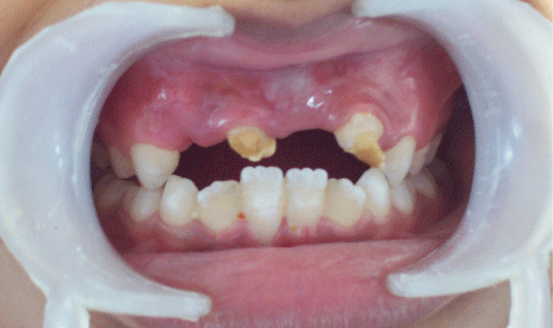
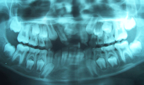
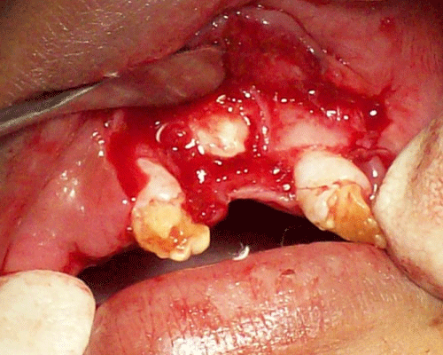
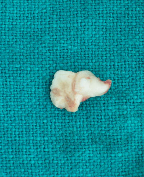
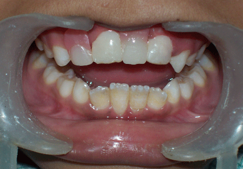
An eight year old girl visited the Department of Pediatric Dentistry, Kothiwal Dental College, Moradabad, India, with a complaint of discolored upper front teeth. Patient's mother gave a history of trauma to the upper front teeth at the age of 16 months due to fall which resulted in intrusion of primary maxillary incisors. She visited the local dentist who kept the patient on wait and watch. There was no regular follow up. The intruded teeth were surgically removed six months after trauma. Medical and family histories were non-contributory. The intraoral clinical examination revealed moderate oral hygiene with class I molar relationship. Only two permanent maxillary incisors were present which were severely deformed and discolored (Figure 1). Panoramic radiographic examination revealed impacted radiopaque calcified mass resembling odontoma like malformation in place of one permanent maxillary central incisor and other permanent maxillary central incisor was missing. Maxillary right permanent lateral incisor had erupted in the position of missing maxillary right permanent central incisor (Figure 2). Both erupted permanent maxillary lateral incisors showed severely hypo plastic crown with immature root. The impacted odontoma like malformation was surgically removed under local anesthesia (Figure 3 and 4). Esthetic composite resin build ups were performed on the hypo plastic crowns and missing teeth were restored with a removable prosthesis (Figure 5).
Figure 1: Intra oral view showing two severely deformed and discolored permanent maxillary incisors.
Figure 2: Panoramic radiograph illustrating impacted odontoma like malformation in place of one permanent maxillary central incisor and other permanent maxillary central incisor is missing.
Figure 3: Impacted odontoma like malformation visible after raising buccal flap.
Figure 4: Impacted odontoma like malformation after surgical removal.
Figure 5: Intra oral view after esthetic and functional rehabilitation of the patient.
Discussion
The traumatic injuries to primary predecessors disturb the formation and maturation of the developing permanent tooth germs because of their intimate relationship with the apices of their overlying primary teeth [3] The peak period for trauma to the primary teeth is preschool-age because in this age children lack the psychomotor skills needed to perform precise and safe movements and, as a result they are susceptible to falls and other injuries [10,11,21,22]. According to the literature, 15-30% of children suffer traumatic injuries to primary teeth [12,16,23] Boys are more likely to sustain primary tooth trauma than are girls, with frequency ratios ranging from 1.8:1 to 1.3:1[10,24]. Maxillary primary incisors have the highest predisposition for traumatic injury.
The age at which the trauma took place explains the sequel noted in the permanent dentition. Formation of the tooth germ of the permanent maxillary central incisors takes place at 20 weeks of gestation, and calcification begins at the age of 3-4 months with completion of enamel at 4-5 years of age [13]. The degree of damage to the permanent successor depends on the stage of crown formation. White discoloration is the result of accelerated deposition of minerals caused by trauma during the maturation stage of enamel development. Yellow-brown discolorations are caused by incorporation of the breakdown products of hemoglobin from bleeding in the periapical area. The destruction of ameloblasts in the active enamel epithelium results in enamel hypoplasia [3]. Andreasen et al [2] also stated that hypo plastic defects may be the result of localized damage to the enamel matrix before the completion of mineralization.
Odontoma-like malformation has been described as a mass of mineralized tissue slightly similar in structure to a normal tooth and may have a relatively normal or rudimentary root [4]. Similar to our case, Yasemin O et al [20] and Shaked L et al [18] reported odontoma like malformation in the permanent dentition caused by the intrusion of primary incisor. Nelson-Filho et al [4] also reported a case of odontoma like malformation in a permanent maxillary central incisor subsequent to traumatic avulsion of the primary incisor predecessor. They stated that the traumatic force probably dislocated the root of the maxillary right primary central incisor to the palatal direction during avulsion, causing alteration in the position of the germ of the maxillary right central incisor which resulted in this kind of malformation.
Odontomas are pseudo-tumoral lesions composed of both epithelial and mesenchymal cells, which appear histologically normal, with a deficit in structural arrangement. Odontomas have been referred to as hamartoma and not as true neoplasm's, and are rarely associated with pathologic development of cysts, such as a calcifying odontogenic cyst or a dentigerous cyst [25]. Odontomas are of two types: complex odontoma, a malformation in which all dental tissues are present, but arranged in a more or less disorderly pattern; and compound odontoma, a malformation in which all the dental tissues are represented in a pattern that is more orderly than that of the complex type. The etiology of odontoma has been attributed to various pathological conditions like local trauma, inflammatory and/ or infectious process, hereditary anomalies (Gardners syndrome, Hermanns syndrome), odontoblastic hyperactivity, and alterations in the genetic component responsible for controlling dental development [26].
Surgical removal of the odontoma like malformation as soon as possible is the optimal treatment. The treatment of yellow brown discoloration of enamel implies restoration with composite resin. When discoloration and enamel defects occupy most of the labial surface, a porcelain jacket crown or veneer may be indicated. Possible reason for the missing permanent central incisor in the present case could be either due to unintended removal of the permanent central incisor tooth germ during surgical removal of intruded primary incisors or agenesis. However, agenesis of permanent central incisor is rare (0.00-0.01%) [27]. Thus, regular follow-up and radiographs are recommended in cases of intrusion injuries in children 1-3 years of age to allow early detection and management of possible developmental disturbances.
References
- Von Arx T. Developmental disturbances of permanent teeth following trauma to the primary dentition. Aust Dent J. 1993; 38: 1-10.Andreasen JO, Andreasen FM, Andersson L. Textbook and color atlas of traumatic injuries to the teeth. 4th ed.Copenhagen: Munksgaard, 2007.
- Andreasen JO, Andreasen FM, Andersson L. Textbook and color atlas of traumatic injuries to the teeth. 4th ed.Copenhagen: Munksgaard, 2007.
- Diab M, elBadrawy HE. Intrusion injuries of primary incisors. Part III: Effects on the permanent successors. Quintessence Int. 2000; 31: 377-384.
- Nelson-Filho P, Silva RA, Faria G, Freitas AC. Odontoma-like malformation in a permanent maxillary central incisor subsequent to trauma to the incisor predecessor. Dent Traumatol. 2005; 21: 309-312.
- do Espírito Santo Jácomo DR, Campos V. Prevalence of sequelae in the permanent anterior teeth after trauma in their predecessors: a longitudinal study of 8 years. Dent Traumatol. 2009; 25: 300-304.
- Turgut MD, Tekçiçek M, Canoglu H. An unusual developmental disturbance of an unerupted permanent incisor due to trauma to its predecessor - a case report. Dent Traumatol. 2006; 22: 283-286.
- Lenzi AR, Medeiros PJ. Severe sequelae of acute dental trauma in the primary dentition--a case report. Dent Traumatol. 2006; 22: 334-336.
- Kuvvetli SS, Seymen F, Gencay K. Management of an unerupted dilacerated maxillary central incisor: a case report. Dent Traumatol. 2007; 23: 257-261.
- Smith RJ, Rapp R. A cephalometric study of the developmental relationship between primary and permanent maxillary central incisor teeth. ASDC J Dent Child. 1980; 47: 36-41.
- Garcia-Godoy F, Garcia-Godoy F, Garcia-Godoy FM. Primary teeth traumatic injuries at a private pediatric dental center. Endod Dent Traumatol. 1987; 3: 126-129.
- Andreasen JO. Etiology and pathogenesis of traumatic dental injuries. A clinical study of ,298 cases. Scand J Dent Res. 1970; 78: 329-342.
- Skaare AB, Jacobsen I. Primary tooth injuries in Norwegian children (1-8 years). Dent Traumatol. 2005; 21: 315-319.
- Sennhenn-Kirchner S, Jacobs HG. Traumatic injuries to the primary dentition and effects on the permanent successors - a clinical follow-up study. Dent Traumatol. 2006; 22: 237-241.
- Rodríguez JG. Traumatic anterior dental injuries in Cuban preschool children. Dent Traumatol. 2007; 23: 241-242.
- Altun C, Cehreli ZC, Güven G, Acikel C. Traumatic intrusion of primary teeth and its effects on the permanent successors: a clinical follow-up study. Oral Surg Oral Med Oral Pathol Oral Radiol Endod. 2009; 107: 493-498.
- Flores MT. Traumatic injuries in the primary dentition. Dent Traumatol. 2002; 18: 287-298.
- Andreasen JO, Sundström B, Ravn JJ. The effect of traumatic injuries to primary teeth on their permanent successors. I. A clinical and histologic study of 117 injured permanent teeth. Scand J Dent Res. 1971; 79: 219-283.
- Shaked I, Peretz B, Ashkenazi M. Development of odontoma-like malformation in the permanent dentition caused by intrusion of primary incisor--a case report. Dent Traumatol. 2008; 24: 395-397.
- Güngörmüs M, Yolcu U, Aras MH, Halicioglu K. Simultaneous occurrence of compound odontoma and arrested root formation as developmental disturbances after maxillofacial trauma: a case report. Med Oral Patol Oral Cir Bucal. 2010; 15: 398-400.
- Ozdemir Y, Akin A, Eden E. Management of multiple sequelaes in permanent dentition: 3 years follow-up. Dent Traumatol. 2011; 27: 67-70.
- Galea H. An investigation of dental injuries treated in an acute care general hospital. J Am Dent Assoc. 1984; 109: 434-438.
- Harrington MS, Eberhart AB, Knapp JF. Dentofacial trauma in children. ASDC J Dent Child. 1988; 55: 334-338.
- García-Godoy F, Morbán-Laucer F, Corominas LR, Franjul RA, Noyola M. Traumatic dental injuries in preschoolchildren from Santo Domingo. Community Dent Oral Epidemiol. 1983; 11: 127-130.
- Borssen E, Holon A-K. Traumatic dental injuries in a cohort of 16-year-olds in northern Sweden. Endod Dent Traumatol. 1997; 13: 276-80.
- Owens BM, Schuman NJ, Mincer HH, Turner JE, Oliver FM. Dental odontomas: a retrospective study of 104 cases. J Clin Pediatr Dent. 1997; 21: 261-264.
- Vengal M, Arora H, Ghosh S, Pai KM. Large erupting complex odontoma: a case report. J Can Dent Assoc. 2007; 73: 169-173.
- Polder BJ, Van't Hof MA, Van der Linden FP, Kuijpers-Jagtman AM. A meta-analysis of the prevalence of dental agenesis of permanent teeth. Community Dent Oral Epidemiol. 2004; 32: 217-226.
