Case Report
Austin J Dent. 2014;1(2): 1006.
Oral Staining and Malodor Secondary to Tobacco Abuse in Southwestern Saudi: A Case Report
Hossam A Eid1* and Manea Musa Musleh2
1Department of Oral medicine & Periodontology, Suez Canal University, Egypt
2Department of Periodontology, Ministry of Health, Saudi Arabia
*Corresponding author: Hossam A Eid, Department of Oral medicine & Periodontology, Suez Canal University, Ismailia, Egypt
Received: July 02, 2014; Accepted: July 28, 2014; Published: July 30, 2014
Abstract
Smokeless tobacco (ST) chewing has detrimental effects on oral tissues including hard and soft tissues; it is often associated with gingivitis/periodontitis, impaired healing, dental caries, and oral mucosal lesions. This case report describes a 25 year-old male patient, who presented to King Khalid University, College of Dentistry (KKUCOD) dental clinic, with a chief complaint of oral malodor and staining. The clinical examination revealed heavy brown stains of the lower lip vermilion border and the facial aspect of the mandibular anterior dentition and localized gingival recession and areas of fenestrations at the attached gingiva of the mandibular central incisors. The patient admitted to an eight year history of cigarette smoking and smokeless tobacco. The heavy staining was noted by the patient to be observable two years ago. The staining was a social embarrassment and the most important issue for the patient.
Keywords: Smokeless tobacco; Gingival recession; Gingival fenestration; Pigmentation
Introduction
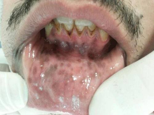
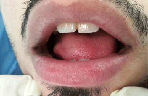
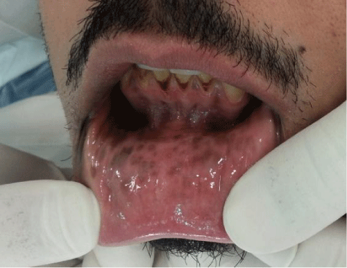
Smokeless tobacco (ST) effects on the prevalence and severity of periodontal disease have been established with several systemic hazards. In the past 20 years, the use of smokeless tobacco has almost tripled. Considering the widespread use of ST products globally, the effects of such products on the periodontal tissues may be important [1]. Smokeless tobacco (ST) is an extremely addictive substance with a high rate of use in certain demographic groups, specially adolescents and young adults; it is available in two forms [2]. Snuff is a finely ground tobacco which is either dry (inhaled or sometimes placed in the mouth) or moist (placed in the mouth). Smokeless tobacco comes in three forms: loose, leaf, plug or twist. All forms of chewing tobacco are held or chewed in the mouth. There are 2,550 known compounds in processed tobacco in addition to nicotine [3]. Smokeless tobacco contains at least 30 metals including nickel and a radioactive compound called polonium-210. Formaldehyde and nitrosamines are also found in smokeless tobacco. All of these compounds have been known to cause cancer [4,5]. Snuff and chewing tobacco contain high concentrations of sodium (salt), swallowing tobacco juice containing sodium salt may contribute to the risk of high blood pressure. High blood pressure has been found to be a problem for a number of smokeless tobacco users [6,7]. This increase may give you a feeling of preparedness. However, the elevated blood pressure and heart rate actually decreases your heart's performance and thereby reduces your overall stamina [8]. Several kinds of sugar are found in unprocessed chewing tobacco and added during its processing which may cause dental cavities specially root caries frequently associated with gingival recession [7]. Cessation of smokeless tobacco use associated with withdrawal symptoms, including: irritability, impatience, anxiety, tension, poor concentration, sleep problems, changes in appetite, and craving [8]. The A seer region of southwestern Saudi Arabia is known for both a high rate of Khat chewing and tobacco use. The purpose of this paper was to report a case of 25 year old Saudi male who presented with oral malodor, gingival pigmentation and unaesthetic appearance due to unique intensifying pigmentation on lower lip mucosa and vermillion border (Figure 1&2).
Figure 1: Preoperative view showing pigmentation of labial mucosa & gingival and teeth staining.
Figure 2: Preoperative view showing pigmentation of lower lip vermillion border.
Case Presentation
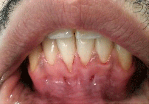
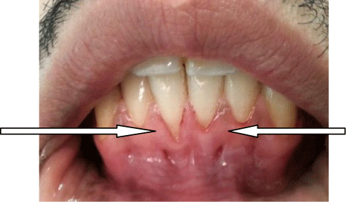
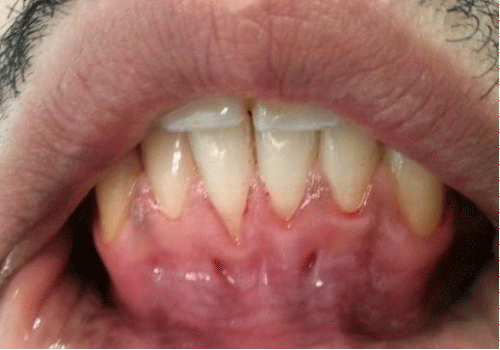
A 25 years old male was referred form the Division of Diagnostic specialty clinics to the Division of Periodontics specialty clinics at King Khalid University College of Dentistry, Abha, Saudi Arabia, presenting with oral malodor and unaesthetic appearance due to unique heavy dark brown stains on lower lip vermillion border and mucosa, labial surfaces of lower anterior teeth with localized gingival recession and areas of fenestrations at attached gingiva of lower central incisors (Figure3,Figure 4). The main complaint was related to general social handicap and embarrassment due to bad oral smell and unaesthetic appearance with psychological stress due to phobia of failure in future marriage plan. The patient has history of cigarette smoking accompanied with smokeless tobacco chewing since 8 years ago. Clinical examination of this individual revealed that: there is oral malodor exceeding socially accepted level (360 ppb) was measured by using Halimeter devicez [9] (following manufacturer instructions). Average gingival recession was 3.5 mm measured using calibrated periodontal probe graduated in millimeters (University of Michigan '0' probe with William's markings; Hu Friedy, Chicago, USA, under a standard dental light with patient seated in a semi-supine position in a standard dental chair) in the lower central incisors, besides two areas of gingival fenestration (window like defect at attached gingival) at attached gingiva of lower central incisors. Heavy dark brown/black pigmentation score 3 on labial mucosa and vermillion border of lower lip and gingival of lower central incisors Figure 5 (according to Dummetts oral pigmentation: Score 0 (Pink tissue-no clinical pigmentation) suited the clinical assessment of non-pigmented and the score 3 (Deep brown or blue/black tissue -heavy clinical pigmentation) [10,11]. After thorough discussion with the individual a treatment plan was scheduled as following: 1.The patient requires a tobacco cessation program resulting in the elimination of tobacco utilization. 2. Patient oral hygiene education and instruction. 3. Hygiene appointment for prophylaxis and the removal of tooth enamel stains. 4. Consideration for gingival surgery to correct gingival recession. 5. Consideration for de-pigmentation laser therapy (Figure 6).
Figure 3: Gingival fenestration.
Figure 4: Gingival recession.
Figure 5: Labial mucosa & gingival pigmentation score 3.
Figure 6: After first visit of scaling & polishing.
Discussion
Nowadays, ST chewing habit spread among youth in both developed and undeveloped countries with almost same percentage. In Sri Lanka various forms of smokeless tobacco products, 15.8% used smokeless tobacco products and its use is three-fold higher among men compared to women. Some 8.6% of the youth and adolescents are current users of smokeless tobacco [12]. Notably, in Saudi Arabia 35.4% of these oral cancers were referred from one province - Jizan in south western of Saudi Arabia when compared to the whole of the KSA population. These data suggest that there is a relationship between the factors smokeless tobacco product (shamma), where shamma is common [13]. Several cross-sectional studies showed a higher prevalence rate of leukoplakia among smokers, with a dose-response relationship between tobacco use and oral leukoplakia, and intervention studies show a regression of the lesion after stopping the smoking habit [14]. In a Swedish study [15] stated positive correlation between tobacco use and leukoplakia, frictional white lesion, coated tongue, hairy tongue and excessive melanin pigmentation, while a negative correlation was observed for geographic tongue and aphthous ulcers. Approximately 70% of the lesions were associated with local irritants as dentures, tobacco [16,17]. The excessive use of tobacco products has been associated with various lesions in the oral cavity. Tobacco-associated lesions include tooth/ restorative materials stains, gingival/ tooth abrasions, smoker's melanosis, acute necrotizing ulcerative gingivitis and other periodontal conditions, burns and keratotic patches, black hairy tongue, nicotinic stomatitis, palatal erosions, soft tissue erythema, leukoplakia, epithelial dysplasia and squamous-cell carcinoma [18,19]. Tobacco use affects the surface epithelium, resulting in changes in the appearance of the tissues. The changes may range from an increase in pigmentation to thickening of the epithelium (white lesion). Tobacco use can also irritate the minor salivary glands on the hard palate decreasing ability to taste and smell [21,22]. Nearly three-quarters of the patients with the tobacco habit had oral mucosal lesions, ST users tend to have more severe gingival recession (REC) and clinical attachment loss (CAL) and a greater proportion of sites with higher values of REC and CAL compared with never-users. The greatest increase in severity of CAL was found to be localized to sites on mandibular teeth, buccal surfaces, anterior and molars, which may be a result of the retention of the ST product in the oral cavity [22]. This description is supported by the findings in the reported case of this work in which the patient has gingival recession & clinical attachment loss in lower centrals besides the unique finding in this case which is the unique heavy pigmentation in labial mucosa and vermillion border of lower lip and attached gingival along with bad smell. Here it is worth to mention that the gingival fenestration is also a unique characteristic finding in this case.
Conclusion
Public health programs towards smokeless tobacco use habit are needed and to be intensified and targeted to vulnerable younger age groups in the community.
References
- Anand PS, Kamath KP, Bansal A, Dwivedi S, Anil S. Comparison of periodontal destruction patterns among patients with and without the habit of smokeless tobacco use--a retrospective study. See comment in PubMed Commons below J Periodontal Res. 2013; 48: 623-631.
- International Agency for Research on Cancer. Smokeless Tobacco and Some Tobacco-SpecificN-Nitrosamines. Lyon, France: World Health Organization International Agency for Research on Cancer. IARC Monographs on the Evaluation of Carcinogenic Risks to Humans Volume 89. 2007.
- National Cancer Institute. Smokeless Tobacco or Health: An International Perspective. Bethesda, MD: National Cancer Institute; 1992. Smoking and Tobacco Control Monograph 2.
- Richter P, Hodge K, Stanfill S, Zhang L, Watson C. Surveillance of moist snuff: total nicotine, moisture, pH, un-ionized nicotine, and tobacco-specific nitrosamines. See comment in PubMed Commons below Nicotine Tob Res. 2008; 10: 1645-1652.
- Djordjevic MV, Doran KA. Nicotine content and delivery across tobacco products. See comment in PubMed Commons below Handb Exp Pharmacol. 2009; 61-82.
- [No authors listed]. Women and smoking: a report of the Surgeon General. Executive summary. See comment in PubMed Commons below MMWR Recomm Rep. 2002; 51: 1-4.
- NIH State-of-the-Science Panel. National Institutes of Health State-of-the-Science conference statement: tobacco use: prevention, cessation, and control. See comment in PubMed Commons below Ann Intern Med. 2006; 145: 839-844.
- The Clinical Practice Guideline Treating Tobacco Use and Dependence 2008 Update Panel, Liaisons, and Staff. A clinical practice guideline for treating tobacco use and dependence: 2008 update. A U.S. Public Health Service report. American Journal of Preventive Medicine. 2008; 35:158-176.
- Salako NO, Philip L. Comparison of the use of the Halimeter and the Oral Chroma™ in the assessment of the ability of common cultivable oral anaerobic bacteria to produce malodorous volatile sulfur compounds from cysteine and methionine. See comment in PubMed Commons below Med Princ Pract. 2011; 20: 75-79.
- Dummett CO. First symposium on oral pigmentation. J Periodontol 1960; 31: 345.
- Dummett CO, Sakumura JS, Barens G. The relationship of facial skin complexion to oral mucosa pigmentation and tooth color. See comment in PubMed Commons below J Prosthet Dent. 1980; 43: 392-396.
- Somatunga LC, Sinha DN, Sumanasekera P, Galapatti K, Rinchen S, Kahandaliyanage A, et al. Smokeless tobacco use in Sri Lanka. See comment in PubMed Commons below Indian J Cancer. 2012; 49: 357-363.
- Allard WF, DeVol EB, Te OB. Smokeless tobacco (shamma) and oral cancer in Saudi Arabia. See comment in PubMed Commons below Community Dent Oral Epidemiol. 1999; 27: 398-405.
- Sujatha D, Hebbar PB, Pai A. Prevalence and correlation of oral lesions among tobacco smokers, tobacco chewers, areca nut and alcohol users. See comment in PubMed Commons below Asian Pac J Cancer Prev. 2012; 13: 1633-1637.
- Salonen L, Axéll T, Helldén L. Occurrence of oral mucosal lesions, the influence of tobacco habits and an estimate of treatment time in an adult Swedish population. See comment in PubMed Commons below J Oral Pathol Med. 1990; 19: 170-176.
- Mirbod SM, Ahing SI. Tobacco-associated lesions of the oral cavity: Part I. Nonmalignant lesions. See comment in PubMed Commons below J Can Dent Assoc. 2000; 66: 252-256.
- Taybos G. Oral changes associated with tobacco use. See comment in PubMed Commons below Am J Med Sci. 2003; 326: 179-182.
- Christen AG, McDonald JL, Olson BL, Christen JA. Smokeless tobacco addiction: a threat to the oral and systemic health of the child and adolescent. See comment in PubMed Commons below Pediatrician. 1989; 16: 170-177.
- Robertson PB, Walsh MM, Greene JC. Oral effects of smokeless tobacco use by professional baseball players. See comment in PubMed Commons below Adv Dent Res. 1997; 11: 307-312.
- Winn DM. Tobacco use and oral disease. See comment in PubMed Commons below J Dent Educ. 2001; 65: 306-312.
- Anand PS, Kamath KP, Bansal A, Dwivedi S, Anil S. Comparison of periodontal destruction patterns among patients with and without the habit of smokeless tobacco use--a retrospective study. See comment in PubMed Commons below J Periodontal Res. 2013; 48: 623-631.
- Meleti M, Vescovi P, Mooi WJ, van der Waal I. Pigmented lesions of the oral mucosa and perioral tissues: a flow-chart for the diagnosis and some recommendations for the management. See comment in PubMed Commons below Oral Surg Oral Med Oral Pathol Oral Radiol Endod. 2008; 105: 606-616.
