
Case Report
Austin J Dent. 2023; 10(1): 1173.
Application of Dental Magnetic Attachments to a Root-Retained Complete Overdenture after Repairing a Failed Maxillary Prosthesis: A Case Report
Yasuda H¹; Akita D¹*; Kumabe T²; Ito T³; Tsukimura N¹; Okubo T¹; Inoue J4; Kato I4; Suzuki A4; Hagiwara Y1
1Department of Partial Denture Prosthodontics, Nihon University School of Dentistry, Japan
2Department of Oral and Maxillofacial Surgery, Nihon University School of Dentistry, Japan
3Department of Complete Denture Prosthodontics, Nihon University School of Dentistry, Japan
4Division of Applied Oral Sciences, Nihon University Graduate School of Dentistry, Japan
*Corresponding author: Akita D Department of Partial Denture Prosthodontics, Nihon University School of Dentistry, Tokyo 101-8310, Japan. Tel: +81-3-3219-8144; Fax: +81-3-3219-8350 Email: akita.daisuke10@nihon-u.ac.jp
Received: May 29, 2023 Accepted: June 10, 2023 Published: June 17, 2023
Abstract
The aim of prosthodontic treatment is to restore oral function as quickly as possible. However, the stability and retention of removable dentures strongly depend on the condition of the remaining alveolar bone. Accordingly, the treatment period for tissue-supported removable dentures tends to be long. Moreover, the treatment period for tissue-supported removable dentures varies according to the residual ridge remodeling after extraction. Tooth-supported overdentures transmit occlusal forces to the alveolar bone through the periodontal ligament, thereby suppressing residual ridge resorption. Currently, dental magnetic attachments are used globally to improve the stability of dentures by close approximation of a magnetic assembly with a keeper for receiving the magnet.
An 84-year-old woman was referred to our department because of detachment of an extensive maxillary prosthesis due to root fracture. We performed pre-prosthetic treatment including dental magnetic attachments for the upper abutment teeth and inserted the repaired prosthesis as an interim denture after tooth extraction. Finally, we transferred the magnetic assemblies from the repaired prosthesis to a new horseshoe-shaped complete overdenture. Therefore, we were able to prevent alveolar bone resorption and rapidly restore the patient’s oral function.
Magnetic attachments enhance the support, stability, and retention of overdentures and increase the treatment options for the rehabilitation of oral function.
Keywords: Complete overdenture; Preventive prosthodontics; Retention; Stability; Dental magnetic attachment
Case Presentation
Patient Information
An 84-year-old woman was referred to the Dental Hospital at Nihon University School of Dentistry. The patient’s chief complaint was dislodgement of a maxillary fixed partial denture installed at a previous clinic. Regarding medical history, the patient had bronchial asthma which was controlled with medication. Except for the above, the patient had a good physical condition, no bleeding disorders, and no history of smoking.
Extraoral examination revealed no facial asymmetry or abnormal findings in the temporomandibular joints.
Intraoral examination revealed no abnormalities in the frenula attachments and mucous membrane covering the residual ridge. In the upper jaw, teeth 16, 15, 14, 11, 25, and 26 were missing, and all remaining teeth were nonvital with endodontic fillings. The probing depths of teeth 17, 13, 22, 23, and 24 were approximately 2–3 mm, and no pathological mobility was observed. However, the probing depth and mobility of teeth 15 and 27 were approximately 9mm and grade I, respectively. In the mandible, tooth 47 was missing. The probing depths of all mandibular teeth were approximately 2–3 mm, and no pathological mobility was observed. The occlusion was stable, but a deep bite was observed.
Radiographic examination revealed root fractures in teeth 15 and 27 (Figure 1a and 1b).
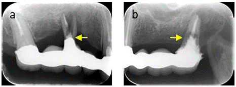
Figure 1: Preoperative radiographs. A) Radiographic image of the maxillary right molars. The yellow arrow indicates root fracture. B) Radiographs of the left maxillary molars. The yellow arrow indicates root fracture.
Initially, the patient was keen to preserve the teeth. However, we recommended an implant-supported bridge or a complete overdenture with magnetic attachments to the retained roots after extraction of teeth with root fracture. Eventually, the patient consented to tooth extraction and treatment with complete overdentures with magnetic attachments.
Clinical Procedure
First, we repaired the detached extensive maxillary prosthesis using a self-curing resin (UNIFAST; GC Dental Co. Ltd., Tokyo, Japan) after extracting teeth 15 and 27 (Figure 2a-e).
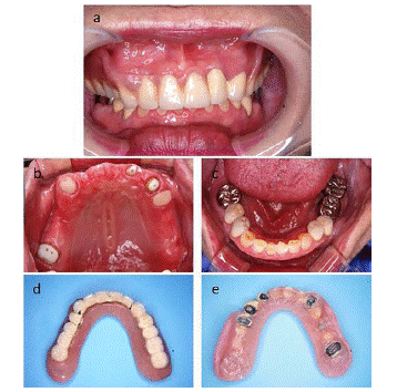
Figure 2: Intraoral photographs and images of the maxillary prosthesis after tooth extraction. A) Frontal view. B) Maxillary occlusal view. C) Mandibular occlusal view. D) Repaired and polished maxillary. E) Intaglio surface of the repaired maxillary prosthesis.
Thereafter, abutment teeth 17, 13, 22, 23, and 24 were prepared, gingival retraction (SURE-Cord plus; YOSHIDA Co. Ltd., Tokyo, Japan) was performed, and silicone rubber impressions (EXAHIFLEX; GC Dental Co. Ltd., and SILDE FIT; SHOFU INC., Kyoto, Japan) were made.
Wax patterns with an appropriate path of insertion were fabricated for the copings and keeper housings and cast using a gold-silver-palladium alloy. The upper surfaces of the keepers (GIGAUSS; GC Dental Co. Ltd.,) embedded in the copings were positioned in accordance with the occlusal plane (Figure 3a). Thereafter, magnet assemblies of various sizes (GIGAUSS C300, C600, D1000; GC Dental Co. Ltd.,) were fixed to the interim denture (Figure 3b).
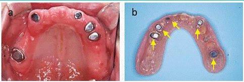
Figure 3: Prosthodontic pretreatment. A) Intraoral image shows copings embedded with keepers of various sizes on all abutment teeth. B) Intaglio surface of the repaired prosthesis used as an interim overdenture. Yellow arrows indicate the magnetic assemblies.
After observing the recovery of oral function, including esthetics, speech, and comfort, for several months, we proceeded with the fabrication of the definitive overdenture.
In the maxillary arch, a two-step impression, including muscle trimming using an impression compound (PERI COMPOUND; GC Dental Co. Ltd.,), was made using an individual dental tray (Figure 4a and 4b), and the previously described impression materials. Two weeks later, horizontal and vertical maxillo–mandibular relationships were recorded using record bases with occlusion rims and transferred to a semi-adjustable articulator. The vertical occlusal dimensions of the fabricated denture were approximately the same as those of the interim denture (Figure 4c).
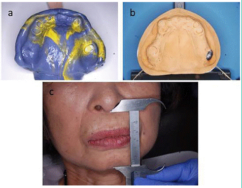
Figure 4: A) Maxillary impression using silicone impression material. B) Maxillary working model. C) The occlusal vertical dimensions are measured using calipers. The occlusal vertical dimension for producing the new overdenture was determined to be the same as that of the interim maxillary prosthesis.
After verification of the trial denture, the maxillary denture was completed. The mucosal and occlusal surfaces of the definitive denture were checked using pressure-indicating paste (FIT CHECKER; GC Dental Co. Ltd.,) and articulating paper (Mutsumi Chemical Industry, Mie, Japan), respectively, and equilibrated using dental carbide bars. Approximately four weeks later, the five magnetic assemblies were transferred from the interim denture to the definitive denture using a metal adhesive primer (METAL PRIMER Z; GC Dental Co. Ltd.,) and self-curing resin to fix the magnet after the dentures were placed in the maxilla. The overdenture was removed after polymerization of the resin, recontoured, and polished (Figure 5a and 5b).
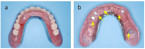
Figure 5: The images of maxillary definitive prosthesis. A) Polished surface of the definitive denture. B) Intaglio surface of the definitive denture. Yellow arrows indicate transferred magnetic assemblies.
Denture conformity, occlusion, and the periodontium of the remaining teeth were reevaluated at the one-year follow-up (Figure 6a-e).
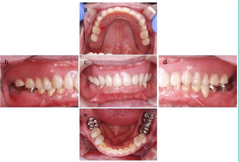
Figure 6: Postoperative intraoral photographs. A) Maxillary occlusal view. B) Right lateral view. C) Frontal view. D) Left lateral view. E) Mandibular occlusal view.
Discussion
Tooth loss caused by periodontal disease, dental caries, root fracture, tumors, or trauma leads to serious impairment of masticatory function, esthetics, and speech. The primary objective of prosthodontic treatment is to restore the function as much as possible. After tooth loss, the residual ridge plays a pivotal role in supporting dental prostheses in partially and completely edentulous patients [1]. Treatment of residual ridges with dentures improves oral function, including mastication, esthetics, and speech. However, many patients who use dentures are dissatisfied due to the instability of the prosthesis and impediments to speech and mastication [2,3]. Partial or complete overdentures overlying the abutment teeth and/or implants prevent alveolar bone resorption and exhibit improved retention and stability [4-13]. Further, various attachments, including bars, studs, and magnets, have been developed to augment the retention and stability [14]. Specialized prosthetic dentists should use appropriate attachments according to the clinical situation [15]. Generally, stud attachments are used to protect the exposed root dentin from the oral environment. Magnetic dental attachments are widely used in clinical practice because of their simplicity, compactness, and continuous force of attraction [16-18]. Currently, the principal components of dental magnetic attachments are samarium-cobalt (Sm-Co) or neodymium-iron-boron (Nd-Fe-B) alloys [19-26]. Conventionally, most dental magnetic attachments comprise a keeper-embedded coping within the abutment tooth root or dental implant and a magnet assembly incorporated into the overdenture. Although dental magnetic attachments are extremely small, a strong force of attraction is observed on approximating the keeper and magnetic assembly. Therefore, over-dentures with dental magnetic attachments are a promising treatment option for partially or completely edentulous patients. Previously, our department reported that differences in size influenced the force of attraction between various keepers and magnetic assemblies [27]. However, the application of magnetic dental attachments of various sizes in clinical practice has not been reported from our department. In this study, we repaired a failed prosthesis in the upper jaw. Next, we performed preprosthetic treatment using dental magnetic attachments of various sizes for the upper abutment teeth, and used the repaired prosthesis as an interim denture after tooth extraction necessitated by root fracture. Finally, a new horseshoe-shaped complete overdenture was fabricated and the magnetic dental assemblies were transferred from the interim denture. Herein, we emphasized the usefulness of magnetic dental attachments in prosthetic dentistry.
The stability and retention of a removable prosthesis are highly dependent on the condition of the remaining alveolar bone [1]. Several treatment options are available for the rehabilitation of missing molars, including removable prostheses or implant-retained fixed prostheses. However, dental implants may not always be feasible owing to tissue deficiency or underlying medical problems such as blood, heart, or liver disease.
Currently, most dental magnetic attachments consist of a keeper fixed to the abutment teeth/implant and a magnet assembly incorporated into the prosthesis. The magnetic attraction force is determined by the proximity of the keeper to the magnetic assembly. An overdenture with a dental magnetic attachment is a promising treatment option for abutment teeth with chronic periodontal disease because it improves poor clinical crown-to-root ratios and relieves the lateral stress on the abutment teeth [25,28-34]. Magnetic dental attachments can also be used for root-retained overdentures.
In this patient, we used the dental magnetic attachment GIGAUSS, which consists of a keeper made of stainless steel and a magnetic assembly made of Nd-Fe-B alloys. The greatest advantage of this hybrid device is that it exhibits a small and robust force of attraction. The size of the elliptical or circular magnetic attachments in this device is determined based on the dimensions of the abutment teeth [27]. The smallest dental magnetic attachment has a diameter of 3 mm, a force of attraction of approximately 400 gf, and is applied on the roots of the anterior teeth. Conversely, the largest dental magnetic attachment is 4.9mm in diameter, has a force of attraction of approximately 1000 gf, and is applied on the roots of the molar teeth. Hence, we used dental magnetic attachments of various sizes with different forces of attraction on the abutment teeth.
In this patient, prosthetic retreatment using a fixed prosthesis was ruled out because root fractures were observed in teeth 15 and 27. Although treatment using a denture after tooth extraction was planned, we had to reduce the patient's anxiety regarding the form and occlusion of the denture. Therefore, we repaired and used the failed fixed prosthesis as an interim denture, and the patient reported no discomfort due to occlusion. Additionally, we used five dental attachments with small and robust forces of attraction on the repaired interim denture and abutment root surfaces. As the stability and retention of the interim denture increased, we were able to resolve the patient’s discomfort by repairing the denture modestly.
Subsequently, a definitive horseshoe-shaped complete overdenture was fabricated to replicate the form of the interim denture. As minimum equilibration was required, the magnetic assemblies were transferred from the interim denture early. Therefore, the treatment period, including tooth extraction was only approximately seven months.
The procedures described in this report may increase the treatment options for partially or completely edentulous patients, and overdentures with dental magnetic attachments may contribute to rehabilitation of oral function.
Author Statements
Author Contributions
Conceptualization, H.Y.; Methodology, H.Y., D.A., T.K., and Y.H.; Validation, T.I., N.T., and T.O.; Investigation, J.I., I.K., and A.S.; Resources, T.N.; Data curation, D.A., T.I., and Y. H.; Writing—original draft, H.Y and D.A.; Writing — review and editing, D.A.; Visualization, H.Y.; Supervision, Y.H.; Project administration, D.A.; Funding acquisition, H.Y.
All authors have read and agreed to the published version of the manuscript.
Funding
This research was supported in part by JSPS KAKENHI (grants 23K09282) and Nihon University School of Dentistry Sato Funds (2023).
Acknowledgments
The authors thank Giko Co., Ltd., Fukuoka, Japan for the technical support and Editage (www.editage.com) for English language editing.
Conflicts of Interest
The authors declare no conflicts of interest. The funders had no role in the study design; collection, analyses, or interpretation of data; writing of the manuscript; or decision to publish the results.
References
- Kaku M, Akiba Y, Akiyama K, Akita D, Nishimura M. Cell-based bone regeneration for alveolar ridge augmentation - Cell source, endogenous cell recruitment and immunomodulatory function. J Prosthodont Res. 2015; 59: 96-112.
- Ellis JS, Pelekis ND, Thomason JM. Conventional rehabilitation of edentulous patients: the impact on oral health-related quality of life and patient satisfaction. J Prosthodont. 2007; 16: 37-42.
- Turkyilmaz I, Company AM, McGlumphy EA. Should edentulous patients be constrained to removable complete dentures? The use of dental implants to improve the quality of life for edentulous patients. Gerodontology. 2010; 27: 3-10.
- Morrow RM, Feldmann EE, Rudd KD, Trovillion HM. Tooth-supported complete dentures: an approach to preventive prosthodontics. J Prosthet Dent. 1969; 21: 513-522.
- Morrow RM, Powell JM, Jameson WS, Jewson LG, Rudd KD. Tooth-supported complete dentures: description and clinical evaluation of a simplified technique. J Prosthet Dent. 1969; 22: 415-424.
- Dodge CA. Prevention of complete denture problems by use of “overdentures”. J Prosthet Dent. 1973; 30: 403-411.
- Lord JL, Teel S. The overdenture: patient selection, use of copings, and follow-up evaluation. J Prosthet Dent. 1974; 32: 41-51.
- Reitz PV, Weiner MG, Levin B. An overdenture survey: preliminary report. J Prosthet Dent. 1977; 37: 246-258.
- Rissin L, House JE, Manly RS, Kapur KK. Clinical comparison of masticatory performance and electromyographic activity of patients with complete dentures, overdentures, and natural teeth. J Prosthet Dent. 1978; 39: 508-511.
- Crum RJ, Rooney GE Jr. Alveolar bone loss in overdentures: a 5-year study. J Prosthet Dent. 1978; 40: 610-613.
- Fenton AH, Hahn N. Tissue response to overdenture therapy. J Prosthet Dent. 1978; 40: 492-498.
- Ettinger RL, Taylor TD, Scandrett FR. Treatment needs of overdenture patients in a longitudinal study: five-year results. J Prosthet Dent. 1984; 52: 532-537.
- Parel SM. Implants and overdentures: the osseointegrated approach with conventional and compromised applications. Int J Oral Maxillofac Implants. 1986; 1: 93-99.
- Langer Y, Langer A. Root-retained overdentures: Part I--Biomechanical and clinical aspects. J Prosthet Dent. 1991; 66: 784-789.
- Al-Jallad W. Tooth supported overdenture for partially edentulous denture using magnetic attachment -a case report. J Dental Health Oral Res. 2020; 1: 1-18.
- Gillings BR. Magnetic retention for complete and partial overdentures. Part I. J Prosthet Dent. 1981; 45: 484-491.
- Akin H, Coskun ME, Akin EG, Ozdemir AK. Evaluation of the attractive force of different types of new-generation magnetic attachment systems. J Prosthet Dent. 2011; 105: 203-207.
- Khoo HD, Chai J, Chow TW. Prosthetic outcome, patient complaints, and nutritional effects on elderly patients with magnet-retained, implant-supported overdentures-a 1-year report. Int J Oral Maxillofac Implants. 2013; 28: 1278-1285.
- Becker JJ. Permanent magnets. Sci Am. 1970; 233: 92–100
- Strnat KJ. The hard-magnetic properties of rare earth-transition metal alloys. IEEE Trans Magn. 1972; 8: 511–516.
- Tawara Y, Strnat KJ. Rare earth-cobalt permanent magnets near the 2–17 composition. IEEE Trans Magn. 1976; 12: 954–958.
- Sagawa M, Fujimura S, Yamamoto H, Matsuura Y, Hiraga K. Permanent magnet materials based on the rare earth-iron-boron tetragonal compounds. IEEE Trans Magn. 1984; 20: 1584–1589.
- Croat JJ, Herbst JF, Lee RW, Pinkerton FE. Pr-Fe and Nd-Fe-based materials: a new class of high-performance permanent magnets(invited). J Appl Phys 1984; 55: 2078–2082.
- Sagawa M, Fujimura S, Togawa N, Yamamoto H, Matsuura Y. New material for permanent magnets on a base of Nd and Fe (invited). J Appl Phys. 1984; 55: 2083–2087.
- Riley MA, Walmsley AD, Harris IR. Magnets in prosthetic dentistry. J Prosthet Dent. 2001; 86: 137-142.
- Anupam P, Anandakrishna GN, Vibha S, Suma J, Shally K. Mandibular overdenture retained by magnetic assembly: a clinical tip. J Indian Prosthodont Soc. 2014; 14: 328-333.
- Hasegawa M, Umekawa Y, Nagai E, Ishigami T. Retentive force and magnetic flux leakage of magnetic attachment in various keeper and magnetic assembly combinations. J Prosthet Dent. 2011; 105: 266-271.
- Sasaki H, Kinouchi Y, Tsutsui H, Yoshida Y, Karv M, Ushita T. Sectional prostheses connected by samarium–cobalt magnets. J Prosthet Dent. 1984; 52: 556–558.
- Obatake RM, Collard SM, Martin J, Ladd GD. The effects of sodium fluoride and stannous fluoride on the surface roughness of intraoral magnet systems. J Prosthet Dent. 1991; 66: 553–558.
- Akaltan F, Can G. Retentive characteristics of different dental magnetic systems. J Prosthet Dent. 1995; 74: 422–427.
- Van Waas MA, Kalk W, van Zetten BL, van Os JH. Treatment results with immediate overdentures: an evaluation of 4.5 years. J Prosthet Dent. 1996; 76: 153–157.
- Matsumura H, Kawasaki K. Magnetically connected removable sectional denture for a maxillary defect with severe undercut: a clinical report. J Prosthet Dent. 2000; 84: 22–26.
- Maeda Y, Nakao K, Yagi K, Matsuda S. Composite resin root coping with a keeper for magnetic attachment for replacing the missing coronal portion of a removable partial denture abutment. J Prosthet Dent. 2006; 96: 139–142.
- Boeckler AF, Morton D, Ehring C, Setz JM. Mechanical properties of magnetic attachments for removable prostheses on teeth and implants. J Prosthodont. 2008; 17: 608–615.