Research Article
Austin J Dent. 2023; 10(1): 1174.
Clinical Relevance of Dental Parameters and Symptoms in Patients Suffering from TMD and Tinnitus and a Randomised Trial Measuring Symptom Relief after Using a Relaxation Device
Marie Tullberg, DDS¹*; Mattias Billing DDS, MSc¹; Göran Laurell MD, PhD²
1Specialist in Orofacial Pain and Function and Periodontology, Brahekliniken Stockholm, Sweden
2Professor at the Department of Surgical Sciences, Otorhinolaryngology and Head and Neck Surgery, Uppsala University, Sweden
*Corresponding author: Marie Tullberg, DDS Brahekliniken, Brahegatan 36, 114 37 Stockholm, Sweden. Email: marie.tullberg@brahekliniken.se
Received: June 02, 2023 Accepted: July 01, 2023 Published: July 08, 2023
Abstract
Aim: Tinnitus is a common symptom in dental patients presenting with Temporomandibular Disorders (TMD). In this study, 66 patients were examined and underlying variations in dental appearance, jaw position, jaw joint health, radiographic findings, and symptoms were recorded. The patients were randomly placed into either an intervention or a control group to assess the effectiveness of a relaxation device aiming to reduce tinnitus.
Method: 100 patients referred for TMD problems and suffering from tinnitus were asked to participate in the study. 66 patients completed two questionnaires before the first consultation with a specialist in orofacial pain and function and 46 of them completed the randomised trial.
Results: TMD patients most likely to be suffering from tinnitus had experienced stress, felt tension in the jaw or presented with neck problems. Clinical dental examinations revealed that these patients displayed a deep bite, had a click or scrape sound from their jaw joints and/or had parafunctional habits (grinding or clenching their teeth). Patients in the relaxation device group reported significant improvement at p<0.01 compared with control group after 4 months treatment.
Conclusion: This study presents general clinical signs and criteria that also non-dental medical professionals can use as a guide for referring tinnitus patients for specialist dental care. Patients under stress who are clenching or grinding their teeth, have missing teeth, midline shift and experience sounds from their jaw joints could benefit from examination by a dental specialist and the use of a relaxation device.
Keywords: Tinnitus; Temporomandibular joint; Risk factor; Symptoms; Treatment
Introduction
Previous dental studies have indicated that certain developmental positions of the upper and lower jaw and the teeth as well as dental and orthodontic therapeutic interventions can cause certain painful or uncomfortable symptoms in patients later in life. These are known as Temporomandibular Disorders (TMD) [1–3]. Other studies have reported that the pathological position of the jaws or disadvantageous of occlusal patterns do not because TMD, instead it is reported that the long-term effect of mastication and dysfunctional habits leading to stress that is a major source [4–6]. In this study we assess the subjective variations in the occlusion and bite in tinnitus patients presenting with TMD.
Reference
Dental (7), surgical (8) and orthodontic (9) textbooks teach us that the adult position and function of the upper and lower jaw depends on the skeletal development and on factors such as digit sucking habits, missing teeth (both aplasia and extractions), breathing challenges and trauma, previous dental or orthodontic intervention as well as parafunctional activity of the muscles and ligaments and the positioning of the condyle heads and discs in the temporomandibular joint. Many patients do not experience problems although they present with the most severe functional deviation and bite discrepancies. However, other patients with minor discrepancies can experience debilitating pain and symptoms possibly indicating that some interpersonal sensitivity to stress and pain influence on the patient’s experience [10,11]. Several studies demonstrate that tinnitus is more common among patients with TMD, which could be related to an increased muscular tonus caused by parafunction through teeth grinding and clenching of the jaws, particularly at night [12,13]. There are also suggested links between the presence of tinnitus in stressed patients with TMD [14].
Previous estimates suggest that approximately 10–20% of the European population suffer from tinnitus [15]. However, a study by Hasson et al. of approximately 9,756 individuals [16] found the prevalence of tinnitus in Sweden to be as high as 25%. Tinnitus is defined as an individual hearing a noise without external stimuli [14]. In most cases, tinnitus is reported by patients with sensorineural hearing loss such as presbycusis, acquired hearing loss after noise trauma, or use of ototoxic medications. The pathophysiology behind tinnitus is still unknown and there are reasons to believe that the site of pathophysiological mechanisms can vary and relate to lesions at different sites in the cochlea, central auditory pathways, or structures in auditory cortex [17–21]. Functional tests using SPECT and MR have also found that several locations in the CNS are involved in patients with chronic tinnitus [22,23].
Somatosensory tinnitus is when the tinnitus can be modulated by somatic stimulation or movement by for instance, the eyes or jaws. Somatic modulation has been reported to be observed in up to 83% of tinnitus patients, which could indicate that in some patients somatic or somatosensory tinnitus has its origin in disharmony and tension in the jaws and that this tension could be reversed by adjusting the lower jaw to a relaxed and comfortable position [24–27].
In this study, we first wanted to assess if there is an association between symptoms and certain dental parameters or jaw deviations in patients presenting with TMD and suffering from tinnitus. We also wanted to investigate the effectiveness of a jaw relaxation device in these TMD patients and see whether it could provide symptomatic tinnitus relief and could be investigated in future research and tinnitus referral frameworks or used as a comparison test in treatment outcomes.
Material and Methods
Patients with a combination of TMD and chronic tinnitus (chronic referring to duration of over 6 months) were examined by a dental specialist in orofacial pain and function in a specialist dental clinic in the centre of Stockholm, with the aim to investigate the association between TMD and tinnitus.
To participate in the study, the patients must have previously undergone a consultation with an ENT specialist or audiologist and been unable to receive tinnitus treatment from healthcare services.
Patients were excluded if they were undergoing any therapy that could affect the outcome of the present intervention, such as Cognitive Behavioural Therapy (CBT), dental treatment or soft tissue laser muscle treatment, or physiotherapy. Patients received two questionnaires via post before attending the clinic – a HADS questionnaire [28] and a non-validated study-specific tinnitus questionnaire. They also received written information about the study.
The HADS scale was used to determine whether the patients were affected by anxiety and/or depression. In contrast, the study-specific tinnitus questionnaire asked patients to specify the variety of symptoms they had, their onset, and complaints. There were also questions covering previous dental history and history of specialist dental treatments.
Patients participating in the randomised study of the relaxation device were asked to complete a follow-up questionnaire both at the start and upon completion of the study. This questionnaire asked about the symptoms of the tinnitus sounds they heard in terms of pitch, modulation, and impact.
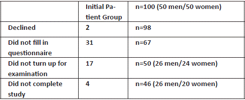
100 patients were offered appointments to participate in the study, and 98 agreed. 32 patients did not fill in the forms correctly, failed to show up for a clinical examination or declined treatment. We were then left with 66 patients suitable for assessment, all having consented to the study. These patients underwent a clinical evaluation of their teeth, bite, and jaw relationship. Clinical photographs of the patient’s teeth and face were taken during this examination, and measurements relating to a deviation from a neutral Class I bite were recorded. An OPG radiograph was also taken to exclude any underlying dental pathology that could interfere with the study or needed immediate treatment.
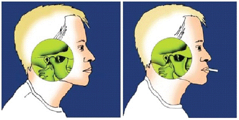
The patients were randomly assigned to an intervention group and a control group by terms of lottery before they came to see the specialist for the assessment. Patients in the intervention group received a bag of 5 relaxation pegs (Gapnap®, Sweden), to use to relieve the muscular tension in the jaws, head, and neck region. These patients received written instructions on how to use the device but received no verbal information to keep the study as non-biased as possible and patients were asked to fill in a diary to measure compliance. They were also advised to use the peg several hours per day, especially when reading, driving, working on the computer or online, watching television and even playing golf. The control group received no treatment. Figure 1 shows the placement of the relaxation device between the lips as per initial instructions.

Figure 1: Showing the movement of the condyle head to an anterior-inferior position.

Chart 1:

Chart 3: Participant flow diagram.
The study was approved by the regional ethical committee in Stockholm under 2010/1415-31/3.
Results
Tinnitus Questionnaire and HADS
The study-specific tinnitus questionnaire found that although 66% of patients reported that they could hear their tinnitus during all waking hours, only 50% complained of tinnitus intervening with their daily life but rose to 75% who reported their tinnitus was more severe in the evening. This may explain the 50% of the patients who also reported that tinnitus could affect sleep and why 10% of patients had to take sleeping tablets to help them fall asleep.
When questioned about what the patients thought caused the onset of their tinnitus, 62% reported suffering from tension in their jaws, 57% were under stress, 52% had neck problems, and interestingly only 32% thought that traumatic noise was the reason. It was also noted that 67% of patients could modulate the sound by moving their lower jaw or stimulate their face in other ways, indicating some form of somatosensory tinnitus or other form of tinnitus. Tinnitus characteristics are shown in Table 1.
    Whizzing

    42(63%)


    Rippling

    2(3%)


    Roaring

    31(46%)


    Ringing

    17(26%)


    Pulsing

    16(24%)


    Always the same

    21(31%)


    Varying

    39(58%)


    Can disappear

    10(15%)


    Always present

    44(66%)
Table 1: Tinnitus characteristics (more than one alternative was possible).
From the responses, we found that over 50% of the patients reported hyperacusis, jaw locking, fatigue, facial pain and clicking from the jaw joints. This was supported by the patients naming their parafunctional habits presented in Table 2. More than half of the participants reported that when stressed, they would grind their teeth, tense their jaw muscles, and report more tooth activity.
    Facial pain

    40(60%)


    Headache

    26(39%)


    Pain when    moving the jaws

    23(35%)


    Pain when    opening mouth

    21(32%)


    Fatigue

    42(64%)


    Clicking in the jaws

    37(56%)


    Crepitation

    21(32%)


    Sensitive teeth

    16(24%))


    Vertigo

    23(35%)


    Hyperacusis

    47(71%)


    Locking

    43(65%)
Table 2: Clinical features, symptoms, and characteristics.
The HADS scale showed that 51% of patients were more than moderately anxious and 30% of our patients were depressed. A third of the latter individuals also admitted being depressed to the extent that they needed treatment (Tables 3 and 4).
    1 not so    much

    33(49 %)


    2 moderate

    19(29%)


    3 very    much

    15(22%)
Table 3: HADS scale (anxiety).
    1 not depressed

    47(70%)


    2 depressed

    14(21%)


    3 may need treatment

    6(9%)
Table 4: HADS scale (depression).
Clinical Assessment of Bite and Dental Occlusion (n=50)
50 of the 66 patients were able to attend the clinical evaluation. The group consisted of 26 men and 24 women, and the average age was 47 years.
Clinically, half of the 50 patients showed an overjet of more than 2mm horizontally compared to the lower front teeth. Interestingly, 31 patients (62%) presented with a vertically deep bite of more than 2mm and 32 patients (64%) demonstrated a click or crepitation from the jaw joints. The latter is normally caused by pressure and tension in the area around the disc complex, as the condyle head projects upwards and backwards towards the disc, forcing it to move due to the lack of posterior height in the bite. We also noted that 41 patients (82%) presented with a midline shift and 21 patients (42%) were missing one or more premolars (Table 5).
    Clinical findings

    n=50


    Max opening


    <40 mm

    4

    8%


    >40 mm

    46

    92%


    Deep bite >2 mm

    31

    62%


    Overjet >2 mm

    25

    50%


    Discrepancy of Midline right or left

    41

    82%


    Any tooth missing including wisdom teeth

    44

    88%


    Missing 1–4 premolars

    21

    42%


    Pain in TMJ

    19

    38%


    Clicking or crepitation

    32

    64%
Table 5: Jaw discrepancies and tooth anomalies in 50 TMD patients with tinnitus.
Effect of Relaxation Peg use N=50
In total, 21 individuals in the intervention group and 25 in the control group completed the randomised study. There were 10 men and 11 women left in the relaxation peg group and 16 men and 9 women in the control group. The mean age of men in the intervention and control group was identical at 40 years, and the mean age of the women in the two groups was 50 years. Nine out of 21 patients (48%) using the relaxation device reported positive effects and experienced reduced severity of tinnitus. The outcome was proven significant in a chi-square test (p<0.01) and confirmed with the Fisher Exact test for small patient groups. Perhaps even more important, the likelihood for the tinnitus to be more severe after 4 months was three times as high in the control group as in the treatment group and significant by chi-square test (p<0.05).
Discussion
Although the heterogeneity of tinnitus aetiology is well established, there is controversy whether temporomandibular joint problems can cause a subtype of this phantom perception. 66 patients with TMD and tinnitus entered a randomised study aimed to evaluate the effect of a relaxation device on tinnitus. At baseline the patients answered two questionnaires. A main finding of our study revealed that the majority of tinnitus patients with TMD and tinnitus were aware of stress and holding tension in their jaws, something that has also been reported in earlier studies [29–32]. The HADS scale found that 50% of the patients reported anxiety and 30% depression which were expected, as most patients had been living with tinnitus for over 3 years. An interesting observation was that the majority could modulate the tinnitus sound by moving their lower jaw or stimulating their face in other ways, which demonstrated that they were suffering from the somatosensory tinnitus variant. Recent European guidelines have also mentioned referring such patients to dentists or orofacial pain and function specialists [33].
In the second part of the study, we investigated factors related to the bite and occlusion that could be attributed to the presence of tinnitus in TMD patients, as earlier studies have indicated that certain occlusal and jaw relationships were more prevalent among TMD patients [34–37]. Over 50% of the patients presented with an overjet greater than 2mm, a deep bite with anterior overlap of 2mm, a midline shift or crepitation form the jaw joints. Interestingly, as the average age of our patients was 47, only 18% of patients were suffering from hearing loss whereas 71% reported having hyperacusis. If we compare the low number of patients with hearing loss with the high level of patients presenting with jaw joint clicks or scraping sounds, it would be recommended that these signs of tinnitus at a younger age should also require a referral to a dental specialist or clinician working in this field. It is worth mentioning that this practice based study was conducted in 2010/11 and despite the examiner not having been calibrated to use the RDC-TMD, the standard format for orofacial pain and related disorders, the results of this study still remains the same for TMD patients as a cohort.
It is known that increased activity in the masseter and temporalis muscles can cause increased pressure between the upper and lower jaws and could lead to temporary or permanent displacement of the disc due to laxity in the surrounding ligaments and muscles. It has been reported that tinnitus is more common in these patients [38–40]. It should also be noted that the duration of the force when clenching the jaws during night can be longer than during daytime parafunction due to the lack of inhibitive awareness [41]. This may be why sleep bruxism has regularly been quoted as a contributing factor for TMD.
Thirdly, we wanted to evaluate if a relaxation peg could help to relieve chronic tinnitus. Among the patients in the intervention group, nine patients reported a reduction in tinnitus after 4 months; nine were unchanged and three patients had felt an increase in symptoms. In the control group, only one patient reported an improvement of tinnitus, 13 were unchanged and 11 experienced an increase in symptoms. It would be of clinical interest to perform a large-scale randomised study to compare peg use with active physiotherapy [42] or CBT [43] or noise modulation therapy [44].
Finally, from a socio-economic point [45], an aspect often forgotten in clinical discussions, we found that that in the group of patients receiving relaxation peg treatment, the percentage of patients on sick leave decreased from 38% to 21%.
There is an abundance of literature available pointing in various directions regarding the aetiology and possible treatments of tinnitus in patients with TMD. The present study further supports that no opportunity should be missed to help this patient group, as tinnitus can be an extremely debilitating condition.
Conclusion
The clinical implication of this study points to several general symptoms and visual clinical parameters that a non-dental medical professional can use as a guide for to referring patients with tinnitus to a dental specialist in orofacial pain and function, for further investigations. Although the small final sample size may give a weighted result and further studies on larger patient cohorts could give us better statistical evidence, our results show a clinical significance between the control group and the treatment group who used a relaxation peg.
Key Findings
Patients under stress who were aware of clenching and grinding their teeth, presented with missing teeth, had a midline shift, and experienced clicking or scraping sounds from their jaw joints, could benefit from a thorough dental examination to investigate their orofacial and functional jaw relationships to determine who would benefit from the use of the relaxation device shown here, to reverse their parafunctional habits and alleviate their tinnitus symptoms.
Author Statements
Author Contributions
Marie Tullberg. Study design, Patient examination, data analysis, ethical approval; Mattias Billing.
Manuscript writing, Data analysis; Göran Laurell. Data analysis, approval of final manuscript
Disclosures
Competing interests: Marie Tullberg has a financial interest in the Gapnap® relaxation device. However, the data presented has been reviewed by the other authors to avoid any unintentional weighting or bias in favour of the device.
Funding
Marie Tullberg received an initial grant from Kvinnliga Tandläkarklubben in 2009, however self-funded by the other authors.
References
- Lundh H, Westesson PL. Long-term follow-up after occlusal treatment to correct abnormal temporomandibular joint disk position. Oral Surg Oral Med Oral Pathol. 1989; 67: 2-10.
- John ZAS, Shrivastav SS, Kamble R, Jaiswal E, Dhande R. Three-dimensional comparative evaluation of articular disc position and other temporomandibular joint morphology in Class II horizontal and vertical cases with Class I malocclusion. Angle Orthod. 2020; 90: 707-14.
- Tervahauta E, Närhi L, Pirttiniemi P, Sipilä K, Näpänkangas R, et al. Prevalence of sagittal molar and canine relationships, asymmetries, and midline shift in relation to temporomandibular disorders (TMD) in a Finnish adult population. Acta Odontol Scand. 2022; 80: 470-80.
- Aboalnaga AA, Amer NM, Elnahas MO, Salah Fayed MM, Soliman SA, et al. Fahim malocclusion and temporomandibular disorders: verification of the controversy. J Oral Facial Pain Headache. Fall 2019; 33: 440-50.
- Manfredini D, Lombardo L, Siciliani G. Temporomandibular disorders and dental occlusion. A systematic review of association studies: end of an era? J Oral Rehabil. 2017; 44: 908-23.
- Stone JC, Hannah A, Nagar N. Nathan Nagar Dental occlusion and temporomandibular disorders. Evid Based Dent. 2017; 18: 86-7.
- Herbert T. Shillingburg Fundamentals of Fixed Prosthodontics Quintessence Publishing (IL). 4th rev ed 2012.
- William Arnett G, Richard P. McLaughlin facial and dental planning for orthodontists and oral surgeons. Mosby. 2004.
- Proffit W, Fields H, Larson B, Sarver D. Contemporary orthodontics. 6th ed. Mosby. 2018.
- Slade GD, Ohrbach R, Greenspan JD, Fillingim RB, Bair E, et al. Painful temporomandibular disorder: decade of discovery from OPPERA studies. J Dent Res. 2016; 95: 1084-92.
- Janal MN, Lobbezoo F, Quigley KS, Raphael KG. Stress-evoked muscle activity in women with and without chronic myofascial face pain. J Oral Rehabil. 2021; 48: 1089-98.
- Fernandes G, van Selms MK, Gonçalves DA, Lobbezoo F, Camparis CM. Factors associated with temporomandibular disorders pain in adolescents. J Oral Rehabil. 2015; 42: 113-9.
- Beddis H, Pemberton M, Davies S. Sleep bruxism: an overview for clinicians. Br Dent J. 2018; 225: 497-501.
- Edvall NK, Gunan E, Genitsaridi E, Lazar A, Mehraei G, et al. Impact of temporomandibular joint complaints on tinnitus-related distress. Front Neurosci. 2019; 13: 879.
- Schlee W et al. Towards a unification of treatments and interventions for tinnitus patients: the EU research and innovation action UNITIProg. Brain Res. 2021; 260: 441-51.
- Hasson D, Theorell T, Wallén MB, Leineweber C, Canlon B. Stress and prevalence of hearing problems in the Swedish working population. BMC Public Health. 2011; 11: 130.
- Lv H, Zhao P, Liu Z, Li R, Zhang L, et al. Abnormal resting-state functional connectivity study in unilateral pulsatile tinnitus patients with single etiology: A seed-based functional connectivity study. Eur J Radiol. 2016; 85: 2023-9.
- Lee AC, Godfrey DA. Godfrey DA Current view of neurotransmitter changes underlying tinnitus. Neural Regen Res. 2015; 10: 368-70.
- Chen GD, Sheppard A, Salvi R. Noise trauma induced plastic changes in brain regions outside the classical auditory pathway. Neuroscience. 2016; 315: 228-45.
- Pilati N, Large C, Forsythe ID, Hamann M. Acoustic over-exposure triggers burst firing in dorsal cochlear nucleus fusiform cells. Hear Res. 2012; 283: 98-106.
- Gauvin DV, Yoder JD, Tapp RL, Baird TJ. Small compartment toxicity: CN VIII and quality of life: hearing loss, tinnitus, and balance disorders. Int J Toxicol. 2017; 36: 8-20.
- Farhadi M, Mahmoudian S, Saddadi F, Karimian AR, Mirzaee M, et al. Functional brain abnormalities localized in 55 chronic tinnitus patients: fusion of SPECT coincidence imaging and MRI. J Cereb Blood Flow Metab. 2010; 30: 864-70.
- Ueyama T, Donishi T, Ukai S, Yamamoto Y, Ishida T, et al. Alterations of regional cerebral blood flow in tinnitus patients as assessed using single- photon emission computed tomography. PLOS ONE. 2015; 10: e0137291.
- Schiffman E, Ohrbach R, Truelove E, Look J, Anderson G, et al. Diagnostic Criteria for Temporomandibular Disorders (DC/TMD) for Clinical and Research Applications: recommendations of the International RDC/TMD Consortium Network* and Orofacial Pain Special Interest Group†. J Oral Facial Pain Headache. Winter 2014; 28: 6-27.
- Ward J, Vella C, Hoare DJ, Hall DA. Subtyping somatic tinnitus: A cross-sectional UK cohort study of demographic, clinical and audiological characteristics. PLoS One. 2015; 10: e0126254.
- Levine RA, Nam EC, Oron Y, Melcher JR. Evidence for a tinnitus subgroup responsive to somatosensory based treatment modalities. Prog Brain Res. 2007; 166: 195-207.
- Won JY, Yoo S, Lee SK, Choi HK, Yakunina N, et al. Prevalence and factors associated with neck and jaw muscle modulation of tinnitus. Audiol Neurootol. 2013; 18: 261-73.
- Bjelland I, Dahl AA, Haug TT, Neckelmann D. The validity of the Hospital Anxiety and Depression Scale. An updated literature review. J Psychosom Res. 2002; 52: 69-77.
- Björne A. Assessment of temporomandibular and cervical spine disorders in tinnitus patients. Prog Brain Res. 2007; 166: 215-9.
- Pezzoli M, Ugolini A, Rota E, Ferrero L, Milani C, et al. Tinnitus and its relationship with muscle tenderness in patients with headache and facial pain. J Laryngol Otol. 2015; 129: 638-43.
- Kang JH, Song SI. Autonomic and psychologic risk factors for development of tinnitus in patients with chronic temporomandibular disorders. J Oral Facial Pain Headache. 2019; 33: 362-70.
- Edward F. Wright Otologic symptom improvement through TMD therapy. Quintessence Int. 2007; 38: 564-71.
- Cima RFF, Mazurek B, Haider H, Kikidis D, Lapira A, et al. A multidisciplinary European guideline for tinnitus: diagnostics, assessment, and treatment. HNO. 2019; 67: 10-42.
- Ingervall B. Thilander B. Activity of temporal and masseter muscles in children with lateral forced bite. Angle Orthod. 1975; 45: 249-58.
- Mohlin B, Kopp S. A clinical study on the relationship between malocclusions, occlusal interferences and mandibular pain and dysfunction. Swed Dent J. 1978; 2: 105-12.
- Bilgiç F, Gelgör IE. Prevalence of temporomandibular dysfunction and its association with malocclusion in children: an epidemiologic study. J Clin Pediatr Dent. 2017; 41: 161-5.
- de Kanter RJAM, Battistuzzi PGFCM, Truin G-J. Temporomandibular disorders: occlusion matters! Pain Res Manag. 2018; 2018: 8746858.
- Buergers R, Kleinjung T, Behr M, Vielsmeier V. Is there a link between tinnitus and temporomandibular disorders? J Prosthet Dent. 2014; 111: 222-7.
- Porto De Toledo I, Stefani FM, Porporatti AL, Mezzomo LA, Peres MA, et al. Prevalence of otologic signs and symptoms in adult patients with temporomandibular disorders: a systematic review and meta-analysis. Clin Oral Investig. 2017; 21: 597-605.
- Manfredini D, Olivo M, Ferronato G, Marchese R, Martini A, et al. Prevalence of tinnitus in patients with different temporomandibular disorders symptoms. Int Tinnitus J. 2015; 19: 47-51.
- Lee SKY, Salinas TJ, Wiens JP. The effect of patient specific factors on occlusal forces generated: best evidence consensus statement. J Prosthodont. 2021; 30: 52-60.
- Delgado de la Serna P, Plaza-Manzano G, Cleland J, Fernández-de-Las-Peñas C, Martín-Casas P, et al. Effects of Cervico-mandibular manual therapy in patients with temporomandibular pain disorders and associated somatic tinnitus: A randomized clinical trial. Pain Med. 2020; 21: 613-24.
- Fuller T, Cima R, Langguth B, Mazurek B. Johan Ws Vlaeyen, Derek J Hoare Cognitive behavioral therapy for tinnitus. Cochrane Database Syst Rev. 2020; 1: CD012614.
- Schoisswohl S, Arnds J, Schecklmann M, Langguth B, Schlee W, et al. Amplitude modulated noise for tinnitus suppression in tonal and noise-like tinnitus. Audiol Neurootol. 2019; 24: 309-21.
- Maes IH, Cima RF, Vlaeyen JW, Anteunis LJ, Joore MA. Tinnitus: a cost study. Ear Hear. 2013; 34: 508-14.