Research Article
Austin J Dent. 2023; 10(2): 1175.
The Visibility and Affecting Factors of Bifid Mandibular Condyles on Panoramic Radiography
Ayse Zeynep Zengin¹*; Ahmet Eren Karabiyik²; Kübra Cam²; Hande S Dogruer²; Lale Rizeli²; Ayse Pinar Sumer³
1Associate Professor, Department of Dentomaxillofacial Radiology, Faculty of Dentistry, Ondokuz Mayis University, Turkey
2Research Assistant, Department of Dentomaxillofacial Radiology, Faculty of Dentistry, Ondokuz Mayis University, Turkey
3Professor, Research Assistant, Department of Dentomaxillofacial Radiology, Faculty of Dentistry, Ondokuz Mayis University, Turkey
*Corresponding author: Ayse Zeynep Zengin Associate Professor, Department of Dentomaxillofacial Radiology, Faculty of Dentistry, Ondokuz Mayis University, Turkey. Email: dtzeynep78@yahoo.com.tr
Received: September 15, 2023 Accepted: October 06, 2023 Published: October 13, 2023
Abstract
Aim: The aim of this study was to determine the visibility of Bifid Mandibular Condyle (BMC) and affecting factors on panoramic radiography compared with Cone Beam Computed Tomography (CBCT).
Material and Methods: Radiologically depression or notch on superior condylar surface or duplication of condylar head with continuous cortex on CBCT images was considered as BMC. Characteristics of BMC such as location, type, groove type, depth, and horizontal angle on CBCT images were noted. Panoramic radiographs of 63 BMC and 65 normal mandibular condyles confirmed on tomographic images were evaluated by three groups of observers with different experiences.According to the results, the common correct and incorrect estimations were compared.
Results: There was no statistically significant difference between the results of the observers.
Despite there was no statistically significant difference between the characteristics of BMC according to whether the estimations were correct or incorrect. (p>0,050), it was determined that the horizontal angle of the BMC that observers stated correctly was 20.5% less than the cases with bifid condyles that they stated incorrectly.
Conclusion: In conclusion, experience of clinicians had no effect on the visibility of BMC. Panoramic radiographs frequently misread bifidity and bifidity was only confirmed by the CBCT. However, it can be estimated that when the angle between the condylar head and the horizontal plane become closer, BMC is more likely to be detected on panoramic radiographs. Further studies are required to determine which factors have effect on visibility of BMC on panoramic images.
Keywords: Bifid mandibular condyle; Panoramic radiography; Cone beam computed tomography
Introduction
Bifid Mandibular Condyle (BMC) appeared as a double condylar head characterized by a vertical depression, notch or deep cleft in the center of the condylar head [1]. They are usually diagnosed by a panoramic radiography during a routine examination [2]. The panoramic radiography is useful for providing a broad overview of the Temporomandibular Joint (TMJ) and surrounding structures. Gross osseous changes in the condyles can be identified, such as asymmetries, extensive erosions, large osteophytes, tumors, or fractures. However panoramic radiographs can misread bifidity by the overlapping of anatomical structures or inherent distortion. They can either under or overestimate bifidity [3]. When a detailed assessment of bony structures of TMJ is needed, the panoramic view should be supplemented with advanced imaging techniques [1].
To avoid excessive radiation, clinicians can prefer Cone Beam Computed Tomography (CBCT) rather than other tomographic techniques. Cone Beam Computed Tomography (CBCT) can produce thin slices that allow the structures of the joints for determining the presence and extent of ankylosis and neoplasms, imaging fractures, and examining for heterotopic bone growth be assessed without superimposition of surrounding anatomy. The joints can be viewed in coronal and sagittal planes, corrected along the long axes of the condylar heads [1].
In 2005, Mawani et al. [4]. investigated condylar shape analysis using panoramic radiograph and conventional tomography. In 2006, Schmitter et al [5]. study about assessment of the reliability and validity of panoramic imaging for assessment of mandibular condyle morphology using both MRI and clinical examination as the gold standard. In 2007, Honey OB et al [6]. examined accuracy of CBCT imaging of the temporomandibular joint compared with panoramic radiolography and linear tomography. In 2020, Arayapisit et al [7]. studied on understanding mandibular condyle morphology on panoramic radiography compared with CBCT. In this study, we evaluated the efficiency of panoramic radiography in the visibility of BMC compared with CBCT imaging.
This study was aimed to evaluate the visibility of BMC on panoramic radiographs and to determine the factors affecting the visibility of BMC on panoramic radiograph compared with CBCT.
Material and Methods
The study was reviewed and approved by the Institutional Review Board of Ondokuz Mayis University (OMU KAEK 2022/231).
Tomographic Imaging
63 mandibular condyle showed bifidity on CBCT images were included in the present study. Radiologically depression or notch on superior condylar surface or duplication of condylar head with continuous cortex on CBCT images was considered as BMC [1]. CBCT evaluation was accepted as gold standard for BMC.
Presence of space occupying lesions within the temporomandibular joint area and low-quality CBCT images with motion blurring or imaging artifacts that could adversely affect the evaluation were excluded in the study. The CBCT evaluation was carried out in the axial, coronal, sagittal, cross-sectional, and tangential views using a standardized approach in viewing the CBCT scans (distance of 40 cm, dimly-lit room). The same CBCT scanner (Sirona Dental Systems, Bensheim, Germany), operating at 98kVp, 15-30 mA was used in all examinations. Voxel and FOV sizes were 0.25 mm3 and 15x15 cm. Exposure time of 2-6 seconds and scanning time was 14 seconds. Assessments were performed in 1 mm thickness slices by using “distance tool bar” feature of the SIDEXIS XG 2.56 (Sirona Dental Inc., Bensheim, Germany) image analysis program. All examinations were performed under light illumination at 3.7 MP, 68 cm, 2560 x 1440 resolution, 27-inch color LCD display (The RadiForce MX270W, Eizo Nanao Corporation, Ishikawa, Japan).
Image Analysis
Two dentomaxillofacial radiologists independently examined the CBCT for the presence of BMC. They came to a consensus in cases of disagreements.
The Examined Radiologic Properties of BMC were
¾Localization: right-left mandibular condyle.
¾Type of BMC:
• Mediolateral (ML) bifidity was assessed using coronal images parallel to the long axis of the condyle, mediolateral cases appears as “heart” shape in coronal images.
• Anteroposterior (AP) bifidity was assessed using lateral images perpendicular to the long axis of the condyle. Anteroposterior case appears as two condyles, one anterior to other, in sagittal reformat [8].
• Trifid/ Multiheaded: The formation of more than two condyles can be named Multi-Headed Condyles (MHC) [9].
¾The type of groove: The BMC divided into two parts by sulcus with variable depth. This splitting can range from shallow groove to deep groove [9].
• TyType 1 deep and narrow groove
• TyType 2 wide and shallow groove
¾The BMC depth: It was measured by the shortest distance from the line connecting the two highest points of the condyles to the lowest point of the condyles.
¾Horizontal angulation of each condyle were determined by measuring the angle between the long axis of the condyle in the axial cross-section with the largest ML dimension and an imaginary horizontal line [10].
After giving information about the radiographic appearance of BMC on panoramic views (that is divided into two parts of more or less equal size by a deep groove [11]), observers independently were asked to evaluate 128 mandibular condyles (63 BMC, 65 normal) on panoramic radiographs for presence or absence of BMC and signed in a special form. Three groups of observers consisting of four PhD students in dentomaxillofacial radiology [two of them were two-year asistants and two of them were one-year asistants] and two dentomaxillofacial radiology specialist [with at least 10 years’ experience] evaluated images separately. According to the answers of the observers from panoramic radiographs, the common correct and incorrect estimations were compared.
SPSS software version 23.0 (IBM Corp., Armank, NY, USA) was used to analyze the data. Suitability for normal distribution was evaluated by Kolmogorov-Smirnov. Kappa testt was used to compare categorical variables according to groups. The Mann-Whitney U test was used to compare the data that were not normally distributed according to the paired groups. The Kruskal Wallis test was used to compare the data that were not normally distributed according to groups of three or more, and multiple comparisons were analyzed with the Dunn test. Analysis results were presented as frequency (percentage) for categorical data, and as mean ± Standard Deviation, and median (minimum – maximum) for quantitative data. Significance level was taken as p<0.05.
Results
All BMC were in multiheaded and ML orientation. AP orientation could not found.
There was no statistically significant difference between the results of the experts, the results of the two-y-year asistants and the one-year asistants (p=0.261). Statistically good agreement was obtained between the three observer groups (K=0.640; p<0.001).
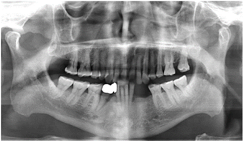
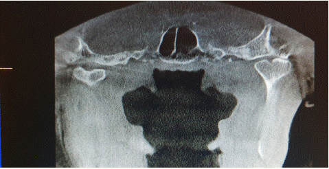
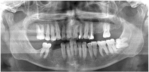
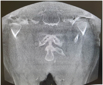
Considering the knowledge from the tomography and results of the observers, the rate of correct estimations was 51.4% (Figure 1a & 1b), while the rate of incorrect estimations (Figure 2a & 2b) was 48.6%. There was no statistically significant difference between localization, type of BMC, type of groove, BMC depth and horizontal angle of BMC values according to whether the estimations were correct or incorrect (p>0,050) (Table 1 & 2).
Estimations
Total
p
Incorrect
Correct
Right-left
Right
Left
8(44.4)
10(55.6)
10(52.6)
9(47.4)
18(48.6)
19(51.4)
0,866*
Type of BMC
Mediolateral
Multiheaded
8(44.4)
10(55.6)
13(68.4)
6(31.6)
21(56.8)
16(43.2)
0,255*
The type of groove
Deep
Shallow
8(44.4)
10(55.6)
8(42.1)
11(57.9)
1,000*
*Yates Correction
Table 1: Comparison of knowledge from the tomography and the distributions of the variables according to the estimations made jointly by the observers.
    Groups


      p


    Incorrect

    Correct


    Mean±std. dev.

    Mean(min-max)

    Mean±std.dev.

    Mean(min-max)


    Depth of BMC

    2.25±1.26

    1.9(0.7 -5)

    2.08±0.72

    2.2(0.7–3.5)

    0.625*


    Horizontal angle of BMC

    19.55±6.8

    20.1(2.2-29)

    15.55±12.28

    18.6(-18.6–34.5)

    0.232*


    *Independent Samples t-Test
Table 2: Comparison of quantitative variables according to the CBCT and estimates made jointly by the observers.

Figure 1a: Panoramic radiograph of bilateral BMC of patient A.

Figure 1b: CBCT coronal image of bilateral BMC of patient A.

Figure 2a: Wrong estimation of panoramic radiograph of patient B. ( bilateral normal condyles) They look like divided into two parts of more or less equal size by a deep groove.

Figure 2B: CBCT coronal image of bilateral normal condyles of patient B.
A statistically moderate agreement was obtained between the answers given by the observers and the findings obtained in the tomography. (K=0,436; p<0,001). In fact, it was seen that the rate of saying absent in the observers which are normal is higher than the rate of saying present in the observers which actually have BMCs. That is, observers detect non-bifid (normal) condyles better than those with BMCs (Table 3).
    Observer Responses

    Total

    Kappa/p


    Absent

    Present


    CBCT findings


    Apsent

    39(67.2)

    2(10.5)

    41(53.2)

    0.436/<0.001


    Present

    19(32.8)

    17(89.5)

    36(46.8)
Table 3: Examining the compatibility of tomographic findings with the answers given by the observers.
Although there was no statistically significant difference between the values, it was determined that the horizontal angle of the BMCs that observers stated correctly was 20.5% less than the cases with bifid condyles that they stated incorrectly.
Discussion
Bifid Mandibular Condyle (BMC) is an uncommon anomaly, characterized by a division of the mandibular condylar head. It is considered to be developmental although it has also been related to infection, trauma, condylar fractures or condylectomy [12-14]. Multiheaded or trifid condyles are rare disorders of the mandible. Their etiology and pathogenesis are unclear. They can be associated with temporomandibular joint disorders or can be diagnosed incidentally on routine radiographic examination [15]. Several epidemiological studies have been carried out on BMC. Szentpétery et al. [16]. investigated skulls and reported an incidence rate of 0.34% in 2077 condyles. In many studies [11,17,18], BMC prevalence was evaluated by panoramic radiography. Because they are usually detected accidentally during routine dental radiographic examinations, more commonly on panoramic views taken for other dental purposes [2]. Menezes et al. [17] found nine (0.018%) cases of BMC from 50.080 panoramic radiographs in a Brazilian population. Miloglu et al. [11] and Sahman et al. [18] examined panoramic radiographs in Turkish subjects and reported the prevalence of BMC as 0.31% and 0.52%, respectively.
Panoramic radiography is a 2D imaging method, it may not always be an appropriate diagnostic tool in the diagnosis of a BMC. On the other hand, panoramic radiographs can misread bifidity by the overlapping of anatomical structures or inherent image distortion [11,17]. They can either under- or overestimate bifidity. Also the rift between the duplicated condylar heads may be partial and partial BMC’s are hard to detect on panoramic views [8]. Three-dimensional (3D) imaging methods are essential for the clinician, especially when evaluating the condyle head that is grooved in the ML direction [14]. Also it should be supplemented by CT or Magnetic Resonance Imaging (MRI), especially in cases when surgery is planned. Daniels et al. [19]. reported a case of bifid condyle associated with TMJ ankyloses. The ankylosed joint was seen on panoramic examination but bifidity was only confirmed by the CT scan. This is because the lateral condyle had obscured the medial condyle in the panoramic radiograph. Further more, 3D imaging allows for a more accurate assessment of hypoplastic, hyperplastic, or degenerative changes of the condyle. Differential diagnosis of a BMC with degenerative changes such as cysts, tumors, metastatic lesions, or condylar fractures is essential, and the ideal method for imaging BMC morphology is three-dimensional imaging [20].
There are very few studies in the literature evaluating BMCs using CT or CBCT. In a study of Sahman et al. [21] using CTs, the prevalence of a BMC was found to be lower (1.82%). Accordingly, Sampaio et al. [22] published a retrospective study which found a 1.1% prevalence of BMC in asymptomatic patients by using CT meanwhile only half of them could be diagnosed by checking previous panoramic radiographs. In accordiance with this study, our study showed the rate of correct estimations was 51.4% on panoramic radiographs.
CT machines have limitations in dentistry because of their high cost, large footprint and high radiation exposure. CBCT is more advantageous in evaluating the bone structures in the dentomaxillofacial region due to the lower radiation. The image is obtained in a shorter scanning time, and bone resolution is high [8]. Also CBCT is an excellent imaging modality for the assessment of BMC. It allows detailed visualization of condylar morphology without osseous superimposition. CBCT can make optimal evaluation of morphological aspects like the condyle shape and size, orientation of condylar angle, joint position, depth of glenoid fossa [1].
Khojastepour et al. [10], reported in their retrospective study that the prevalence of BMC (4.53%) seemed to be higher using CBCT than that found in the studies performed with panoramic radiographs. Sahman et al. [21] speculated that BMC might be a more frequent condition in the Turkish population. They also stated that the difference between CT-based and panoramic image-based studies in the same Turkish population might have occurred from misinterpretations of the panoramic radiographs. Miloglu et al. [11] concluded that due to the widespread use of new diagnostic techniques, the prevalence of BMC is likely to be higher than has been previously reported.
This study examined panoramic radiographs consisting of both normal morphology and BMCs to assess which factors effect the visibility of BMC on panoramic images. We could not find any statistically significant difference between the results of the experts, the results of the two-y-year asistants and the one-year asistants but observers detect non-bifid (normal) condyles better than those with BMCs. We could not find any statistically significant difference between the rift indicators such as localization and type of BMC, type of groove, BMC depth, and horizontal angle values of BMC according to whether the estimations were correct or incorrect. However when the angle between the condylar head and the horizontal plane become closer, ML bifidity or multiheaded BMC may more likely to be detected on the panoramic radiography. In conclusion, within the limitations of the study, panoramic radiographs can misread bifidity and CBCT is the proof of choice to establish a correct diagnosis of BMC. Additional studies should be undertaken to further elucidate which factors have role on the visibility bifidity on panoramic radiographies.
References
- White SC, Pharoah MJ. Oral radiology principles and interpretation. 7th ed Elsevier: Mosby. Inc. 2014: 503-4.
- Borrás-Ferreres J, Sánchez-Torres A, Gay-Escoda C. Bifid mandibular condyles: A systematic review. Med Oral Patol Oral Cir Bucal. 2018; 23: e672-80.
- Cho BH, Jung YH. Nontraumatic bifid mandibular condyles in asymtomatic and symtomatic temporomandibular joint subjects. Imaging Sci Dent. 2013; 43: 25-30.
- Mawani F, Lam EWN, Heo G, McKee I, Raboud DW, Major PW. Condylar shape analysis using panoramic radiography units and conventional tomography. Oral Surg Oral Med Oral Pathol Oral Radiol Endod. 2005; 99: 341-8.
- Schmitter M, Gabbert O, Ohlmann B, Hassel A, Wolff D, Rammelsberg P, et al. Assessment of the reliability and validity of panoramic imaging for assessment of mandibular condyle morphology using both MRI and clinical examination as the gold standard. Oral Surg Oral Med Oral Pathol Oral Radiol Endod. 2006; 102: 220-4.
- Honey OB, Scarfe WC, Hilgers MJ, Klueber K, Silveira AM, Haskell BS, et al. Accuracy of cone-beam computed tomography imaging of the temporomandibular joint: comparisons with panoramic radiology and linear tomography. Am J Orthod Dentofacial Orthop. 2007; 132: 429-38.
- Arayapisit T, Ngamsom S, Duangthip P, Wongdit S, Wattanachaisiri S, Joonthongvirat Y, et al. Understanding the mandibular condyle morphology on panoramic images: A conebeam computed tomography comparison study. Cranio. 2023; 41: 354-61.
- Koenig LJ. Bifid condyle. In: Koenig LJ, editor. Diagnostic imaging: oral and maxillofacial. 1st ed. Vol. II; 4. Canada: Amirsys. 2012; 28.
- Güven O. A study on etiopathogenesis and clinical features of multi-headed (bifid and trifid) mandibular condyles and review of the literature. J Craniomaxillofac Surg. 2018; 46: 773-8.
- Khojastepour L, Kolahi S, Panahi N, Haghnegahdar A. Cone beam computed tomographic assessment of bifid mandibular condyle. J Dent (Tehran). 2015; 12: 868-73.
- Miloglu O, Yalcin E, Buyukkurt MC, Yilmaz A, Harorli A. The frequency of bifid mandibular condyle in a Turkish patient population. Dento Maxillo Fac Radiol. 2010; 39: 42-6.
- Antoniades K, Hadjipetrou L, Antoniades V, Paraskevopoulos K. Bilateral bifid mandibular condyle. Oral Surg Oral Med Oral Pathol Oral Radiol Endod. 2004; 97: 535-8.
- Rehman TA, Gibikote S, Ilango N, Thaj J, Sarawagi R, Gupta A. Bifid mandibular condyle with associated temporomandibular joint ankylosis: A computed tomography study of the patterns and morphological variations. Dento Maxillo Fac Radiol. 2009; 38: 239-44.
- Balaji SM. Bifid mandibular condyle: A study of the clinical features, patterns and morphological variations using CT scans. J Maxillofac Oral Surg. 2010; 9: 38-41.
- Sezgin ÖS, Kayipmaz S. Trifid mandibular condyle. Oral Radiol. 2009; 25: 146-8.
- Szentpétery A, Kocsis G, Marcsik A. The problem of the bifid mandibular condyle. J Oral Maxillofac Surg. 1990; 48: 1254-7.
- Menezes AV, de Moraes Ramos FM, de Vasconcelos-Filho JO, Kurita LM, de Almeida SM, Haiter-Neto F. The prevalence of bifid mandibular condyle detected in a Brazilian population. Dento Maxillo Fac Radiol. 2008; 37: 220-3.
- Sahman H, Sekerci AE, Ertas ET, Etoz M, Sisman Y. Prevalence of bifid mandibular condyle in a Turkish population. J Oral Sci. 2011; 53: 433-7.
- Daniels JS, Ali I. Posttraumatic bifid condyle associated with temporoman¬dibular joint ankylosis: report of a case and review of the literature. Oral Surg Oral Med Oral Pathol Oral Radiol Endod. 2005; 99: 682-8.
- Corchero-Martín G, Gonzalez-Terán T, García-Reija MF, Sánchez- Santolino S, Saiz-Bustillo R. Bifid condyle: case report. Med Oral Patol Oral Cir Bucal. 2005; 10: 277-9.
- Sahman H, Sisman Y, Sekerci AE, Tarim-Ertas ET, Tokmak T, Tuna IS. Detection of bifid mandibular condyle using computed tomography. Med Oral Patol Oral Cir Bucal. 2012; 17: e930-4.
- Sampaio-Neves F, Ricardina Ramírez-Sotelo L, Roque-Torres G, Lopes Resende Barbosa G, Haiter-Neto F, Queiroz de Freitas D. Detection of bifid mandibular condyle by panoramic radiography and cone beam computed tomography. Braz J Oral Sci. 2013; 12: 16-9.