
Case Report
Austin J Dent. 2024; 11(1): 1177.
An Uncommon Management of Fractured Narrow Implant: Clinical Case Report
Starosta M*
Department of Dentistry, Faculty of Medicine, University of Ostrava, Czech Republic
*Corresponding author: Starosta M Department of Dentistry, Faculty of Medicine, University of Ostrava, 703 00 Ostrava, Czech Republic. Email: martin.starosta@osu.cz, mstarosta@seznam.cz
Received: December 26, 2023 Accepted: January 19, 2024 Published: January 26, 2024
Abstract
In most cases, a fracture of the implant is an indication for its removal. However, if the fragments do not completely separate, the implant can be preserved. In this case report, we describe the use of a reinforcement ring without the need to remove the entire implant.
Keywords: Dental implant fracture; Treatment failure; Reinforcement ring
Case Presentation
In January 2018, an 18-year-old woman was referred for a consultation for implantation. The main problem was agenesis of the upper lateral incisors. At that time, she was already finalizing orthodontic treatment with opening the gaps in the area where the implant was to be placed. The interdistal distance in both areas was borderline (6,5 mm). The overall health was without problems. After completing clinical and diagnostic evaluations, an optimal treatment plan was formulated to place 2 narrow implants in the area of the maxillary left and right agenetic lateral incisors to support a fixed partial dentures (Figure 1). In May 2018, the Bioniq 2.9 x 14 mm implants (Lasak co, CZ) were implanted in the respective areas. Healing was uneventful and prosthetic rehabilitation was completed in September. All-ceramic screw - retained crowns were used for the rehabilitation. The fixation screws of the crowns were tightened to 20 N/cm, according to the manufacturer's instructions. The prosthetic restoration was satisfactory both functionally and aesthetically. The patient was instructed on hygiene around the implants and scheduled for a follow-up appointment in one year (Figure 2,3 & 4).
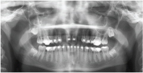
Figure 1: Review X-ray (OPG X-ray) of a patient before implantation.
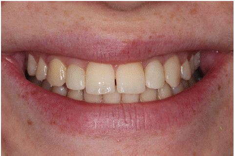
Figure 2: A clinical appearence after implants reconstruction.
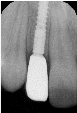
Figure 3: X- ray of implant reg. 12.
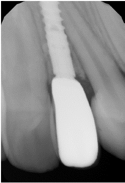
Figure 4: X-ray of implant reg 22.
In October 2019, a check of the implants was carried out. Subjectively, she reported feeling a wiggle crown reg 22. On clinical examination, the tissues around the implant were free of inflammation, and both crowns were firmly fixed. Intraoral radiograph showed evidence of coronal disintegration of the coronal part at implant 22 (Figure 5). The patient was scheduled for a follow-up appointment in six months . In March 2020, the patient presents with a loose crown from the reg 22 implant. According to the i.o. X-ray, there is a longitudinal fracture in the coronal part of the implant (Figure 6). Due to the position of the implant in this situation, the proximity of the surrounding teeth and the alveolar structure, we abandoned explantation and agreed on a solution using a reinforcement ring placed in the area of the fracture. Subsequently, in agreement with Lasak co., titanium rings were fabricated with different diameters (3.1 mm, 3.2 mm, 3.3 mm and 3.4 mm), 2,5 mm height and 0.2 mm width from grade 4 titanium (Figure 7).
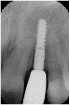
Figure 5: Bone resorption around neck of the implant reg. 22.
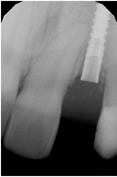
Figure 6: Implant fracture.
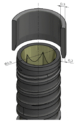
Figure 7: Scheme of titanium reinforcement ring.
In May 2020, a surgical procedure was performed to place a ring in the area of the implant fracture.A ring with a diameter of 3.3 mm was selected for passive adaptation. At the same time, the crown was fixed with tightening of the fixation screw to 15 N/cm (Figure 8,9 & 10). Subsequently, follow-ups were performed every 2 months until June 2020. The prosthetic work remained firm, and the patient was free of problems. Further check-ups took place every six months. At the last follow-up in June 2023, the patient was subjectively uncomfortable, the work was aesthetically and functionally satisfactory, and the intraoral radiograph showed no progression of alveolar loss in the neck implant area (Figure 11,12 & 13).
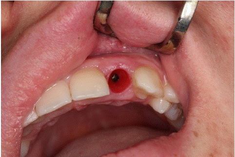
Figure 8: The situation after losing the crown.
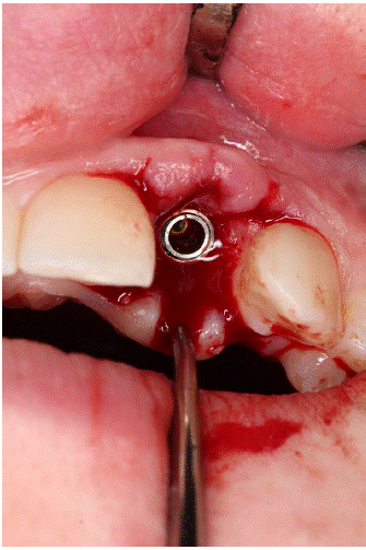
Figure 9: The ring adaptation.
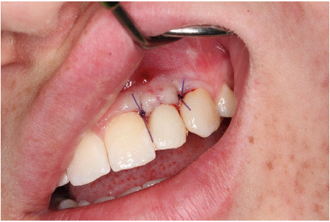
Figure 10: The situation after crown fixation.
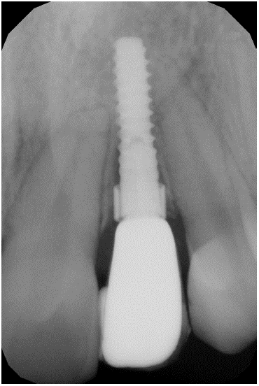
Figure 11: X-ray after surgery.
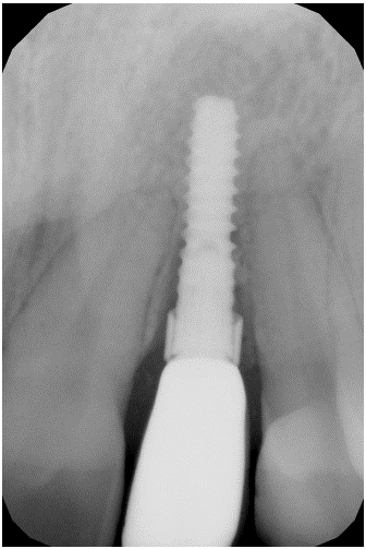
Figure 12: X- ray three years later.
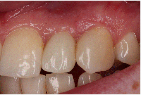
Figure 13: Clinical view three years later.
Currently, both implants have been in function for less than five years. The implant in area 22 is 3.5 years after intervention without any problems.
Discussion
Fracture of the implant body is one of the unpleasant complications of this type of treatment. It is usually a late complication, i.e. it occurs after prosthetic reconstruction and functional loading of the implant [1]. Balshi [2] gives three reasons for this complication: 1, problems related to implant material and design 2, imperfect connection between implant and abutment 3, parafunction (bruxism). In addition to chronic overloading during parafunctions, single trauma exceeding the strength of the implant may also occur [3]. Parafunctions associated with implant overload and inaccurate connection between the implant and abutment often lead to prosthetic failure (loosening) before implant fracture occurs [4]. If this situation occurs, the attending physician should pay close attention to the causes of overloading(articulation ratios) and check the quality of the connection between the implant and abutment. This may prevent further complications associated with implant fracture. If the implant fracture already occurs, 3 treatment methods are described in the literature [5,6], 1, removal of the entire implant (usually using a suitable trepan) 2, removal of only the coronal fractured part of the implant in the area of the abutment and creation of a new base for the abutment using a suitable preparation set 3, removal of only the coronal fractured part of the implant and leaving the rest of the implant in the alveolus (possible if no subsequent implantation is considered).
If we proceeded explantation of implant in this situation, it would be associated with the risk of damage to the surrounding teeth (due to the borderline dimension of the implant gap), loss of alveolus leading to the need for subsequent bone augmentation in the area of future implantation and, of course, the introduction of a new implant and fabrication of its new prosthetic restoration. After considering all these aspects, we chose a minimally invasive and maximally effective solution. This treatment method cannot be considered absolutely reliable but it can be considered a suitable alternative in the given situation.
Conclusion
In this case report, we demonstrate an unusual method of implant fracture treatment considering a minimally traumatic procedure.
References
- Manor Y, Oubaid S, Mardinger O, Chaushu G, Nissan J. Characteristics of early versus late implant failure: a retrospective study. J Oral Maxillofac Surg. 2009; 67: 2649-52.
- Balshi TJ. An analysis and management of fractured implants: a clinical report. Int J Oral Maxillofac Implants. 1996; 11: 660-66.
- Flanagan D. External and occlusal trauma to dental implants and a case report. Dent Traumatol. 2003; 19: 160-64.
- Romeo E, Storelli S. Systematic review of the survival rate and the biological, technical, and aesthetic complications of fixed dental prostheses with cantilevers on implants reported in longitudinal studies with a mean of 5 yearsfollow-up. Clin Oral Implants Res. 2012; 23: 39-49.
- Mendonça G, Mendonça DB, Fernandes-Neto AJ, Neves FD. Management of fractured dental implants: a case report. Implant Dent. 2009; 18: 10-6.
- Gargallo Albiol J, Satorres-Nieto M, Puyuelo Capablo JL, Sánchez Garcés MA, Pi Urgell J, et al. Endosseous dental implant fractures: an analysis of 21 cases. Med Oral Patol Oral Cir Bucal. 2008; 13: E124-8.