
Research Article
Austin J Dent. 2024; 11(1): 1180.
Etching and Bonding between Glass Ionomer Cement and Resin Based Composite Material- Does it Help?
Abu-Younes A¹; Stroianu A¹; Baruch N¹; Garoushi S²; Lassila L²; Zilberman U¹*
¹Pediatric Dental Clinic, Barzilai Medical University Center, Ashkelon, affiliated to Ben Gurion University of the Negev, Beer-Sheva, Israel
²Department of Biomaterials Science and Turku Clinical Biomaterial Center-TCBC, Institute of Dentistry, University of Turku, Turku, Finland
*Corresponding author: Uri Zilberman Pediatric Dental Clinic, Barzilai Medical University Center, Ashkelon, affiliated to Ben Gurion University of the Negev, Beer-Sheva, Israel. Email: uriz@bmc.gov.il
Received: April 10, 2024 Accepted: May 10, 2024 Published: May 17, 2024
Abstract
The use of tooth-colored materials in restorative dentistry raised due to high esthetic demand from the patients. The main material used today is resin-based composite, but glass ionomer cements are more biocompatible to the dentin and can remineralize affected dentin. So, the sandwich technique can answer both issues- biocompatibility and esthetic results. The question raised for this research was if the resin-based composite can be bonded to the glass-ionomer cement so no micro leakage will occur between the two components. Shear Bond Strength (SBS) analyses at (TCBC) Turku Clinical Biomaterial Centre (Finland) and at Pediatric dental clinic at Barzilai Medical Center (Israel) showed that the bonding between resin-based composite and glass-ionomer cement after etching and bonding of glass-ionomer cement happens and increases with time. The conclusion was that the sandwich technique is an optional treatment for deep carious lesions at sites with high esthetic demands.
Keywords: Resin-based composites; Glass-ionomer cements; San witch technique; Deep carious lesions
Introduction
Dental restorative procedures remain the cornerstone of dental practice. Whilst non restorative strategies can arrest early dental caries, many caries lesions progress to cavitation and require restorative intervention. Moreover, many restorative procedures are the result of failed restorations; replacements accounts for more than half of the restorations placed by dental practitioners [1]. Many different tooth-colored materials have been developed and marketed. They can be classified into following groups:
Resin-based materials set by polymerization. These materials are used with an adhesion technique. Polymerization initiation by light requires blue light-emitting curing units.
Glass ionomer cements, set exclusively by an acid-base reaction. The attachment to dental enamel and dentin is developed by a chemical bond between the carboxyl groups of the glass ionomer and the calcium ions of enamel and dentin. The glass-ionomer material can also release fluoride to its vicinity, enamel and dentin [2].
Resin materials combined with components of glass ionomer cements (compomers). The material relies on polymerization and it is used with an adhesive system [3].
A new material of an ion-releasing (calcium) fiber-reinforced flowable composite (Bio-SFRC) that promote mineralization at the interface and inside the organic matrix of demineralized dentin was developed [4].
Class II cavities are often with deep subgingival extensions of the approximal cavity floor. These variables must be considered when choosing a suitable material. The extension of the cavity floor into the gingival sulcus generates challenges to adequate moisture control. This may be a problem for moisture and contamination of sensitive materials such as adhesives required for resin composites [5]. Laboratory studies found more biofilm formation on resin-based materials compared to amalgam, which may contribute to increased secondary caries around resin composites and influence the health of the neighboring periodontal tissues [6]. Moreover, resin-based materials and dental adhesives are cytotoxic and cause an intracellular redox imbalance [7,8], that may influence the periodontal tissues. Therefore, special techniques have been described, such as the open sandwich technique, to overcome the problems [5].
The problem begins when the contact area between the glass ionomer cement placed in the box and the composite material on the occlusal is not tightly connected and secondary caries may affect the dentin layer between the materials. Shear Bond Strength (SBS) of composite to glass-ionomer cement using self-etch and total-etch adhesives was tested and the showed results between 2-28 Mpa [9].
The aim of the study was to analyze in vitro the effect of etching on glass-ionomer cement and the SBS of composite to glass-ionomer cements and to Bio-SFRC after 1 and 24H and after 40 days. The SBS results were compared to composite material bonded to enamel after etching and bonding after 1 hour and 1 week.
Materials and Methods
SEM Analyses
A bulk of glass-ionomer cement (EQUIA Forte, GC Europe) was prepared and left for self-cure for 15 minutes. 37% phosphoric acid was applied on the surface for 20 seconds, washed with copious amount of water and dried. The surface of the glass-ionomer cement was photographed under SEM before and after etching (Figure 1 & 2).
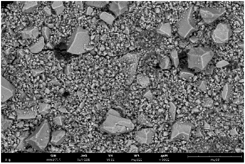
Figure 1: GIC (EQUIA FORTE by GC Europe) before etching (magnification 2300).
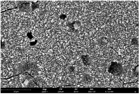
Figure 2: GIC after etching with 37% phosphoric acid (magnification 2200).
On the glass-ionomer cement a flowable composite material (Flow-it ALC by Pentron, USA) was applied after etching (Scotch bond Universal etchant by 3M/ESPE) and bonding (Single Bond Universal by 3M/ESPE) and light-cured for 40 seconds. The bulk of glass-ionomer and composite was sliced using an Isomet 100. The slices were examined under SEM to analyses the contact line between the composite and the glass-ionomer cement (Figure 3).
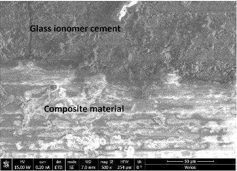
Figure 3: The contact line between Composite (bottom) and GIC after etching and bonding (magnification 250).
SBS (Shear bond strength). Intact premolars extracted for orthodontic reason were used. On the buccal surface a cavity was prepared using a 330-carbide bur (Figure 4). The cavity was filled with a high viscosity glass-ionomer cement (EQUIA Forte by GC Europe-Figure 5). At Turku Clinical Biomaterial Centre (TCBC), 3 groups of 10 premolars were examined after an hour of self-setting, after 24 hours and after 40 days, total of 90 premolars. For each group, on 10 teeth the glass ionomer cement was covered with injectable composite material without etching and bonding, on 10 teeth the composite material was bonded to the glass ionomer after 20 sec etching and bonding and on 10 teeth a bio liner (Bio-SFRC) filled the cavity and the composite material applied on top without etching and bonding (Figure 6). At the pediatric dental unit at Barzilai Medical University center a similar study design was performed. 14 premolars with cavities filled with high-viscosity Glass-Ionomer Cement and composite bonded after 20 sec etching and bonding were examined for SBS. 5 premolars with composite bonded to GI were examined after 1 hour and 24 hours, and 4 premolars after 1 week. 5 premolars with only composite bonded to intact enamel after 20 sec etching and bonding were examined as control after 1H and 1 week. All teeth (both at (TCBC) Turku Clinical Biomaterial Centre and at Barzilai clinic) were kept in artificial saliva before tests and the SBS tests were performed using LR10K plus by LLOYD with a preload stress of 0.005 kN and speed of 1 mm/min. The fracture type (cohesive or adhesive) was examined under a light microscope.
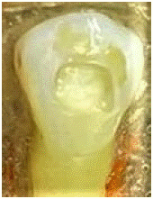
Figure 4: The cavity prepared on the buccal surface of the premolar.
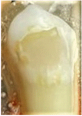
Figure 5: GIC filling of the cavity.
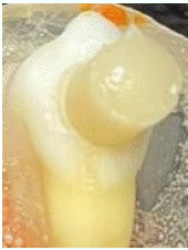
Figure 6: Composite or bioliner cylinder bonded to GIC.
Results
SEM analysis of Glass ionomer before and after etching (Figure 1 & 2). The large particles of the glass ionomer cement were dissolved by the etchant. After bonding and composite material application, there is a very close contact between the two materials (Figure 3).
The SBS results from (TCBC) Turku Clinical Biomaterial Centre lab are presented in Figure 7. The SBS of composite to glass ionomer cement without bonding was very low, but after etching and bonding the SBS improved significantly and the SBS after 40 days was higher in comparison to 1 and 24 hours. The SBS of composite Bonded to Ion-releasing fiber-reinforced flowable composite (Bio-SFRC) was twice as higher than that of composite bonded to glass ionomer cement. The failure type of the composite bonded to GIC without bonding was cohesive (Figure 8) in 4 cases and adhesive (Figure 9) in 6 cases after 1 hour and adhesive in all 10 teeth after 24 hours. Composite bonded to GIC after etching and bonding showed cohesive fracture in 5 cases and adhesive fracture in 5 cases after 1 hour and after 24 hours 2 cohesive fractures and 8 adhesive fractures. Composite bonded to Bio-SFRC showed cohesive fracture in 4 cases and adhesive fracture in 6 cases after 1 hour and after 24 hours 10 adhesive fractures.
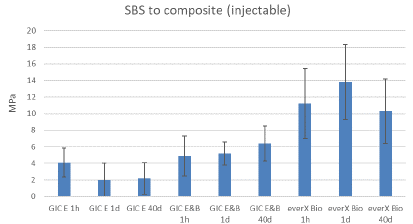
Figure 7: SBS between GIC and composite (without etching and bonding-first three columns on the left, and after etching and bonding- columns 4-6) and between everX bio and composite after 1 and 24 hours and after 40 days (Turku Lab).
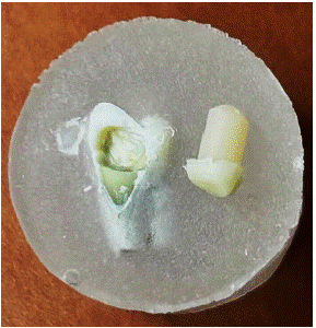
Figure 8: Cohesive fracture.
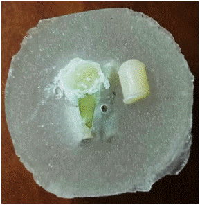
Figure 9: Adhesive fracture.
The SBS results from Barzilai clinic are presented in Figure 10. The SBS of composite bonded to enamel was higher than the SBS of composite bonded to GIC but the differences were smaller after 1 week. The SBS of composite bonded to GIC increased from 1 day to 1 week. The failure of composite bonded to enamel were cohesive in 2 cases and adhesive in 3 cases after 1 hour and cohesive in 3 cases and adhesive in 2 cases after 1 week. When the composite was bonded to GIC the failure was adhesive in all cases after 1 hour, adhesive in 2 cases and cohesive in 3 cases after 1 day and after 1 week.
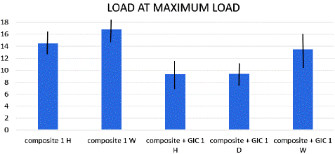
Figure 10: SBS of composite bonded to enamel after 1 hour (1H) and 1 week (1W) and of composite bonded to GIC after 1 hour (1W), one day (1D) and 1 week (1W).
Discussion
The results of this in vitro research showed that composite material can be bonded to glass-ionomer cement and to new ion-releasing fiber-reinforced flowable composite (Bio SFRC). Similar results regarding glass ionomer cement bond to composite resin were published before [10-12]. The SEM analyses showed a close contact between composite resin and GIC after etching and bonding. Glass ionomer cement and composite resin showed similar clinical performance in class II restorations in primary teeth, except for secondary carious lesions, in which GIC presented superior performance [13]. But in the era of high demand of esthetic restorations, the sandwich technique for Class II restorations in permanent teeth can answer both problems- minimal secondary carious lesions due to the effect of glass-ionomer cements in the box and the esthetic requirement using the composite resin on the occlusal surface. The issue of the interface between the two restorative materials was analyzed already some 40 years ago, but the recommendation was to apply the composite resin on GIC after 24 of maturation [13]. This procedure required an additional clinical visit. The SBS results of bonding between composite to glass-ionomer cement after etching and bonding were similar to results published by Hight et al. and Manihani et al. [9,10], and showed improvement after 1 week and 1 month in comparison to the first 24 hours. SBS of composite resin to the new Bio SFRC material showed better results than composite resin to GIC during the first day but was similar to the results at Barzilai Clinic after a week. Composite resin bonded to enamel after etching and bonding showed higher SBS results in comparison to composite resin bonded to GIC but the differences were small after a week. Many of the failures observed in the groups of composite resin bonded to GIC were of cohesive type showing that the bond between the materials was stronger than the bond of GIC to the dentin and enamel. In Class II restorations the bond between GIC and dentin and enamel in the box is not affected by the masticatory forces, so cohesive failure of the restoration due to debonding of GIC from the bottom of the restoration cannot occur.
References
- Elhahlah D, Lynch CD, Chadwick BL, Blum IR, Wilson NHF. An update on the reasons for placement and replacement of direct restorations. J Dent. 2018; 72: 1-7.
- Zilberman U. Ion exchanges between glass-ionomer restorative material and primary teeth components- an in vivo study. Oral Biol and Dent. 2014: 2.
- Schmalz G, Schwendicke F, Hickel R, Platt JA. Alternative direct materials for dental amalgam: A concise review on an FDI policy statement. Int Dent J. 2023; 11: 1-8.
- Garoushi S, Vallittu P, Lassila L. Development and characterization of ion-releasing fiber-reinforced flowable composite. Dent Materials. 2022; 38: 1598-1609.
- Ismail HS, Ali AI, Mehesen RE, Juloski J, Garcia-Godoy F, Mahmoud SH. Deep proximal margin rebuilding with direct esthetic restorations: a systematic review of marginal adaptation and bond strenght. Restor Dent Endod. 2022; 47: e15.
- Maktabi H, Ibrahim MS, Balhaddad AA, Alkhubaizi Q, Garcia IM, Collares FM, et al. Improper light curing of bulkfill composite drives surface changes and increases S. mutans biofilm growth as a pathway for higher risk of recurrent caries around restorations. Dent J. 2021; 9: 83.
- Krifka S, Seidenader C, Hiller KA, Schmalz G, Schweikl H. Oxidative stress and cytotoxiciy generated by dental composites in human pulp cells. Clin Oral Investig. 2012; 16: 215-24.
- Demirci M, Hiller KA, Bosl C, Galler K, Schmalz G, Schweikl H. The induction of oxidative sress, cytotoxicity, and genotoxicity by dental adhesives. Dent Mater. 2008; 24: 362-71.
- Mahinani AK, Mulay S, Beri L, Shetty R, Gulati S, Dalsania R. Effect of total-etch and self-etch adhesives on bond strength of composite to glass-ionomer cement/resin-modified glass-ionimer cement in the sandwich technique- A systematic review. Dent Res J. 2021; 18: 72.
- McLean JW, Powis DR, Prosser HT, Wilson AD. The use of glass ionomer cements in bonding composite resin to dentine. Br Dent J. 1985; 158: 410-414.
- Knight GM, McIntyre JM, Mulyani. Bond strengths between composite resin and auto cure glass ionmer cement using the co-cure technique. Austr Dent J. 2006; 51: 175-179.
- Francois P, Vennat E, Ruscassier N, Attal JP, Dursun E. Shear bond strength and interface analysis between a resin composite and a recent high-viscous glass ionomer cement bonded with various adhesive systems. Clin Oral Investig. 2019; 23: 2599-2608.
- Dias AGA, Magno MB, Delbem ACB, Cuhna RF, Maia LC, Pessan JP. Clinical performance of glass ionomer cement and composite resin in Class II restorations in primary teeth: Asystematic review and meta-analysis. J Dent. 2018; 73: 1-13.