Research Article
Austin J Dent. 2024; 11(1): 1181.
Effect of Laser and Fluoride Gel Application on Microroughness of Permanent Teeth
Amer N¹; Toema S²; Mekled S³*
¹Research Center, Cairo University, Egypt
²Deartment of Pediatric, Temple University, USA
³Department of Restorative, Temple University, USA
*Corresponding author: Mekled S Department of Restorative, Temple University, 3223 N Broad St, Philadelphia, PA 19140, USA. Email: Salwa.Mekled@temple.edu
Received: May 28, 2024 Accepted: June 11, 2024 Published: June 18, 2024
Abstract
Objective: Fluoride plays an important role to reminalize white lesions and to prevent the progression of caries. Studies show that using laser and topical fluoride is effective to increase enamel resistance and reduce acid solubility.
Purpose: To evaluate the effect of laser irradiation and fluoride on microroughness of enamel of permanent lower molars.
Methods: fifteen freshly extracted, third molars were selected, then Immediately immersed in 0.09% saline. The crowns were separated from the roots then sectioned longitudinally in a mesiodistal direction. The specimens were randomly divided into three groups (n=10) as follows: white spot lesions group, fluoride gel group, and Er-YAG laser group. Then a surface roughness test was done for all samples. Data were analyzed using one-way ANOVA followed by Tukey’s post hoc test.
Results: Microroughness increased for teeth treated with ER-YAG (mean 4.50mm) compared to teeth treated with fluoride (mean 1.35mm). The results were statistically significant between all groups (p>0.05). Discussion: Although some studies recommend laser irradiation after fluoride application, our study proves that using fluoride alone improved surface microroughness.
Conclusion: Fluoride treatment improved white spot lesions microroughness. More research is needed to investigate the effect of laser on WSL microroughness.
Keywords: Fluoride; Laser; Microroughness; White lesions
Introduction
The main objective of dentistry is to prevent decay. Enamel demineralization is the initial step to inhibit decay [1-3]. Enamel demineralization occurs due to long-term bacterial plaque accumulation on enamel surfaces. When demineralization starts, enamel loses translucency to appear chalky white [3-5]. Initial demineralized enamel areas can be remineralized either physiologically through minerals found naturally in saliva [6] or externally by topical application of remineralizing agents [7-8]. Topical remineralizing agents include fluoride, bioglass paste, and low viscosity resin “infiltrate” [3].
Topical Fluoride plays a major role preventing decay; as it improves acid resistance of the enamel, enhances remineralization of incipient lesions and, in addition, it interferes with micro-organisms by inhibiting bacterial metabolism and enzymatic process [9-10]. The preventive mechanism of action of fluoridated agents involves deposition of a large amount of Calcium Fluoride (CaF2) on the enamel surface, which is consequently transformed into fluorapatite crystals [11-12].
The use of lasers accompanied with fluoride application has recently been used to arrest enamel demineralization [3]. There are many types of lasers available with different wavelengths, such as diode lasers with wavelengths ranging from 810 nm to 980 nm. Additionally, erbium lasers such as Er:YAG and Er,Cr:YSGG have proven their efficacy in dental enamel remineralization, increasing acid resistance and decreasing dental enamel solubility [3,13-14]. Lasers interact with enamel photo thermally and photochemically, resulting in recrystallization of enamel crystallites, decomposition of organic matrix, and carbonate loss. Thus, increase acid resistance and reduce enamel permeability allow sealing the enamel surface [3,15-23].
The aim of our study is to evaluate the impact of a fluoridate gel and Er:YAG irradiation on enamel micro-roughness of permanent teeth.
Materials and Methods
We collected fifteen freshly extracted lower third molars without anatomical defects. Teeth were immersed in 0.09% saline [29]. A low-speed diamond disc was used to separate the crowns of the collected teeth. Additionally, sectioning of the samples was done in a mesiodistal direction to make thirty working surfaces. Each specimen was coated with nail polish as an acid resistant varnish except for 3×3 mm exposed enamel was covered with acid-resistant adhesive tape. Then the tapes were removed, and the surfaces were cleaned with damped cotton.
Data Collection
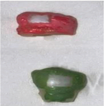
Thirty specimens were divided into three groups (n=10), then specimens were immersed in demineralization solution for 21 days to form enamel lesions. In this study, the demineralization solution is composed of 2.2 mM monopotassium phosphate, 2.2 mM calcium chloride and 0.05 mM acetic acid having pH adjusted to 4.4 using 1 M potassium hydroxide (Figure 1).

Figure 1: Shows marking the white spot lesions.
Study Design
The specimens were divided into three groups (n=10) as follows: Group 1: white spot lesion control group (WNL) did not receive either Fluoride application or irradiation, group 2 received application of APF gel; and group 3 received Irradiation with Er: YAG laser (2940 nm).
Acidulated Phosphate Fluoride Application
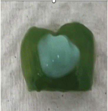
Application of an acidulated phosphate fluoride gel (Ionite fluoride gel, dharma research, USA) containing 1.23% fluoride ions on the samples of group 2. Fluoride gel was applied for four minutes using a cotton swab, then deionized water was used to wash the samples for one minute (Figure 2).

Figure 2: Shows Fluoride application
Er:YAG Laser Irradiation
The irradiation conditions for Er:YAG laser (Light Touch) was: 2940 nm wavelength, pulse energy of 50 mJ; 0.5 W power; frequency of 10 Hz; in a pulse mode of pulse duration of 600 msec. Laser irradiation was performed by sweeping in a uniform motion both horizontally and vertically. A non-contact HO2 hand piece H14 was used with cylindrical tip 0.4 mm at a 1mm distance with air & water spray. Time of exposure was 10 sec [30].
The samples were kept in deionized water between treatment processes. All surfaces were polished then a surface roughness test was conducted using non-contact optical profilometry.
Data Collection
Surface micro-roughness test: Stylus profilometry was used to analyze the surface profile in the center of the delineated area. In this study, three readings were taken for each sample, then the average was calculated.
Statistical Analysis
Both mean and standard deviation values were calculated, then data distribution was evaluated using the Shapiro-Wilk test. One -way ANOVA and Tukey’s post hoc test were used to normally distributed and analyzed data. The significance level was set at p = 0.05 within all tests. Statistical analysis was performed with R statistical analysis software version 4.1.3 for Windows (www.R-project.org/).
Results
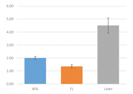
Micro-roughness of white lesions increased for teeth treated with ER: YAG (mean 4.50 μm) and decreased in teeth treated with Fluoride gel (mean 1.35μm) compared to teeth with white spot lesions did not receive any treatment (mean 2.10μm). The results were statistically significant between all groups (p>0.05) (Bar Chart 1, Table 1).

Bar Chart 1: Shows Mean Micro roughness comparison between white spot lesion group, Fluoride gel group, and Laser group.
Discussion
Our study investigates the effectiveness of laser and fluoride in prevention of dental decay and treatment of white spot lesions by measuring the enamel micro-roughness. We compared the use of Er:YAG laser and topical application of fluoride gel.
Fluoride decreases the incidence of decalcification by deposits in hydroxyapatite to form fluorapatite, thus initiates mineralization process [31]. We emphasize that all the methods investigated in our research have been used in previous studies and we had similar results. Our results found that applying Fluoride gel decreased the micro-roughness of white lesions, while the use of ER:YAG increased the micro-roughness.
Da Silva Barbosa et al., suggested that increased fluoride uptake can affect greater crystal surface area for the binding of ions, such as fluoride and calcium ions [32]. Limited research exists on the effect of fluoride on white spot lesion surface micro-roughness. A study assessing surface microroughness of bleached enamel surface after fluoride gel treatment showed statistically significant decrease in surface micro-roughness [33]. This outcome coincides with our results that confirm the effectiveness of fluoride treatment on decreasing surface roughness.
Oksayan R. explained that the Er:YAG laser irradiation with 1 W average power injures the hard tissue leading to high demineralization scores. Many researchers also have concerns about enamel irradiation with Er: YAG lasers can create carious lesions [33]. Rodrigez-Vilchis et al. used SEM to observe the changes in the enamel; that led to craters and cracks on the enamel surface [34]. This result may explain that the average power over 0.50 W might have a negative effect on the enamel of the teeth [33], thus agrees with our results leading to increased micro-roughness of surface enamel.
A study showed a significant increase in surface roughness when Er:YAG was used at 200 mJ and 4 Hz on enamel for 12.5 seconds (50 pulses) [30]. Another study investigated the effect of using 7.5 and 12.7 J/cm2 Er:YAG laser at 100 mJ, and 7 Hz, for 13 seconds on enamel surface roughness to find that surface roughness increases proportionally with the energy density [32]. In this study, a lower energy level was used at 50 mJ; 0.5 W power; frequency of 10 Hz; in a pulse mode of pulse duration of 600 msec for 10 seconds. Our results find that Er:YAG laser increases enamel surface roughness even when using lower energy level.
There are some limitations in our study due to small sample size, and the use of stylus profilometry, which is technique sensitive compared to the optical profilometry. The use of different laser parameters can explain slight difference between our results and other studies. In the prior studies, low-power diode lasers with different parameters were used, some of them used photo-absorber creams. Considering these controversies, further studies on different effects of Er:YAG on enamel microhardness are warranted.
Conclusion
In the present study, enamel surface micro-roughness of white spot lesions decreased by using 1.23% APF gel. Er:YAG laser irradiation increased the enamel surface micro-roughness when used at 50 mJ; 0.5 W power; frequency of 10 Hz; in a pulse mode of pulse duration of 600 msec for 10 seconds. 1.23% APF can improve the aesthetics and caries susceptibility of WSLs. Fluoride gel can help to reduce decay in both children and adults. The easy application would help to form calcium fluoride deposits and prevent decay progression. Thus, will help dental providers to improve preventative treatment for the patients, offer conservative treatment option for the patients and reduce the financial cost of treatments. Our study did not find that Er: YAG laser could reduce the microroughness which in tun can reduce decay. Further studies on different effects of various types of lasers on white spot lesions is warranted.
References
- Mansour YJ, Hanno AG, El Tekeya M, Nagui DA. The effect of combined treatment by laser and fluoride on the acid resistance of enamel in primary teeth (in-vitro study). Alexandria Dental Journal. 2021; 46: 160–65.
- Kamel E, Hassouna DM, Geith ME. In vitro study to evaluate the effect of 45S5 bioglass paste and Er,Cr:YSGG 2780 nm laser on re-mineralization of enamel white spot lesions (energy dispersive x-ray analysis and stereomicroscopic assessments). Egyptian Dental Journal. 2021; 67: 301–14.
- Ciiftci ZZ, Hanimeli S, Karayilmaz H, Gungor O. The efficacy of resin infiltrate on the treatment of white spot lesions and developmental opacities. Niger J Clin Pract. 2018; 21: 1444–1449.
- Yagci A, Seker ED, Demirsoy KK, Ramoglu SI. Do total or partial etching procedures effect the rate of white spot lesion formation? A single-center, randomized, controlled clinical trial. Angle Orthod. 2019; 89: 16–24.
- Belikov AV, Altshuler GB, Grishin VV. Method and apparatus for tooth rejuvenation and hard tissue modification. United States patent application: US 11/868,449. 2008.
- Taha AA, Patel MP, Hill RG, Fleming PS. The effect of bioactive glasses on enamel remineralization: a systematic review. J Dent. 2017; 67: 9–17.
- Bakry AS, Abbassy MA. Increasing the efficiency of CPP-ACP to remineralize enamel white spot lesions. J Dent. 2018; 76: 52–57.
- Vitale MC, Zaffe D, Botticell AR, Caprioglio C. Diode laser irradiation and fluoride uptake in human teeth. Eur Arch Paediatr Dent. 2011; 12: 90–92.
- Villena RS, Tenuta LM, Cury JA. Effect of APF gel application time on enamel demineralization and fluoride uptake in situ. Braz Dent J. 2009; 20: 37–41.
- Delbem ACB, Cury JA, Nakassima CK, Gouveia VG, Theodoro LH. Effect of Er: YAG laser on CaF2 formation and its anti-cariogenic action on human enamel: an in vitro study. J Clin Laser Med Surg. 2003; 21: 197–201.
- Jeng YR, Lin TT, Wong TY, Chang HJ, Shieh DB. Nano-mechanical properties of fluoride-treated enamel surfaces. J Dent Res. 2008; 87: 381–85.
- De Sant’Anna R, Paleari GSL, Duarte DA, Brugnera Jr A, Pacheco-Soares C. Surface morphology of sound deciduous tooth enamel after application of a photoabsorbing cream and infrared low-level laser irradiation: an in vitro scanning electron microscopy study. Photomed Laser Surg. 2007; 25: 500–507.
- Moghadam NCZ, Seraj B, Chiniforush N, Ghadimi S. Effects of laser and fluoride on the prevention of enamel demineralization: an in vitro study. J Lasers Med Sci. 2018; 9: 177–82.
- Abufarwa M, Noureldin A, Campbell PM, Buschang PH. The longevity of casein phosphopeptide–amorphous calcium phosphate fluoride varnish’s preventative effects: Assessment of white spot lesion formation. Angle Orthod. 2019; 89: 10–15.
- El Mansy MM, Gheith M, El Yazeed AM, Farag DB. Influence of Er, Cr: YSGG (2780 nm) and nanosecond Nd:YAG laser (1064 nm) irradiation on enamel acid resistance: morphological and elemental analysis. Open Access Maced J Med Sci. 2019; 7: 1828–1833.
- Serdar-Eymirli P, Turgut MD, Dolgun A, Yazici AR. The effect of Er, Cr: YSGG laser, fluoride, and CPP-ACP on caries resistance of primary enamel. Lasers Med Sci. 2019; 34: 881–91.
- Ramezani K, Ahmadi E, Etemadi A, Kharazifard MJ, Omrani LR, Akhoundi MS. Combined Effect of Fluoride Mouthwash and Sub-ablative Er: YAG Laser for Prevention of White Spot Lesions around Orthodontic Brackets. The Open Dentistry Journal. 2022; 16.
- Kawasaki K, Tanaka Y, Takagi O. Crystallographic analysis of demineralized human enamel treated by laser irradiation or remineralization. Arch Oral Biol. 2000; 45: 797–804.
- Hossain M, Nakamura Y, Kimura Y, Yamada Y, Kawanaka T, Matsumoto K. Effect of pulsed Nd:YAG laser irradiation on acid demineralization of enamel and dentin. J Clin Laser Med Surg. 2001; 19: 105–108.
- Tsai CL, Lin YT, Huang ST, Chang HW. In vitro acid resistance of CO2 and Nd-YAG laser treated human tooth enamel. Caries Res. 2002; 36: 423–29.
- Fried D. Laser processing of dental hard tissues [invited paper]. SPIE Proceedings, Volume 5713, Photon Processing in Microelectronics and Photonics IV (2005 April 12). https://doi.org/10.1117.12.598149
- Ana PA, Bachmann L, Zezell DM. Lasers effects on enamel for caries prevention. Laser Physics. 2006; 16: 865–75.
- Maung NL, Wohland T, Hsu CY. Enamel diffusion modulated by Er: YAG laser:(Part1)-FRAP. J Dent. 2007; 35: 787–93.
- Esteves-Oliveira M, Zezell DM, Meister J, Franzen R, Stanzel S, Lampert F, et al. CO2 Laser (10.6 μm) parameters for caries prevention in dental enamel. Caries Res. 2009; 43: 261–68.
- Allam GG, Aziz AFA. Comparing topical fluoride application, laser irradiation and their combined effect on remineralisation of enamel. Future Dental Journal. 2018; 4: 318–23.
- Poosti M, Ahrari F, Moosavi H, Najjaran H. The effect of fractional CO2 laser irradiation on remineralization of enamel white spot lesions. Lasers Med Sci. 2014; 29: 1349–1355.
- Ceballos-Jiménez AY, Rodríguez-Vilchis LE, Contreras-Bulnes R, Alatorre JA, Scougall-Vilchis RJ, Velazquez-Enriquez U, et al. Chemical changes of enamel produced by sodium fluoride, hydroxyapatite, Er:YAG laser, and combined treatments. Journal of Spectroscopy. 2018.
- Yassaei S, Motallaei MN. The effect of the Er: YAG laser and MI Paste Plus on the treatment of white spot lesions. J Lasers Med Sci. 2020; 11: 50–55.
- de Sant’anna GR, dos Santos EA, Soares LE, do Espírito Santo AM, Martin AA, Duarte DA, et al. Dental enamel irradiated with infrared diode laser and photo absorbing cream: Part 1 -- FT-Raman Study. Photomedicine and laser surgery. 2009; 27: 499–507.
- Sokucu O, Herguner S, Bektas OO, Babacan H. Shear bond strength comparison of a conventional and a self-etching fluoride-releasing adhesive following thermocycling. World J Orthod. 2010; 11: 6-10.
- da Silva Barbosa P, da Ana PA, Poiate IA, Zezell DM, de Sant’ Anna GR. Dental enamel irradiated with a low-intensity infrared laser and photoabsorbing cream: a study of microhardness, surface, and pulp temperature. Photomedicine and laser surgery. 2013; 31: 439–446.
- Martin JM, de Almeida JB, Rosa EA, Soares P, Torno V, Rached RN, et al. Effect of fluoride therapies on the surface roughness of human enamel exposed to bleaching agents. Quintessence international (Berlin, Germany:1985). 2010; 41: 71-8.
- Oksayan R. In Vitro Evaluation Of Enamel Demineralization Around The Brackets After Er-Yag Laser Irradiation And Flouride Application. Medical Journal of Suleyman Demirel University. 2018; 25: 85-90.
- Rodriguez-Vilchis LE, Contreras-Bulnes R, Sanchez-Flores I, Samano EC. Acid resistance and stru- ctural changes of human dental enamel treated with Er: YAG laser. Photomed Laser Surg. 2010; 28: 207- 11.