
Case Report
Austin J Dent. 2024; 11(2): 1183.
Incision-Free, Coronally Advanced Flap (CAF) with GTR Membrane Placed by the Molar or Canine Access (MOCA) Technique: A New Approach for the Treatment of Gingival Recession: A Case Report
Sharashchandra M Patil¹*; Ambika Y Patil²; Uzma Tarannum¹; Farah Naaz¹; Indira Priyadarshini¹
¹Department of Periodontology and Oral Implantology, S B Patil Institute for Dental Sciences and Research Bidar, India
²Department of Oral Medicine and Radiology, SB Patil Institute for Dental Sciences and Research Bidar, India
*Corresponding author: Sharashchandra M PatilDepartment of Periodontology and Oral Implantology, SB Patil Institute for Dental Sciences and research Bidar, Bidar, Karnataka, 585402, India. Tel: 9916508812 Email: drsharad_004p@yahoo.co.in
Received: May 24, 2024 Accepted: June 21, 2024 Published: June 28, 2024
Abstract
Background: Root coverage procedures are challenging for the management of gingival recession. The procedures are technically sensitive, and the above technique provides a predictable alternative to managing facial gingival recessions.
Case Report: The aim of this report is to demonstrate a case of Cairo RT I gingival recession with a follow-up period of 6 months and proceeded with the MOCA technique for the management of gingival recession defect. Here, the GTR (Guided Tissue Regeneration) membrane is placed via the gingival sulcus of an adjacent tooth with minimal trauma and requires no incisions at the recipient site. Thereby, preserving the local blood supply to promote the healing process and minimizes tension on the Coronally Advanced Flap (CAF) through the use of suspension sutures. Hence, this paper explains the step-by-step MOCA technique for GTR membrane placement in the management of gingival recession defect.
Conclusion: In our study, an optimal root coverage result was achieved, and while the MOCA is a technically sensitive procedure with no incisions at the recipient site, there is no uneven texture or scar formation, and healing proceeds with minimal interruption.
Keywords: Gingival Recession; Root Coverage; Incision-free; Molar or Canine Access plastic surgery; GTR membrane
Introduction
Gingival recession is clinically manifested by an apical displacement of the gingival tissues, leading to root surface exposure [1].
Among existing techniques, a coronally advanced flap using the patient’s own sub epithelial connective tissue provides maximum root coverage and represents the gold standard of grafting. However, even with this technique, the outcome of complete root coverage is not predictable [2].
Incision-less techniques for root coverage include tunneling [3] and Vestibular Incision Subperiosteal Tunnel Access (VISTA) [4]. In these techniques, incisions are minimized by utilizing sulcular incisions to create a tunnel.
However, this intrasulcular approach limits access for both graft placement and flap advancement, increases the risk of traumatizing the sulcular tissue, and therefore increases the risk of an unfavorable outcome [5].
This paper overcomes the limitations of previous tunneling and VISTA techniques by allowing an incision-less approach with remote access points. The technique requires no incisions at the recipient site and thereby preserves the integrity of the local blood supply for the healing process [6].
This approach also facilitates graft placement via the use of the gingival sulcus of an adjacent tooth with minimal tension on the flap by using a suspension suturing technique. Specifically, the technique relies on placing the graft via either molar or canine access, hereby known as the “MOCA technique.”
Molar and Canine gingival margin areas are large and thereby easy entry points for the introduction of more delicate and friable graft tissues into sites of recession. Hence, this paper explains the step-by-step MOCA technique for GTR membrane placement in the management of gingival recession defect.
Case Report
A 27-year-old male patient presented with complaints of root sensitivity and aesthetic concerns associated with gingival recession in the mandible anterior region. Clinical examination revealed Class I (RT1) gingival recession on the facial aspect of w.r.t. 43 with 2mm clinical attachment loss with probing depths within normal limits and no signs of active periodontal disease. After obtaining the institutional ethical committee permission, following the Helsinki rule of Declaration, informed consent was taken from the patient, the patient was scheduled for surgical treatment using the MOCA technique. Pre-surgical procedures like oral prophylaxis and root planning were done.
Surgical Procedure
Under local anesthesia (lidocaine 1:80000), a small access tunnel was created at the mucogingival junction distal to the mandibular canine w.r.t. 43 using a half Hollenbach carver (Figure 1). The tunnel was extended apically and coronally to facilitate access to the recession defects on teeth 44 and 42 (Figure 2). Subsequently, a split-thickness flap was raised coronally to the defect margins without full-thickness flap reflection. Care was taken to preserve the interdental papillae and avoid vertical releasing incisions. The exposed root surfaces were thoroughly debrided. A resorbable collagen GTR membrane (@ Advanced Biotech Healiguide-collagen membrane) was then trimmed to fit the recession defects, adapted over the exposed root surfaces (Figure 3), and adjusted in its intended position (Figure 4). The flap was coronally repositioned to cover the membrane and secured in place using sutures with polyglactin 910 (@ Vicryl 4-0) and secured in place with a composite button on the buccal aspect of the tooth (Figures 5 & 6). Postoperative instructions and oral hygiene measures were provided to the patient. The patient was prescribed oral analgesics and antibiotics. For the first 3 weeks, the patient was advised not to brush at the surgical site, avoid hard food, and rinse once daily with 0.2% chlorhexidine digluconate mouthwash, emphasizing gentle brushing and avoidance of trauma to the surgical site.
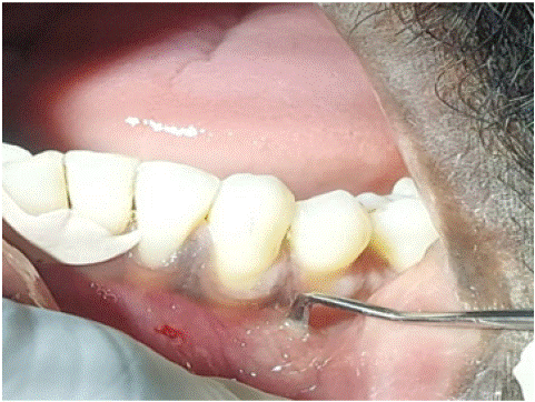
Figure 1: Tunnel preparation at w.r.t 43 using a half hollenbach carver.
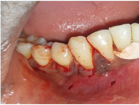
Figure 2: Tunnel for recipient site to facilitate access.
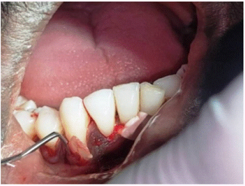
Figure 3: Root surface debridement followed by GTR membrane placed.
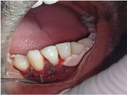
Figure 4: GTR membrane adjusted in its intended position.
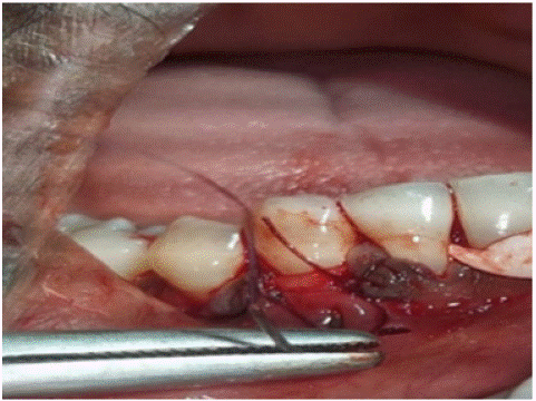
Figure 5: The flap was coronally repositioned to cover the memb membrane.
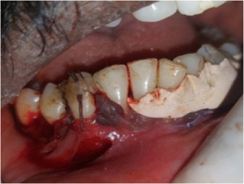
Figure 6: Flap secured with a composite button.
Outcome
At the 6-month follow-up visit, clinical evaluation revealed significant root coverage of the recession defects with regenerated gingival tissues, resulting in improved aesthetics and alleviation of root sensitivity (Figures 7 & 8). Probing depths remained stable, and there were no signs of inflammation or recession recurrence. The patient reported minimal discomfort during the postoperative period and expressed satisfaction with the outcome of the treatment.
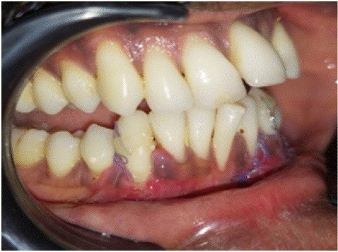
Figure 7: 7 days post operative.
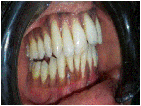
Figure 8: 6 months post operative.
Discussion
The outcome of a graft (soft tissue grafts) depends on a variety of factors, such as the type of surgical procedure, the number and type of incisions, which include vertical incisions, horizontal incisions, de-epithelialization and partial removal of the adjacent papillae, and split-thickness incisions at the base of pedicle flaps [7,8]. Before the establishment of graft vascular supply, the graft survives through plasmatic, transudate, and diffusive circulation for the duration of the first 3 days, but the condition is that the recipient site’s circulation has not been interrupted [9].
CAF with SCTG has been reported to achieve mean root coverage values of 64.5% to 97.3% and complete root coverage of 10%–66% in teeth with Miller class I or II recession (at least 2-mm root recession) [10,11-13]. Regarding the tunneling technique with subperiosteal incision, teeth with a denuded root length of 1–3 mm showed 95% mean coverage and those with a denuded root length of 4–6 mm showed 73% mean root coverage [14]. However, in few studies shown the success rate of CAF or the tunneling (envelope) technique with the SCTG procedure in the anterior mandible, with a denuded root length of >3 mm. The tunneling technique is a procedure without coronal displacement of the mucogingival junction, as described by Raetzke [15], but for teeth with a denuded root length above 3 mm, the graft is exposed in the tunneling technique.
A critical area of the graft is the portion that covers the denuded root surface. The circulation in this area of the graft can be maximized by the MOCA technique as there is no incision and gains access to place the graft via tunneling at the recipient site, which provides optimal clinical results as there is no uneven texture or scar formation, and healing proceeds with minimal interruption. A case series done in 13 cases has shown positive healing in the management of gingival recession [16].
Conclusion
The MOCA technique represents a promising alternative for the treatment of gingival recession, offering the advantages of minimally invasive surgery, enhanced tissue regeneration, and favorable aesthetic outcomes.
Further research and long-term clinical studies are warranted to validate the efficacy and long-term stability of this approach in a larger patient population. Nevertheless, the present case report demonstrates the feasibility and potential benefits of the MOCA technique in addressing gingival recession with minimal surgical trauma and a satisfactory result.
References
- Lavu V, Gutknecht N, Vasudevan A, Balaji SK, Hilgers RD, Franzen R. Laterally closed tunnel technique with and without adjunctive photobiomodulation therapy for the management of isolated gingival recession-a randomized controlled assessor-blinded clinical trial. Lasers Med Sci. 2022; 37: 1625-1634.
- Kashani H, Vora MV, Kuraji R, Fathi-Kelly H, Nguyen T, Tamraz B, Tran C, et al. Incision-free, coronally advanced flap with subepithelial connective tissue graft placed by the molar or canine access (MOCA) technique: 13 case series. Clin Adv Periodontics. 2023; 13: 11–20.
- Zabalegui I, Sicilia A, Cambra J, Gil J, Sanz M. Treatment of multiple adjacent gingival recessions with the tunnel subepithelia connective tissue graft: a clinical report. Int J Periodontics Restorative Dent. 1999; 19: 199-206.
- Zadeh HH. Minimally invasive treatment of maxillary anterior gingival recession defects by vestibular incision subperiosteal tunnel access and platelet-derived growth factor BB. Int J Periodontics Restorative Dent. 2011; 31: 653-660.
- Oliver RC, Loe H, Karring T. Microscopic evaluation of the healing and revascularization of free gingival grafts. J Periodontal Res. 1968; 3: 84- 95.
- Zucchelli G, Testo T, De Sanctis M. Clinical and anatomical factors limiting treatment outcomes of gingival recession: a new method to predetermine the line of toot coverage. J Periodontol. 2006; 77: 714- 721.
- Jung U-W, Um YJ, Choi SH. Histologic observation of soft tissue acquired from maxillary tuberosity area for root coverage. J Periodontol. 2008; 79: 934-940.
- Sanz M, Simion M. Working Group 3 of the European Workshop on Periodontology. Surgical techniques on periodontal plastic surgery and soft tissue regeneration: consensus report of Group 3 of the 10th European Workshop on Periodontology. J Clin Periodontol. 2014; 41: S92-S97.
- Prato GPP, Franceschi D, Cortellini P, Chambrone L. Long-term evaluation (20 years) of the outcomes of subepithelial connective tissue graft plus coronally advanced flap in the treatment of maxillary single recession-type defects. J Periodontol. 2018; 89: 1290-1299.
- da Silva RC, Joly JC, de Lima AFM, Tatakis DN. Root coverage using the coronally positioned flap with or without a subepithelial connective tissue graft. J Periodontol. 2004; 75: 413-9.
- Borghetti A, Glise JM, Monnet-Corti V, Dejou J. Comparative clinical study of a bioabsorbable membrane and subepithelial connective tissue graft in the treatment of human gingival recession. J Periodontol. 1999; 70: 123-30.
- Cheung WS, Griffin TJ. A comparative study of root coverage with connective tissue and platelet concentrate grafts: 8-month results. J Periodontol. 2004; 75: 1678-87.
- Zucchelli G, Amore C, Sforza NM, Montebugnoli L, De Sanctis M. Bilaminar techniques for the treatment of recession-type defects. A comparative clinical study. J Clin Periodontol. 2003; 30: 862-70.
- Allen AL. Use of the supraperiosteal envelope in soft tissue grafting for root coverage. I. Rationale and technique. Int J Periodontics Restorative Dent. 1994; 14: 216-27.
- Raetzke PB. Covering localized areas of root exposure employing the “envelope” technique. J Periodontol. 1985; 56: 397-402.
- Kashani H, Vora MV, Kuraji R, Fathi-Kelly H, Nguyen T, Tamraz B, et al. Incision-free, coronally advanced flap with subepithelial connective tissue graft placed by the molar or canine access (MOCA) technique: 13 case series. Clin Adv Periodontics. 2023; 13: 11–20.