
Case Report
Austin J Dent. 2016; 3(1): 1031.
Spontaneous Bone Regeneration of the Mandible: In a Case of Osteosarcoma and Literature Review
Bataineh AB*
Department of Dentistry, Jordan University of Science & Technology, Jordan
*Corresponding author: Anwar B. Bataineh, Department of Dentistry, Jordan University of Science & Technology, Irbid, Jordan
Received: March 31, 2016; Accepted: May 05, 2016; Published: May 07, 2016
Abstract
There are rare cases reported in the literature that have demonstrated spontaneous bone regeneration after resection of the mandible in benign pathology. Mandibular defects may result from many conditions such tumors. The tumor presented radiographically as an osteolytic lesion, after biopsy, it was found to be an osteogenic sarcoma, a hemimandibulectomy was performed. Clinically and radiographically a very hard-formed bone could be palpated and seen, also a functional left temporal muscle, which had been completely detached from the resected mandible, could be palpated.
The purpose of this article was to report an unusual malignant case of complete spontaneous bone regeneration of the coronoid process ascending ramus and partial regeneration of the body of the mandible in a case of osteosarcoma.
Keywords: Spontaneous bone regeneration; Hemimandibulectomy; Osteosarcoma
Introduction
Osteosarcoma is a tumor that can occur in any bone and most commonly occurs in the long bones. Some studies found that osteosarcomas accounts for about 20% of all primary bone cancers and about 4%-6% occur in the maxillofacial region, while others found account for 6% to 9% [1,2].
Spontaneous bone regeneration is an uncommon form of bone healing where bone grows into critical size defects. In the mandible, spontaneous bone regeneration has replaced extensive defects up to the entire mandible and bilateral condyles [3-5]. The mechanism of spontaneous bone regeneration is not thoroughly understood, however, various factors have been suggested to influence this event [3,6,7]. The age of the patient, presence of infection, soft tissue protection of the bone defect, immobilization and genetic factors have been suspected as factors controlling spontaneous bone regeneration, but the exact are not yet to be identified [4-6,8-12]. In Sub-Saharan Africa reports of spontaneous bone regeneration are very rare; all previous accounts have come from Nigeria following jaw resection for ameloblastoma [3,4,6]. Spontaneous regeneration of the mandible has been rarely stated in previous studies [3-5,8]. Our review of literature showed only one report of spontaneous regeneration of the whole mandible [8]. This case report followed mandibular resection for Osteosarcoma in a 10-years-old female.
The purpose of this article was to report a complete spontaneous bone regeneration of the coronoid process ascending ramus and partial regeneration of the body of the mandible in a malignant case of osteosarcoma.
Case Presentation
In January 11, 1995 a 10-year-old girl with a 5 months history of hard non movable painless mass in the posterior region of the left mandible was referred to Oral and Maxillofacial clinic at the Jordan University of Science and Technology. Extra-oral examination showed an enlargement of the left mandible, with mild facial asymmetry. A tangible lymph node in the left sub mandibular region was also found. Intraoral examination revealed a minimal expansion of the body extending from the left first premolar to the angle, the buccal sulcus was obliterated. The overlying mucosa was normal, whereas the first, second premolars, the first and second molars were mobile, with no signs of inflammation or infection and without any exudation.
Orthopantomograph showed the presence of an osteolytic lesion in the left side of the mandibular body, extending from the left first premolar to the angle of the mandible and both medial and left lateral cortexes were scalloped (Figures 1). An aspiration biopsy revealed no presence of liquid or pus content into the lesion. Intra-oral incisional biopsy confirmed the diagnosis of osteosarcoma.

    Figure 1:  Preoperative orthopantomogram showing an ill-defined lytic lesion
involving the left side of the mandible both cortexes were scalloped.
The proposed treatment was hemimandibulectomy and chemotherapy and six months later the mandible should be reconstructed with iliac or rib bone graft. Chemotherapy as an adjuvant treatment and the chemotherapeutic agents doxorubicin, cisplatin, adriamycin have been used, when chemotherapy concluded, the patient was sent for follow up with consultation with the oncologist.
In January 24th, 1995 the patient was admitted for hemimandibulectomy of the left side of the mandible. The surgical approach was done through an extra oral and intra oral approach (Figure 2). Sulcular incision extending from the right first premolar to the ascending ramus parallel to the external oblique ridge was made and periosteum was gently elevated from both buccal and lingual sides. The temporalis muscle attachments were released from the coronoid process. The resection of the mandible was done in the bone only posteriorly from the mandibular notch just below the condylar neck to the right incisor mandibular leaving the periosteum. The tumor was resected together with the body, coronoid process and ramus of the mandible leaving condyle only. After copious irrigation with saline solution, a K-wire was used at the time of resection to preserve the space, immobilization, surgical bed and improve the facial contour (Figure 3). The surgical wound was closed intra and extra orally in three-layer tension free. Postoperative recovery was uneventful and the patient was discharged a week after the surgery, the site of the surgery was slightly swollen, painful to palpation, the patient had no episode of fever, and the sutures were removed when the wounds were free of inflammation and infection. The patient was controlled weekly the swelling was going down and the patient was doing well.

Figure 2: Extra oral approach.

    Figure 3:  Orthopantomogram showing a K-wire was used at the time of
resection that simulated the curvature of the mandible to preserve surgical
bed and improve the facial contour was used.
Radiographic follow-up of the patient 2 months after surgery revealed beginning of spontaneous bone regeneration compared to the immediate postoperative x-ray, and K-wire was removed (Figure 4). Because of the spontaneous bone regeneration, it was decided to wait for reconstruction surgery, and the patient was again followed up. The patient did not miss subsequent appointments and showed up annually for reviewing. Radiographic follow-up of the patient after one-year spontaneous bone regeneration involving symphysis, body, ascending ramus and coronoid process of the left mandible (Figure 5).

    Figure 4:  Orthopantomogram two months after surgery showing K-wire was
removed revealed spontaneous bone regeneration in the resected area.

    Figure 5:  Orthopantomogram showing spontaneous bone regeneration
involving symphysis, body, ascending ramus and coronoid process of left
mandible.
Radiographic follow-up of the patient two and half years after surgery revealed spontaneous healing with a well-shaped image mimicking the ascending mandibular ramus, coronoid process and partial bone regeneration body of the mandible (Figure 6). The new bone was similar in appearance (color and texture) to the cortical bone of the proximal mandibular segments although it lacked height.
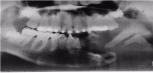
    Figure 6:  Orthopantomogram two and half years revealed a well-shaped
image mimicking the body of the mandible, ascending ramus and coronoid
process.
Radiographic follow-up of the patient six years postoperatively, revealed complete spontaneous bone regeneration of the coronoid process, ascending ramus and the angle of the mandible. Partial bone regeneration of the body of the mandible could be seen as well as bone formation around the end of the segments from the medline (Figure 7). CT scan views also confirmed the evidence of spontaneous bone regeneration of the body of the mandible, coronoid process, ascending ramus and the angle of the mandible (Figure 8).

    Figure 7:  Orthopantomograph of the mandible taken six years postoperatively
showing the completely regenerated bone.

    Figure 8:  CT scan view also confirmed the evidence spontaneous bone
regeneration a well-shaped the body of the mandible, ascending ramus and
coronoid process.
It was observed that the segments and the region of the ascending ramus and the body of the mandible were hard to palpation. During mouth opening, it could be seen and palpated the movements of the ascending ramus as well as function of the left temporalis muscle, which had been detached from the coronoid process during surgery (Figure 9, 10).

Figure 9: The extra oral pictures of the patient after the operation.

Figure 10: Intraoral pictures of the patient after the operation.
After six years of treatment bone graft from the iliac crest to treat the gap between the two segments and to increase the height of this new bone, for future dental reconstruction (Figure 11). In August 2003, after the spontaneous bone regeneration was nearly completed the two fragments of the bone were connected together with a reconstruction plate (Figure 12), with future plan for prosthetic replacement. Radiographic follow up the reconstruction plate and bone continuity was uneventful (Figure 13).

    Figure 11:  Orthopantomograph after six years treatment then proceeded by
grafting iliac compact and cancellous bone.

    Figure 12:  Intraoperatively view the segments were stabilized using a
reconstruction plate to connect the two fragments of the bone together.

    Figure 13:  Radiographic follow up the reconstruction plate and bone
continuity was uneventful.
Discussion
The extensive bone loss of part or the entire mandible, has occasionally been reported in the literature, the reported causes of such have included gunshot, blast injury [13,14], the commonest reported cause has been as a result of enucleation of large cysts [15,16] partial mandibulectomy for the treatment of a tumor or tumor-like condition [4,6,17] and infection [4,6,18]. In this case report was a malignant osteogenic sarcoma, review of literature showed only reported of benign cases of spontaneous regeneration.
Spontaneous bone regeneration is an uncommon phenomenon may take place following bone loss. Osteogenesis is the formation of new bone from osteoprogenitor cells in a wound, it is regarded as responsible for spontaneous bone regeneration from the periosteum, remnants of resected bone and uncommitted connective tissue cells in the region [19]. New bone formation occurs through osteoinduction, osteoconduction and osteogenesis. The committed and uncommitted undifferentiated mesenchymal cells in the periosteum and endosteum contribute to fracture healing. Also, mesenchymal cells in connective tissue can be induced by remaining bone fragments to form new bone and marrow [18,20,21]. New bone was discovered only during routine postoperative clinical and radiographic examinations. A review of the literature showed that a total of 33 reports of spontaneous bone regeneration of the mandible in the English literature including the present case [22].
While the exact explanation for spontaneous bone regeneration is unknown, several factors may influence bone regeneration and several suggestions by various authors have been put forward in an attempt to explain this phenomenon. It was noticed that the age of patients was between 3 and 11 years at the time of bone loss, due to high cellular activity and the availability of abundant mesenchymal cells to form osteogenic tissue [3-,6,23]. However, since not all children experience spontaneous bone regeneration after bone loss, where as noted the high osteogenic potential in young people and in older persons, age may not be the only influencing factor [18,24,25]. In conclusion, the young age of the patient, coupled with the preservation of the periosteum as already suggested by some previous reports [4,6,18,26,27], are essential factors that could contribute to this phenomenon. Soft tissue protection of the bone defect is regarded as essential for new bone growth into critical size defects as seen in spontaneous bone regeneration [10-12]. This can be through preservation of periosteal cover of the defect by suturing it back in position to prevent granulation tissue in growth as was done [3]. Unexpected spontaneous bone regeneration may be explained by the fact that respervation of periosteum with the young ages of patients and infection, possible genetic factors as the source of osteogenic cells, might be responsible for rapid bone regeneration [4,6,28]. Theoretically, infection may have the capacity to stimulate new bone growth, and rapid bone regeneration influenced by infection has been reported [6,8], and infection was believed to encourage periosteal bone regeneration [4,6,8,9]. Other factors mentioned in the literature include maxilla mandibular fixation [5,6], the presence of bony fragments [13,14], and of a temporary reconstruction material such as titanium mesh [5,17] or Kirschner wire [1,2] was employed in the surgery.
The patient presented in the current report was 10 years old with osteosarcoma which is a malignant tumor, a review of the literature showed that this is the only case of spontaneous bone regeneration in malignancy. In the view of the preponderance of spontaneous bone regeneration in young people, the segment was replaced with new bone that was well formed and positioned. Our case had no evidence of frank infection; however, we believe that the absolute absence of infection in jaw tumors is difficult to establish due to the close relationship of the dentate jaw to the polymicrobial oral environment.
The long-term follow-up of regenerated mandible is rare in the literature, and the longest follow-up was for five years [18], while the current case was followed up for 10 years.
In conclusion, a long-term follow-up period of at least one year and more may be advisable for young adults and children, before a decision for reconstruction is made.
References
- Murphey MD, Robbin MR, McRae GA, Flemming DJ, Temple HT, Kransdorf MJ. The many faces of osteosarcoma. Radiographics. 1997; 17: 1205–1231.
- Nissanka EH, Amaratunge EAPD, Tilakaratne WM. Clinicopathologic analysis of osteosarcoma of the jaw bones. Oral Dis. 2007; 13: 82–87.
- Ogunlewe MO, Akinwande JA, Ladeinde AL, Adeyemo WL. Spontaneous regeneration of whole mandible after total mandibulectomy in a sickle-cell patient. J Oral Maxillofac Surg. 2006; 64: 981-984.
- Adekeye EO. Rapid bone regeneration subsequent to subtotal mandibulectomy. Report of an unusual case. Oral Surg Oral Med Oral Pathol. 1977; 44: 521-526.
- Nagase M, Ueda K, Suzuki I, Nakajima T. Spontaneous regeneration of condyle following hemimandibulectomy by disarticulation. J Oral Maxillofac Surg. 1985; 43: 218-220.
- Nwoku AL. Unusually rapid bone regeneration following mandibular resection. J Maxillofac Surg. 1980; 8: 309-315.
- Ruggiero SL, Donoff RB. Bone regeneration after mandibular resection. J Oral Maxillofac Surg. 1992; 49: 647-52.
- Elbeshir EI. Spontaneous regeneration of the mandibular bone following hemimandibulectomy. Br J Oral Maxillofac Surg. 1990; 28: 128-130.
- de Villa GH, Chen CT, Chen YR. Spontaneous bone regeneration of the mandible in an elderly patient: A case report and review of the literature. Chang Gung Med J. 2003; 26: 363-369.
- Boyne PJ. The restoration of resected mandibles in children without the use of bone grafts. Head Neck Surg. 1983; 6: 626-631.
- Lemperle SM, Calhoun CJ, Curran RW, Holmes RE. Bony healing of large cranial and mandibular defects protected from soft-tissue interposition: A comparative study of spontaneous bone regeneration, osteoconduction, and cancellous auto grafting in dogs. Plast Reconstr Surg. 1998; 101: 660-672.
- Hata T, Hosoda M, Hirokawa M, Deguchi H, Nakamura H, Mizukawa A. Spontaneous regeneration of the mandibular bone following marginal resection against chronic diffuse sclerosing osteomyelitis. Case Report. Kawasaki Med J. 1998; 24: 125-130.
- Kisner WH. Spontaneous posttraumatic mandibular regeneration. Plast Reconstr Surg. 1980; 66: 442–447.
- Shuker S. Spontaneous regeneration of the mandible in a child. A sequel to partial avulsion as a result of a war injury. J Maxillofac. 1985; 13: 70–73.
- Chiapasco M, Rossi A, Motta JJ, Crescentini M. Spontaneous bone regeneration after enucleation of large mandibular cysts: a radiographic computed analysis of 27 consecutive cases. J Oral Maxillofac Surg. 2000; 58: 942–948.
- Di Dio M, De Luca M, Cammarata L, Pierazzi G. Bone regeneration after removal of a mandibular cyst and following prosthetic rehabilitation with implants. Case report. Minerva Stomatol. 2005; 54: 373–378.
- Glenda H, de Villa, Chen CT, Chen YR. Spontaneous bone regeneration of the Mandible in an Elderly Patient: A Case Report and Review of the Literature. Chang Gung Med J. 2003; 26: 363–369.
- Budal J. The osteogenic capacity of periosteum. Oral Surg. 1979; 47: 227–229.
- Jelic JS, Perciaccante VJ. Oral and maxillofacial reconstruction. In: Abubaker AO, Benson KJ, editors. Oral and Maxillofacial Surgery Secrets. Philadelphia: Hanley and Belfus; 2001; p. 281.
- Adebayo ET, Fomete B, Ajike SO. Spontaneous bone regeneration following mandibular resection for odontogenic myxoma. Ann Afr Med 2012; 11: 182-185.
- Abdulai AE. Complete Spontaneous Bone Regeneration Following Partial Mandibulectomy. Ghana Med J. 2012; 46: 174–177.
- Ahmad O, Omami G. Self-Regeneration of the Mandible Following Hemimandibulectomy for Ameloblastoma: A Case Report and Review of Literature. J Maxillofac Oral Surg. 2015; 14: 245–250.
- Park HW, Kim HJ, Park BM. Spontaneous regeneration of the lateral malleolus after traumatic loss in a three-year old boy: A case report with seven-year follow-up. J Bone Joint Surg Br. 1997; 79: 66-67.
- Webb D, Petty S, Tsay R, Carrao V. Spontaneous mandibular bone regeneration. Columbia Dent Rev. 2006; 10: 5-7.
- Khodayari A. Khojasteh MT, Kiani A, Nayebi L, Mehrdad M, Vahdatinia. Spontaneous Regeneration of the Mandible after Hemimandibulectomy: Report of a Case. J Dent. 2011; 8: 152-156.
- Throndson RR, Johnson JM. Spontaneous regeneration of bone after resection of central giant cell lesion: a case report. Tex Dent J. 2013; 130: 1201-1209.
- Martins WD, de Castro Avila LF. Partial spontaneous bone regeneration subsequent to mandibulectomy. J Contemp Dent Pract. 2004; 5: 108-120.
- Coen Pramono D. Spontaneous bone regeneration after mandible resection in a case of ameloblastoma: A case report. Ann Acad Med Singapore. 2004; 33: 59–62.