Case Report
Austin J Dent. 2016; 3(2): 1036.
Three Years Follow Up of a Combined Periodontal and Endodontic Therapy in a Patient with Severe Gingival Recession: A Case Report
Akpinar KE1*, Marakoğlu I2 and Akpinar KE3
1Department of Periodontology, Cumhuriyet University, Turkey
2Department of Periodontology, Selçuk University, Turkey
3Department of Endodontic, Cumhuriyet University, Turkey
*Corresponding author: Aysun Akpinar, Department of Periodontology, Cumhuriyet University, 58140-Sivas, Turkey
Received: May 10, 2016; Accepted: June 20, 2016; Published: June 22, 2016
Abstract
In this case report, the treatment of an advanced gingival recession leaving open the lower right central root tip due to trauma with combined endodontic and mucogingival techniques were presented.
After phase I periodontal treatment, endodontic treatment were performed. Gingival recession was treated with lateral sliding flap operation and the patient was taken under observation. At third week after operation, discoloration of tooth was treated with commercial bleaching kit.
After three years following, an optimal gingival health was maintained with the normal sulcus depth and the esthetic pleasure is satisfactory.
Keywords: Gingival recession; Lateral sliding flap; Apical resection
Introduction
Gingival recession is defined as the displacement of the gingival margin to apical from the cementoenamel junction. It is characterized by the loss of periodontal connective tissue fibers, along with tooth cementum and alveolar bone [1,2]. Reports on prevalence range from 14 to 90 percent, depending on the population being studied [3]. The main causes of gingival recession are periodontal disease, improper oral hygiene, frenal pull, bone dehiscence, improper restorations, tooth mal position, sub gingival calculus formation and trauma. Recession of gingival tissue causes root hypersensitivity, poor esthetic appearance and cervical root caries. Gingival recession defects are typically treated by periodontal plastic surgery to correct or eliminate the deformities of the gingival mucosa [1,4,5].
To cover exposed root surfaces, various mucogingival procedures have been used successfully, including pedicle flaps, free soft tissue grafts, and guided tissue regeneration procedures. A review of the literature shows that these procedures may result in mean root coverage between 62% and 89% of the original defect [6]. Lateral sliding flap operation is one of pedicle flaps and it was developed by Gruope and Warran [7] at 1956. This technique is a microgingival surgery technique when used in the condition that there is enough donor tissue at lateral side of the operation area.
Endodontic-periodontal lesions can provide many challenges to clinicians. They are often characterized by extensive loss of periodontal attachment and alveolar bone. It seems that periodontal disease rarely jeopardizes vital functions of the pulp. In teeth with moderate break down of attachment apparatus, the pulp usually remains in proper function. Breakdown of the pulp presumably does not occur until the periodontal disease process has reached a terminal state, i.e. when bacterial plaque involves the main apical foramina [8,9].
In this case report, 3 years follow-up of endodontic-periodontal combined treatment of an advanced level gingival recession with the opening root tip, was presented.
Case Presentation
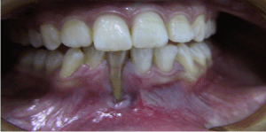
A 16-year-old Caucasian girl referred to Cumhuriyet University School of Dentistry, Department of Periodontology with complaint of localized gingival recession on the buccal aspect of tooth and discoloration of tooth. The patient was in good health with no contraindication for periodontal therapy. In the intra oral examination, it was determined that a gingival recession (Miller class II) with also denuded root point at buccal side of the teeth was due to trauma on 41(st) tooth which was exposed when she was 9 years old (Figure1). Mobility was observed (periotest value of 18) as a result of trauma. Although the calculus formation was seen in some areas, a good oral hygiene of the patient was observed. The entire dentition except traumatic teeth had normal structure and pattern.
After phase I periodontal treatment, temporary splint was made at lingual surfaces between 33(rd) and 43(rd) teeth in order to decrease the mobility. After phase I periodontal treatment, root canal treatment was started immediately with access opening and working length determined, calcium hydroxide dressing was given and patient was recalled after a week, after which obturation was done.
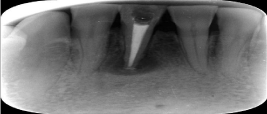
The bleaching agent used in this case is a 38% hydrogen peroxide power bleaching gel. Coronal debridement was established, the guttapercha in the root canal was sealed off from the pulp chamber with conventional glass ionomer cement. Opal dam was placed as a barrier for protection of the gingiva from the bleaching agent. The bleaching agent was applied extra coronally on the labial surface of the tooth and intracoronally in the prepared pulp space. The bleaching agent was applied for 15 minutes and agitated every 5 minutes to activate the agent. This was continued for two more cycles. The patient was recalled after a week. (Figure-2).

Figure 1: Condition prior to any treatment.

Figure 2: Postoperative radiograph.
In order to treat the gingival recession, lateral sliding flap operation was preferred. Laterally positioned flap was accomplished as previously described [2]. After obtaining adequate anesthesia root planning was performed carefully. The recipient bed was prepared. To prepare the donor area, the flap raised (dissected) from the distance 1.5-2 times wider than the recipient area was slid to the opened root surface and sutured (4-0).
A periodontal dressing (Coe Pak) was applied to protect the surgical area from mechanical injury during the initial phase of healing. Patient was instructed not to brush the teeth in the treated area, but to use a 0, 2% chlorhexidine solution two times daily for two weeks. In addition, patient was in structed to avoid excessive muscle traction or trauma to the treated area. Seven days following surgical treatment, the dressing and sutures were removed. The formerly exposed root surface was covered with new tissue. Clinical healing in both the donor and recipient sites was complete 3 weeks post operative.
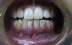
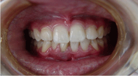
Discoloration of the tooth was bleached using a commercial bleaching kit. The technique of Walking Bleach was applied in this procedure [10]. At the end of the application, the evident color change was observed on the teeth. The patient was recalled once every 3 months until the final examination at three years (Figure 3,4).

Figure 3: Clinical aspects after nine months.

Figure 4: Clinical appearance 3 years later.
Discussion
The main goal of periodontal therapy is to improve periodontal health and there by to maintain patient’s functional dentition throughout his/her life. However, aesthetic represent an inseparable part of today’s oral therapy, and several procedures have been proposed to preserve or enhance patient aesthetics [11].
One of the most frequent indications of periodontal plastic surgery is the treatment of buccal gingival recessions. Many authors reported high success rate and 2-3 mm attachment gain following lateral sliding flap operations. The merits of full- versus splitthickness flaps for the laterally positioned flap procedure are still a controversial. It appears that split-thickness flaps have more vascular disruptions and are more prone to partial or complete necrosis than are full flaps. Reattachment is more apt the occur with full-thickness flaps than with split flaps, and entire healing is faster, with less recession at the recipient site, when a full-thickness flap is used. The only disadvantage of the full-thickness flap is the denuded bone left at the border of the donor site at the completion of the surgery. To prevent the recession on donor area of the patient, the full thickness flap application except marginal gingival tissue was preferred. After the operation, the healing was uncomplicated [12].
In comparing the results between the laterally positioned flap and the coronally positioned flap graft, Caffesse and Guinard [13] verified that both techniques covered 65 to 79 % of the exposed root surfaces and that the results remained stable for 3 years. It was also shown that in laterally positioned flaps recessions of an average of 1 mm were created in the donor teeth. In this case, 85% coverage was achieved on the opened root surface and the result remained stable for 3 years. Additionally, any gingival recession did not occur in the donor area.
Treatment of combined endodontic-periodontal lesions does not differ from the treatment given when the two disorders occur separately. In general, if the root canal system is infected, endodontic treatment should be commenced prior to any periodontal therapy in order to remove the intra-canal infection before any cementum is removed. If there is a periapical lesion on the tip of the root originating from the pulp, improvement of this lesion will be with
the root canal treatment providing complete apical plugging. If the applied canal treatment dos not achieve the leakage, it will be suitable that making corrections for retrograde filling together with the apical surgery on this area [8, 9, 10]. The endodontic treatment was thought as not enough, because of the existence of lateral canals in apical in our case have the root tip opened to the oral cavity.
The root canal system primarily becomes infected as a result of dental caries, traumatic injurius and coronal microleakege [14]. In this case, the trauma at 9 years old caused the pulp necrosis and the gingival recession. The tooth color change and the progressive gingival recession occurred by the time disturbed the patient esthetically and the patient came us for treatment.
Prognosis of periodontal-endodontic lesions depends on the diagnosis, treatment and chronicity of the lesion, as well as the duration of periodontal involvement [15]. A clinical case is presented in which a periodontal-endodontic lesion has been successfully treated with a combination of endodontic therapy and regenerative periodontal surgery. At 3rd year control, the teeth was asymptomatic, her esthetic pleasure was continued. In the radiographic evaluation, there was clear improvement in the periapical tissues and the gingival tissue was maintained in position.
This case demonstrates the value of having effective communication between dental specialties and the necessity to work together to achieve the optimal esthetic result. The timing and order of treatment was critical and needed to be planned in advance, prior to any one specialty beginning treatment. This required constant communication between the providers in order to increase the likelihood of achieving a more effective and efficient result from a multidisciplinary approach.
References
- Carranza FA, Newman GM. Glickman’s Clinical Periodontology. 8th ed. Philedalphia: Saunders Company; 1996: 653-662.
- Cohens ES. Atlas of cosmetic and reconstructive periodontal surgery. 2nd ed. Pennsylvania, 1994: 99-120.
- Kassab MM, Cohen RE. The etiology and prevalence of gingival recession. J Am Dent Assoc. 2003; 134: 220-225.
- Machado AW, Hodges R, Moon W. A multi disciplinary approach to treating a severe gingival recession in the esthetic zone. Dentistry. 2013; 3:175-179.
- Tözüm TF. A promising periodontal procedure for the treatment of adjacent gingival recession defects. J Can Dent Assoc. 2003; 69: 155-159.
- Wennström JL. Mucogingival therapy. Ann Periodontol. 1996; 1: 671-701.
- Groupe J, Warran R. Repair of gingival defects by a sliding flap operation. J Periodontol. 1956; 27: 92-95.
- Abbott P. Endodontic management of combined endodontic-periodontal lesions. J N Z Soc Periodontol 1998; 83: 15-28.
- Lindhe J. Clinical periodontology and implant dentistry. 3rd ed. Munskgaard, Copenhagen; 1998.
- Cohens SC, Burns R. Pathways of the pulp. 8th ed. St Louis: Mosby-Year Book; 2002: 755-757.
- Roccuzzo M, Bunino M, Needleman I, Sanz M. Periodontal plastic surgery for treatment of localized gingival recessions: a systematic review. J Clin Periodontol. 2002; 29: 178-194.
- Ramjford SP, Ash MM. Periodontology and periodontics: Modern theory and practice. St Louis, Tokyo: Ishi yaku Euroamerica Inc. 1989; 305-316.
- Caffesse RG, Guinard EA. Treatment of localized recession. J Periodontol. 1980; 51:167-170.
- Zehnder M, Gold SI, Hasselgren G. Pathologic interactions in pulpal and periodontal tissues. J Clin Periodontol. 2002; 29: 663-671.
- John V, Warner NA, Blanchard SB. Periodontal-endodontic inter disciplinary treatment- a case report. Compend Contin Educ Dent. 2004; 25: 604-606.