
Case Report
Austin J Dent. 2016; 3(3): 1039.
Verrucous Carcinoma in Association with OSMF: A Rare Case Report
Yunus SM1, Gadodia P1, Wadhwani R1, Nayyar AS2*, Patil N1, Kumar V1 and Murgod V1
1Department of Oral Pathology and Microbiology, Saraswati Dhanwantari Dental College and Hospital and Post-Graduate Research Institute, India
2Department of Oral Medicine and Radiology, Saraswati Dhanwantari Dental College and Hospital and Post- Graduate Research Institute, India
*Corresponding author: Abhishek Singh Nayyar, Department of Oral Medicine and Radiology, Saraswati Dhanwantari Dental College and Hospital and Post- Graduate Research Institute, Parbhani, Maharashtra, India
Received: June 15, 2016; Accepted: July 26, 2016; Published: July 28, 2016
Abstract
Verrucous carcinoma (VC) is a distinct variety of epidermoid carcinoma with pathognomonic clinical appearance, behavior and microscopic features. Verrucous carcinoma (VC) usually present with a hyperplastic epithelium with abundant keratin superficially is projecting as exophytic, church-spire keratosis and also, depicting parakeratin plugging which is believed to be characteristic of this tumor. The development of oral squamous cell carcinoma (OSCC) is seen in one-third of the oral sub mucous fibrosis (OSMF) patients but the development of verrucous carcinoma is rare in such patients. Herein, we are presenting a rare case report of verrucous carcinoma in a 26 year old male patient with OSMF who reported to the Outpatient Department with a chief complaint of an intra-oral growth in relation to left side of mouth since 4-5 years. On intraoral examination, an exophytic, broad-based, proliferative, papillary lesion measuring approximately 4x3cm was seen in relation to the left buccal mucosa. Also, blanching with a characteristic marble stone-like appearance was present in relation to right buccal mucosa and labial mucosa of the lower lip.
Keywords: OSMF; Verrucous carcinoma; Ackerman tumor
Introduction
Verrucous carcinoma (VC) is clinico-pathologic entity which was initially described by Ackerman in 1948 and is sometimes also called as Ackerman Tumor. It is a distinct variety of epidermoid carcinoma with a pathognomonic clinical appearance, behavior and microscopic features [1]. The neoplasm is chiefly exophytic and appears papillary in nature with a rough, pebbly surface. The lesion commonly has rugae-like folds with deep clefts between them. Lesions of the buccal mucosa may become quite extensive before the involvement of deeper contiguous structures [2]. Most of the patients give a history of chewing tobacco, may have poorly fitting dentures, poor oral hygiene and/or, carious and jagged teeth [3].
Verrucous carcinoma is a rare tumor representing only 3% to 4% of all oral carcinomas reported with an annual incidence of one to three cases for every 1 million carcinomas diagnosed. 3 Diagnosis of verrucous carcinomas can be difficult and is normally based on histopathological examination of clinically suspicious oral lesions which are characterized by exophytic overgrowths and a locally destructive pushing tendency against the connective tissue without metastatic tendency [4].
Proliferative verrucous leukoplakia (a high-risk pre-cancerous lesion; PVL) represents its precursor although many cases are closely associated with the use of smokeless tobacco or spit-tobacco [5,6]. In terms of tumor biology, verrucous carcinomas are characterized by a locally aggressive growth pattern and rarely, metastasis. Verrucous carcinoma is reported to metastasize to the regional lymph nodes in the immediate vicinity and rarely, to distant sites whereas adjacent structures are often involved with time and growth of the primary tumor [7].
Oral sub mucous fibrosis (OSMF) is a potentially malignant epithelial disorder associated with chronic betel nut chewing habit [8]. The development of OSCC is seen in one-third of the OSMF patients though the reported cases of VC is rare in such patients. Herein, we are presenting a rare case report of verrucous carcinoma in a 26 year old male patient with OSMF who reported with a chief complaint of an intra-oral growth in relation to left side left of mouth since 4-5 years.
Case Presentation
A 26 year old male patient reported to the Outpatient Department with a chief complaint of an intra-oral growth in relation to left side of mouth since 4-5 years. The growth was small and painless initially which gradually increased to the present size. There was a rapid increase although in its size in last 4-5 months. The patient was on ayurvedic medicine since nearly the same duration. The patient also complained of burning sensation on having spicy foods and a gradually decreasing mouth opening. The patient had a habit of chewing gutkha and tobacco with lime 10-15 times a day since time unknown and was an occasional alcoholic. The patient used to keep the quid more commonly in the left buccal vestibule that was the presenting area of his chief complaint. The medical history of the patient was not found to be significant and the patient was not on any systemic drugs.
On extra-oral examination, gross facial asymmetry with an extra-oral diffuse swelling was seen in relation to left side of the face (Figure1). The swelling was approximately 3x2 cm in size, oval in shape and slightly red in color as compared to adjacent skin. On intra-oral examination, an exophytic, broad-based, proliferative, papillary lesion measuring approximately 4x3 cm was seen in relation to the left buccal mucosa (Figure 2). The lesion was extending anteroposteriorly from the left commissural area to the left retro-molar area and supero-inferiorly from the upper left buccal vestibule till the lower buccal vestibular region and was soft to firm in consistency on palpation. On examination of the rest of the oral cavity and in particular, oral mucosa, blanching with a characteristic marble stonelike appearance was present in relation to right buccal mucosa (Figure 3) and labial mucosa of the lower lip (Figure 4).
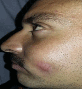
Figure 1: Extra-oral swelling in relation to left side of face.
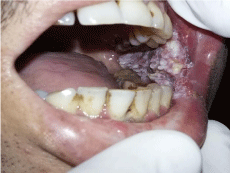
    Figure 2:  Exophytic, broad-based, ulcero-proliferative, papillary growth in
relation to left anterior buccal mucosa.
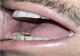
    Figure 3:  White, marble-like, blanched appearance in relation to relation to
right buccal mucosa.
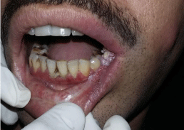
    Figure 4:  Blanched appearance in relation to lower lip mucosa with patchy
areas of erythema and pigmentation.
On palpation, vertical fibrous bands were palpable in relation to both the right and left retro-molar areas with fibrosis in relation to labial mucosa of the lower lip. Based on the above-mentioned clinical examination, a provisional diagnosis of verrucous carcinoma secondary to oral sub mucous fibrosis was arrived-at while proliferative verrucous leukoplakia (a high-risk pre-cancerous lesion; PVL) and verrucous hyperplasia were considered as the differential diagnoses. To rule-out these entities, investigations like routine hematological examinations and incisional biopsy were performed.
Histo-pathological examination: Haematoxylin and Eosin stained sections of the tissues revealed a hyperplastic, parakeratinized, stratified squamous epithelium with highly cellular connective tissue and epithelium showing broad bulbous pushing rete pegs into the subjacent connective tissue (Figure 5). Numerous cleft-like spaces were seen with parakeratin plugging within them (Figure 6). The epithelial cells exhibited increase in the basal cell layer with some cells exhibiting pleomorphism, hyperchromatism and an increased mitotic activity. The underlying connective tissue showed infiltration of numerous darkly staining inflammatory cells. (Figure 4,5) The histo-pathological examination further supported a final diagnosis of verrucous carcinoma in a setting of oral sub mucous fibrosis as thought.
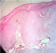
Figure 5: Overall survival, autologous stem cell transplant (ASCT) versus no ASCT (p=0.12).
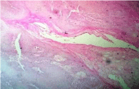
Figure 6: Parakeratin plugging extending from the surface into the epithelium.
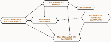
Figure 7:
Discussion
Oral sub mucous fibrosis (OSMF) is a pre-malignant condition which comes in the category of potentially malignant epithelial lesions (PMELs) characterized by fibrosis of the lining mucosa of the upper digestive tract involving the oral cavity, oro-and hypopharynx and the upper third of the oesophagus. The fibrosis involves the lamina propria and the sub mucosa of the tissues and may often extend to the underlying musculature resulting in the deposition of dense fibrous bands which gives rise to limited mouth opening, a hallmark of this disorder [2,8]. The diagnosis of oral sub mucous fibrosis is usually based on clinical signs and symptoms which include oral ulcerations, burning sensation (particularly with spicy foods), marble stone appearance of the affected mucosa with restricted mouth opening [2,6]. The potentially malignant nature of this condition has been well documented. A malignant transformation rate of 7.6% over a period of 10 years was described in an Indian cohort study while the relative risk for malignant transformation was reported to be as high as 397.3. Oral sub mucous fibrosis may cause atrophy of the epithelium increasing carcinogen penetration. Numerous studies suggest that dysplasia is seen in about 25% of the biopsied cases and the rate of transformation to malignancy varies from 3% to 19% [8]. The evidence supporting the malignant potential of OSMF includes: (i) higher prevalence of leukoplakia among OSMF patients; (ii) high frequency of epithelial dysplasia; (iii) histologic diagnosis of carcinoma without clinical signs of carcinoma; (iv) concurrent finding of OSMF among patients with oral cancers; and (v) a higher incidence of oral cancers among patients with OSMF. The case reported here had history of chewing gutkha and tobacco with lime that contained areca nut along with other harmful ingredients which had led to the development of oral sub mucous fibrosis due to the presence of various components including biologically active alkaloids, arecoline, arecaidine, arecolidine, guvacoline, guvacine, flavonoids (tannins and catechins) and copper. Oral sub mucous fibrosis is strongly associated with a risk of oral cancer although the biology underlying this association is still unresolved. Although development of oral squamous cell carcinoma is seen in only onethird of OSMF patients, the occurrence of verrucous carcinoma is said to be extremely rare.
Oral verrucous carcinoma is a rare, specific variant of welldifferentiated oral squamous cell carcinoma with characteristic clinical and histological features. The incidence of verrucous carcinoma varies from 4.5% to 9% or even higher as reported in some centers. In oral cavity, verrucous carcinoma constitutes 2% to 4.5 % of all forms of oral squamous cell carcinoma. The most common areas affected by oral verrucous carcinoma are the buccal mucosa and the lower alveolus with a reported incidence of 61.4% in relation to the buccal mucosa and 11.9% for the lower alveolus. These findings are in lieu with current literature which suggest that verrucous carcinoma has a predilection for the oral cavity, in particular, the buccal mucosa and the lower alveolus [9]. The tumor rarely crosses 10cm in its greatest dimension. The literature depicts that verrucous carcinoma occurs most commonly in males in their 5th-6th decades of life. Use of tobacco in smokeless and inhaled forms has been predominantly reported in the affected patients followed by betel nut chewing and use of alcohol. The oral hygiene is invariably poor in all the cases. The role of human papilloma virus (HPV) in verrucous carcinoma has been a matter of debate. Shear and Pindborg reported that out of 28 patients with verrucous lesions, 24 (86%) used tobacco and one was an areca nut chewer. Tobacco appears to be a major risk factor in the causation of verrucous lesions [10]. In our patient, chewing gutkha and tobacco with lime seemed to be the causative factor.
Verrucous carcinoma is a locally invading tumor and does not go for distant metastasis. If lymph nodes are palpable, they usually present an inflammatory reaction in large secondarily infected lesions. In our case, lymph nodes were not palpable. Verrucous carcinoma usually present with a hyperplastic epithelium with abundant keratin superficially is projecting as exophytic, churchspire keratosis and also, depicting parakeratin plugging which is believed to be the characteristic feature of this tumor [11]. The bulbous, well-oriented, rete ridges show endophytic growth pattern with pushing borders, well-appreciated in our case. Abrupt transition from normal epithelium to endophytic in growth is taken as an important parameter to differentiate verrucous carcinoma from other benign verrucous growths. The epithelium is well-differentiated in all the rete pegs.
A classic case of verrucous carcinoma shows minimal or no pleomorphism of cells and no abnormal mitotic activity above the basal and supra-basal layers of the epithelium. If focal atypia or dysplasia is evident, it must be limited to the basal layer of epithelium. Lympho-plasmacytic inflammatory reaction is marked especially, in cases, where keratin has plunged deep into the connective tissue inducing foreign body granuloma formations. Inadequate biopsy and tangential sectioning often deviates the diagnosis. The basement membrane is usually intact and deep cleft-like spaces lined by thick parakeratin extending from the surface deep into the lesion are evident as were also appreciated in our case.
The most important differential diagnoses of verrucous carcinoma includes: (i) Oral squamous cell carcinoma showing verrucoid features, (ii) Proliferative verrucous leukoplakia (PVL), (iii) epithelial hyperplasia, (iv) pseudo-epitheliomatous hyperplasia, (v) verruca vulgaris, and (vi) keratoacanthoma [12].
Grinspan has divided Verrucous Carcinoma into four types: [13]
Type I A: The type which is characterized by acanthosis, papillomatosis, leukoedema, moderate ortho-or para-keratosis, hypertrophic inter-papillary crests and stratification of the basal layer;
Type I B: which is characterized by cryptic depression of the epithelial surface, invagination of the epithelium and a fistulous tendency;
Type II: characterized by areas with the characteristics of type I A or type I B with hyperchromatic nuclei and atypical mitosis;
Type III: This is characterized by areas of type I or type II with features of oral squamous cell carcinoma with anaplastic cells and metastasis that are frequently observed in this type.
Also, Hybrid Verrucous-Squamous Cell Carcinoma (VC-SCC) is a term that has been used in the literature with most of the cases relating to the transformation of VC into frank SCC usually post-radiation treatments [10]. A review of literature revealed 4 cases of SCC arising within VC with one reported case in oral cavity, one in penis, one in vagina, and the remaining one in skin [14].
The prognosis of verrucous carcinoma is better than that of other kinds of life-threatening malignant tumors. Various treatment modalities include surgery, chemotherapy, radiation, or combination of these with photodynamic therapy which has recently been used. The use of buccal fat pad is increasing owing to its reliability, ease of harvest and relatively low complication rate in the reconstruction purposes to cover the post-surgical defects created after extensive surgeries. It is commonly used as a pedicle or free graft in reconstructing small to medium sized defects intra-orally [11].
Conclusion
Oral sub mucous fibrosis (OSMF) is a condition associated with an increased risk of malignant transformation. Verrucous carcinoma is an indolent, low-grade, carcinoma with benign histologic appearance. There are very few cases of verrucous carcinoma (VC) in cases diagnosed with OSMF reported in literature. It is stated that verrucous carcinoma may arise de novo or it may arise from potentially malignant epithelial lesions (PMELs) including OSMF. The characteristic features of verrucous carcinoma include a slow growth, a lack of distant metastases, its pathognomonic pattern of local invasion and the risk of progression to oral squamous cell carcinoma (OSCC). Surgical resection with adequate margins is the treatment of choice.
References
- Jacobson S, Shear M. Verrucous carcinoma of the mouth. J Oral Pathol. 1972; 1: 66-75.
- Shafer, Hine, Levy. Shafer’s Textbook of Oral pathology. 7th ed. Elsevier: 2012.
- Rocco R Addante, Samuel J McKenna. Verrucous Carcinoma. Oral. Maxillofacial Surg Clin N Am. 2006; 18: 513-519.
- Maraki D, Boecking A, Pomjanski N, Megahed M, Becker J. Verrucous carcinoma of the buccal mucosa: Histopathological, cytological and DNAcytometric features. J Oral Pathol Med. 2006; 35: 633-635.
- Regezi JA, Sciuba JJ, Jordan RCK. Verrucal-papillary lesions. In: Saunders WB, ed. Oral pathology. Clinico-pathologic correlations. 4th ed. St Louis; USA: WB Saunders: 2003.
- Neville BW, Damm DD, Allen CM, Bouquot JE. Epithelial pathology. In: Saunders WB, ed. Oral and Maxillofacial Pathology. 2nd ed. St. Louis; USA: WB Saunders: 2002.
- El-Rouby DH. Association of macrophages with angiogenesis in oral verrucous and squamous cell carcinomas. J Oral Pathol Med. 2010; 39: 559- 564.
- Pundir S, Saxena S, Aggrawal P. Oral sub mucous fibrosis a disease with malignant potential: Report of two Cases. J Clin Exp Dent. 2010; 2: e215-e218.
- Kamala K, Sankethguddad S, Sujith SG. Verrucous Carcinoma of Oral Cavity: A Case Report with Review of Literature. International Journal of Health Sciences and Research. 2015; 5: 330-334.
- Agnihotri A, Agnihotri D. Verrucous carcinoma: A study of 10 cases. Indian J Oral Sci. 2012; 3:79-83.
- Shergill AK, Charlotte M Solomon, Carnelio S, Kamath AT, Aramanadka C, Shergill GS. Verrucous Carcinoma of the Oral Cavity: Current Concepts. International Journal of Scientific Study. 2015; 3: 114-118.
- Asha ML, Vini K, Chatterjee Ingita, Patil P. Verrucous Carcinoma of Buccal Mucosa: A Case Report. International Journal of Advanced Health Sciences. 2014; 1: 19-23.
- Kumar RBV, Raj AC. Verrucous carcinoma of lip: An unusual presentation. Amrita J Med. 2012; 8: 1-44.
- Terada T. Squamous cell carcinoma arising within verrucous carcinoma of the oral cavity: A case report. Int J Clin Exp Pathol. 2012; 5: 363-366.