
Case Report
Austin J Dent. 2016; 3(3): 1040.
Clinical, Radiographic and Biochemical Findings of a Case of Florid Cemento-Osseous Dysplasia: A Disorder of its Own Type in Bone Disorders
Kartheeki B1, Nayyar AS1*, Ravikiran A2, Samatha Y2 and Bhargavi D2
1Department of Oral Medicine and Radiology, Saraswati Dhanwantari Dental College and Hospital, India
2Department of Oral Medicine and Radiology, Sibar Institute of Dental Sciences, India
*Corresponding author: Abhishek Singh Nayyar, Department of Oral Medicine and Radiology, Saraswati Dhanwantari Dental College and Hospital and Post- Graduate Research Institute, Parbhani, Maharashtra, India
Received: June 13, 2016; Accepted: August 10, 2016; Published: August 12, 2016
Abstract
Cemento-osseous dysplasias (CODs) are a group of disorders originating from periodontal tissues. They are classified, depending on their extent and location, into three groups: periapical, florid and focal.
Florid cemento-osseous dysplasias (FCODs) are one of the subgroups of cemento-osseous dysplasias (CODs) which were first described by Melrose, Abrams and Mills in 1976 and predominantly affects the jaws in middle-aged black females. They usually manifest as multiple radiopaque cementum-like masses distributed throughout the jaws or involve few quadrants. Patients do not have specific laboratory or radiological evidence of bone disease in other parts of the skeleton. The exact etiology of this disorder however is yet to be known.
Asymptomatic patients generally do not require treatment. Patients with this disease exhibit poor healing and osteomyelitis may develop even after minor surgical procedures including extraction of teeth in the affected areas. Surgical intervention is required for cases with gross disfigurement.
Complete resection of the lesion is considered to be impractical because the lesion usually occupies larger portion of the jaws. Herein, we are presenting a case of florid cemento-osseous dysplasia in a 45 year old female patient who reported to the Outpatient Department with some other odontogenic complaint while the lesion was detected when radiographs were taken for the same.
Keywords: Florid; Radiopaque; Asymptomatic
Introduction
The classification system of cemento-osseous lesions of the jaws has been a matter of discussion for a long time amongst pathologists and clinicians. Florid cemento-osseous dysplasias (FCODs) are one of the subgroups of cemento-osseous dysplasias (CODs) which were first described by Melrose, Abrams and Mills in 1976 [1,2]. The term florid refers to the extensive and widespread presentation of these lesions [3,4]. A review of the literature shows a wide range of terminologies used by various authors to describe these lesions which seem to be similar [5]. The 1992 classification of cemento- osseous dysplasias, released by the World Health Organization, was based on age, sex and histological, radiographic and clinical characteristics, as well as location of the lesion. FCODs were then again reviewed by the World Health Organization in 2005 as bone-related, non-neoplastic, fibro-osseous lesions [2,3].
The exact etiology of FCODs is still unknown [2]. Most authorities suggest that the pathogenesis of these lesions comes from the periodontal ligament since most of the lesions are seen within the proximity of periodontium and they have similar histopathological features [3,6]. Few authors also believe that the remains of cementum left in the bone post-extractions might be the reason for the development of these lesions [7,8].
Florid cemento-osseous dysplasias are more commonly seen in middle-aged black females, although they might also be seen in the Caucasian and Asian populations. The process may be totally asymptomatic and in such cases, the lesion is detected when radiographs are taken for some other purposes [9,10]. Radiographically, the lesions appear as multiple sclerotic masses, located in more than two quadrants, usually in the tooth-bearing regions. They are often confined within the alveolar bone.
Histologically, these lesions consist of anastomosing trabeculae of bone and layers of cementum-like calcifications embedded in a fibroblastic background [11].
Management of these lesions involves clinical and radiographic follow-up. Herein, we are presenting a case of florid cementoosseous dysplasia in a 45 year old female patient who reported to the Outpatient Department with some other odontogenic complaint while the lesion was detected when radiographs were taken for the same.
Case Presentation
A 45 year old female patient was referred from the Outpatient Department to the Department of Oral Medicine and Radiology with a chief complaint of pain in the lower right back tooth region since a week.
On clinical intra-oral examination, tooth # 46 had a deep carious lesion in relation to the distal proximal aspect. The tooth was nontender on vertical percussion. An intra-oral periapical radiograph (IOPAR) was advised. The intra-oral periapical radiograph showed indistinct lamina dura in relation to all the teeth present in the radiograph and there was mixed radiolucent and radiopaque areas extending from middle third of the root of 46 to the periapical region. Multiple sclerotic masses with radiolucent rims were found, confined within the alveolus corresponding to the roots of the teeth.
An orthopantomograph was advised to delineate the extent of the lesion. Radiograph, at first glance, demonstrated a pagetoid, cottonwool appearance with multiple irregularly shaped radiopaque areas. The radiopaque cloud-like masses, varying greatly in size and shape, dense and disseminated, appeared as generalized radiopacity of the jaws. Some were spherical, whereas others were lobular, suggesting coalescence. On closer examination, well-defined radiolucent rims were seen surrounding most of the radiopaque areas. The radiopaque patterns varied in size and were large, diffuse and continuous throughout the tooth-bearing regions of the jaw. In maxilla, they were multiple and discrete pertaining to the periapical areas of teeth # 13, 14, 15, and 22, 23 region. Root clubbing with hypercementosis was evident. They were seen bilaterally and were almost symmetrically positioned (Figure1). Intra-oral periapical radiographs of all teeth were taken to confirm the involved teeth (Figure 2).
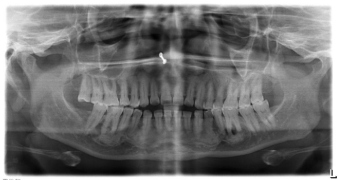
    Figure 1:  Panoramic radiograph showing multiple sclerotic masses with
radiolucent rims in maxilla and mandible.
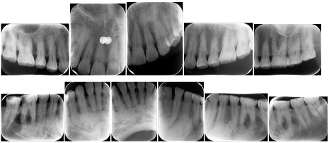
Figure 2: Periapical radiographs showing radiopaque masses confined within the alveoli at a level corresponding to the roots of the teeth.
Tooth vitality test was done with electrical pulp tester and all the teeth were found to be vital. The radiographs taken for the skull and extremities did not show any radiographic alterations. Biochemical analysis of serum calcium and phosphorus as well as serum alkaline phosphatase levels was carried-out and was shown to be within normal limits.
As the patient was completely asymptomatic, biopsy was not performed to prevent unnecessary surgical intervention as no treatment is generally required for asymptomatic cases other than periodic clinical and radiographic follow-ups for any secondary complications that might arise in case of an infection and/or, subsequent surgical procedures that induce iatrogenic damage to the bone structure. Every effort was made to preserve the natural dentition since patients with this disease exhibit poor healing and osteomyelitis might develop even after minor surgical procedures including simple, non-surgical tooth extractions. For a similar reason, biopsy was also not recommended for the patient.
Discussion
FCODs have three developmental stages with different radiographic features depending on the stage at which these lesions are diagnosed. The first or osteolytic stage is characterized by welldefined radiolucencies with loss of lamina dura in relation to the affected teeth. In the second or cementoblastic stage, multiple small radiopacities develop in the radiolucent foci because of the deposition of cementum-like tissue in the dysplastic areas. The last stage is characterized by definite and well-defined radiopaque foci seen in majority of the lesions [12].
The diagnosis of FCODs is largely made by clinical and radiographic features [1,2]. Paget’s disease, Osteopetrosis, Multiple cemento-osseous dysplasia, Familial gigantiform cementoma, and diffuse sclerosing osteomyelitis are the most important conditions to be considered in the differential diagnoses for this disorder [13].
Paget’s disease and Osteopetrosis show involvement of additional bones of the skeleton. Florid cemento-osseous dysplasia has a strong predilection for black females over 30 years of age where as Osteopetrosis is seen in older patients. The most salient feature of this disorder is the exquisite involvement of the jaw bones wherein the radiographs taken for the jaws reveal multiple radiopaque masses rimmed by radiolucent peripheries. These two are the unique features that help in differentiating florid cemento-osseous dysplasia from Paget’s disease and Osteopetrosis [13,14]. Also, osteopetrosis usually involves all the skeletal bones; but in Paget’s disease, there is involvement of five or six bones at most. Florid cemento-osseous dysplasia involves only mandible and maxilla [13].
Multiple cemento-osseous dysplasias are more common and have multiple small lesions of periapical and focal dysplasias distributed throughout the tooth-bearing regions of the jaws. But these lesions remain small, neither cause cortical expansion nor not susceptible to osteomyelitis like florid cemento-osseous dysplasia [13].
Familial gigantiform cementomas are rare. Both genders are affected equally, and patients are usually affected at an earlier age than they are with in case of florid cemento-osseous dysplasia [15].
Diffuse sclerosing osteomyelitis involves only one segment of the jaws. The radiopaque part is more diffuse, and its margins gradually blend into normal bone at the periphery. Clearly, it does not have the radiolucent rim in the periphery that is common in florid cementoosseous dysplasia. Also, it does not show female predilection [16].
Malignant osteopetrosis is given the lowest rank in differential diagnosis because it is almost invariably fatal by the age of 20 years [13].
Florid cemento-osseous dysplasias were initially described by Melrose, Abrams and Mills in 1976 as a dysplastic lesion or developmental abnormality arising in tooth-bearing areas [17]. They exhibited a sclerotic appearance on conventional radiographs [18,19]. Paget’s disease of the bone may also have a cotton-wool like appearance. It affects the alveolus of the entire mandible and shows loss of lamina dura, whereas florid cemento-osseous dysplasia is usually above the inferior alveolar nerve canal and the middle and cervical thirds of roots are normal. Paget’s disease is polyostotic, involving spine, femur, skull, pelvis and sternum and produces serum changes like elevated serum alkaline phosphatase levels. No biochemical changes and other bone involvement were observed in the case reported [19,20].
Another disease that may closely resemble florid cementoosseous dysplasia is chronic diffuse sclerosing osteomyelitis. It usually appears as a localized, poorly delineated radiopaque segment of the jaw bone, whereas florid cemento-osseous dysplasia is seen as multiple radiopaque masses [21,22].
Florid cemento-osseous dysplasia may be familial in some cases with an autosomal dominant inheritance pattern, but there are only a few examples in the literature confirming this inheritance. In the present case, no familial pattern of the disease could be noticed [19].
Asymptomatic patients generally do not require treatment. Patients with this disease exhibit poor healing and osteomyelitis may develop even after minor surgical procedures including extraction of teeth in the affected areas due to poor vascularity [1,2].
Surgical intervention is required for cases with gross disfigurement although complete resection of the lesion is considered impractical because the lesion usually occupies larger portion of the jaws. In extensive lesions, where surgical intervention is indicated, bone remodeling following resection is recommended for esthetic reasons [19].
Conclusion
The diagnosis of florid cemento-osseous dysplasia in the jaws is usually done by clinical and radiographic features. Also, histology plays an important role in confirming the diagnosis of florid cemento-osseous dysplasias although it is not generally recommended because the disease exhibits poor healing and there are high chances of development of osteomyelitis even after minor surgical procedures including simple, non-surgical tooth extractions. The diagnosis, therefore, in case of florid cemento-osseous dysplasias, is largely arrived-at only on the basis of clinical and radiographic features.
This case report is one of the classic examples for stating that “Radiographs play a crucial role not only in diagnosis but also, in treatment planning”. In these situations, where there are chances of delayed wound healing, a treatment plan should focus towards the Devan’s dictum.
References
- Gerlach RC, Dixon DR, Goksel T, Castle JT, Henry WA. Case presentation of florid cemento-osseous dysplasia with concomitatnt cemento-ossifying fibroma discovered during implant explantation. Oral Surg Oral Med Oral Pathol Oral Radiol. 2013; 115: e44-e52.
- Sarmento DJ, Monteiro BV, de Medeiros AM, da Silveira EJ. Severe florid cemento-osseous dysplasia: A case report treated conservatively and literature review. Oral Maxillofac Surg. 2013; 17: 43-46.
- Sanjai K, Kumarswamy J, Kumar VK, Patil A. Florid cemento-osseous dysplasia in association with dentigerous cyst. J Oral Maxillofac Pathol. 2010; 14: 63-68.
- Rao KA, Shetty SR, Babu SG, Castelino RL. Co-occurence of florod cemento-osseous dysplasia and simple bone cyst: A case report. J Oral Maxillofac Res. 2011; 2: e5.
- Young SK, Markowitz NR, Sullivan S, Seale TW, Hirschi R. Familial gigantiform cementoma: Classification and presentation of a large pedigree. Oral Surg Oral Med Oral Pathol. 1989; 68: 740-747.
- Neville BW, Damm DD, Allen CM, Bouquot JE, eds. Bone pathology. In: Neville BW, Damm DD, Allen CM, Bouquot JE., editors. Oral and Maxillofacial Pathology. St.Louis, Missouri: Saunders Elseviers: 2009.
- Peter AR, Philipsen HA. Odontogenic tumors and allied lesions. In: Peter AR, Philipsen HA, editors. Florod cemento-osseous dysplasia . London: Quintessence: 2004.
- Paul MS, Roman C. Maxillofacial fibro-osseous lesions. Curr Diagn Pathol. 2006; 12: 1-10.
- Neville BW, Damm DD, Allen CM, Bouquot JE. Oral and Maxillofacial Pathology. W.B. Saunders Company Philadelphia; Pennsylvania: 1995.
- Kramer IRH, Pindborg JJ, Shear M. Neoplasma and others lesions related to bone. In: World Health Organization (Editors) - Histologic Typing of Odontogenic Tumours. Berlim: Springer-Verlag: 1992.
- White SC, Pharoah MJ. Oral Radiology: Principles and Interpretation. Saint Louis: Mosby: 2000.
- Bhandari R, Sandhu SV, Bansal H, Behl R, Bhullar RK. Focal cemento-osseous dysplasia masquerading as a residual cyst. Contemp Clin Dent. 2012; 3: S60-S62.
- Norman K Wood, Paul W Goaz. Differential Diagnoses of Oral and Maxillofacial Lesions. Mosby. 1997.
- Damm DD, Fantasia JE. Multifocal mixed radiolucencies: Florid cemento-osseous dysplasia. Gen Dent. 2001; 49: 461, 538.
- Toffanin A, Benetti R, Manconi R. Familial florid cemento-osseous dysplasia: A case report. J Oral Maxillofac Surg. 2000; 58: 1440-1446.
- Groot RH, van Merkesteyn JP, Bras J. Diffuse sclerosing osteomyelitis and florid osseous dysplasia. Oral Surg Oral Med Oral Pathol Oral Radiol Endod. 1996; 81: 333-342.
- Melrose RJ, Abrams AM, Mills BG. Florid osseous dysplasia. A clinical-pathologic study of thirty-four cases. Oral Surg Oral Med Oral Pathol. 1976; 41: 62-82.
- Marcelo G, Ronaldo P, Fabio de Abreu A, Carlos Eduardo BL, Andrea G. Clinical, Radiographic, Biochemical and Histological Findings of Florid Cemento-Osseous Dysplasia and Report of a Case. Braz Dent J. 2005; 16: 247-250.
- Said-al-Naief NA, Surwillo E. Florid osseous dysplasia of the mandible: Report of a case. Compend Contin Educ Dent. 1999; 20: 1017-1019.
- Langlais RP, Langland OE, Nortjé CJ, eds. Diagnostic Imaging of the Jaws. Malvern: Williams & Wilkins: 1995.
- Waldron CA, Giansanti JS, Browand BC. Sclerotic cemental masses of the jaws (so-called chronic sclerosing osteomyelitis, sclerosing osteitis, multiple enostosis, and Gigantiform cementoma). Oral Surg Oral Med Oral Pathol. 1975; 39: 590-604.
- Kim JH, Song BC, Kim SH, Park YS. Clinical, radiographic and histological findings of florid cemento-osseous dysplasia: A case report. Imaging Science in Dentistry. 2011; 41:139-142.