
Case Report
Austin J Dent. 2016; 3(3): 1041.
Acute Necrotizing Periodontitis: A Case Report
Assimi S, Abdallaoui L and Ennibi OK*
Department of Periodontology, Mohammed V University in Rabat, Morocco
*Corresponding author: Ennibi Oumkeltoum, Department of Periodontology, Faculty of Dental Medicine, Mohammed V University in Rabat, BP 6212, Rabat, Morocco
Received: June 14, 2016; Accepted: August 22, 2016; Published: August 24, 2016
Abstract
Necrotizing periodontal diseases are the most severe inflammatory periodontal disorders caused by bacterial plaque. The diagnosis is based on clinical and radiological features. Necrotizing periodontitis lesions are confined to periodontal tissues, including gingiva, periodontal ligament, and alveolar bone. The pathognomonic clinical characteristics are the typical punched out appearance and interproximal craters. In this case report, we presented a 20- year old man with necrotizing periodontitis and no systemic disease with a history of tobacco use and intense stress. The case was managed by conservative oral treatment and regular psychological follow up.
Keywords: Necrotizing periodontitis; Diagnosis; Treatment
Introduction
Necrotizing periodontal diseases (NPD) are the most severe inflammatory periodontal disorders caused by bacterial plaque and usually run an acute course [1]. They are classified as necrotizing gingivitis, periodontitis, or stomatitis, and they appear to represent various stages of the same disease [2]. Clinical features in necrotizing periodontitis (NP) are characterized by the presence of punchedout, ulcerated and necrotic lesions that may be covered by a pseudomembrane of necrotic tissue [3]. The ulcerations are extremely painful and show spontaneous bleeding [4]. An important feature of NP is the rapid and severe loss of clinical attachment and alveolar bone within a few days or weeks [1]. Other possible clinical features include the presence of oral halitosis, adenopathies, fever, and/or general discomfort [5].
In developing countries, the prevalence of NPD is higher than in the industrialized countries, however, the prevalence of these diseases has declined significantly during the twentieth century [6]. The disease frequently occurs in children and young adults, military persons and slightly more often among HIV-infected individuals [1,7]. The prevalence of NP seems to be low and decreases with age. Its occurrence in systemically healthy population is difficult to estimate since most studies of necrotic oral lesions have failed to differentiate between necrotizing gingivitis and necrotizing periodontitis based on the presence or absence of attachment loss and bone loss at affected sites [8]. Nonetheless, in HIV-seropositive individuals, the prevalence of NP was about 2% to 6% [9]. This low prevalence may be explained by the introduction of the antiretroviral therapy [10] which makes no difference between this patient’s category and the general population [1].
Factors causing NP are not clearly established. It may be a consequence of necrotizing ulcerative gingivitis or as a history of previous occurring NP [11,12]. However, a combination of etiological factors may play a role in its pathogenesis [13] such as Human Immunodeficiency Virus (HIV), diabetes, leukemia, poor oral hygiene, intense and prolonged psychological stress with insufficient rest and recent illness, nutritional deficiency, alcohol abuse and smoking [2,12,14]. Here, we report a case of an acute necrotizing periodontitis, describing the clinical features, treatment approach, and successful outcomes.
Case Presentation
A 20-year-old male patient was referred to the Clinical Department of Periodontology, at the center of consulting and dental treatment, Ibn Sina hospital in Rabat, Morocco, with complaint of intense and persistent oral pain, gingival bleeding and fetid breath for two weeks. Because of the extremely pain, the patient reported that he didn’t slept for two days and stopped eating for four days. In addition to that, he didn’t have a regular oral hygiene and he stopped tooth brushing since the pain started. He weighed 60 kg and was 182 cm tall and claimed having weight loss since one month (6 Kg).
The patient was otherwise systemically healthy with no medical history of interest. He was a heavy smoker for 6 years (20 cigarettes per day), he also used to chew tobacco and consume alcohol. He declared having had unprotected sex and had been experiencing heavy stress due to family problems.
On extraoral examination, bilateral submandibular lymph nodes were tender on palpation and a rise in body temperature was detected.
On intraoral examination, poor oral hygiene was noticed with plaque, heavy calculus deposits, visible suppuration and severe halitosis. The oral lesions were extremely painful hindering periodontal probing. Examination of the gingiva revealed a thin whitish film (pseudo-membrane) that covered a part of the attached gingiva with bleeding on slight stimulation. Ulcerations and tissue necrosis were almost generalized and severe dividing the papillae into separate edges one facial and one lingual portion with an interposed necrotic depression producing considerable tissue destruction and formation of characteristic punched out crater like depressions with exposition of the interdental bone (Figure 1A,B and C).
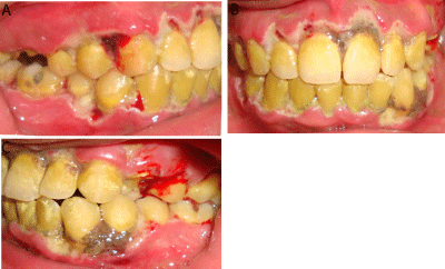
    Figure 1:  Day 1; intra oral view [(a) Wright side, (b) front, (c) left side] showing
severe acute necrotizing ulcerative periodontitis with the characteristic
punched-out eroded gingival margin with surface pseudo membrane and
bone sequestration.
One week later, a full mouth periodontal probing was possible. It showed no attachment loss expect for the lower and upper incisors.
A periapical radiographic examination showed a slight horizontal bone loss in the lower anterior region (Figure 2).
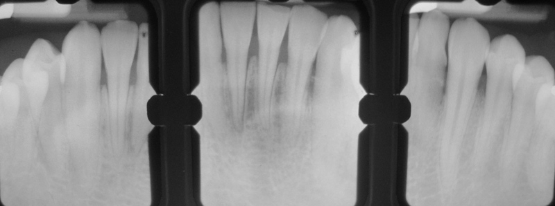
    Figure 2:  A periapical radiographic examination showed slight horizontal
bone loss in the lower anterior region.
Based on the anamnesis, the clinical and radiographic findings, a diagnosis of necrotizing periodontitis was made.
At the first day visit, 3% hydrogen peroxide was gently applied to the lesions using sterile swabs to remove the pseudo membranes and non-attached surface debris. Then, the area was cleaned with warm water and povidone-iodine solution (Figure 3A). The superficial big calculus was removed using ultrasonics devices. Patient was prescribed 250 mg of Metronidazole tree times per day for 7 days, an analgesic (Paracetamol associated with Codeine) every 6 hours and vitamin complexes. An alcohol free mouth rinse based on 0.2% Chlorhexidine was recommended twice a day for 7 days. The patient was instructed to avoid smoking, tobacco chewing, alcohol consumption, and spicy and acidic food. Advises to take adequate rest and proper diet were given.
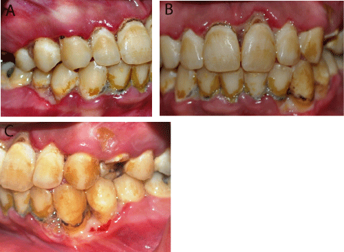
    Figure 3:  Day 3; intra oral view [(a) Wright side, (b) front, (c) left side] showing
the persistence of erythema, but the clinical condition was greatly improved.
That situation allowed supragingival scaling.
After three days the patient was re-examined (Figure 3B) and supragingival scaling was performed (Figure 3C). Proper oral hygiene instructions were given with prescription of soft tooth brush. Based on the weight loss, oral features and patient’s habits (unprotected sex, smoking and chewing tobacco and alcohol), biochemical blood analysis and HIV serology were requested.
After seven days the patient was almost symptom free (Figure 4). The results of the biochemical blood analysis were normal and the HIV sero-status was negative. Thus, complete periodontal debridement was done. Oral hygiene instructions were reinforced with prescription of interdental brushes and dental floss to perform mechanical plaque control. Additionally, he was advised to use Chlorhexidine mouth rinse 0.12% twice a day for further 10 days. Patient was referred to a smoking cessation center and to a psychologist to rule out his stress due to his family problems.
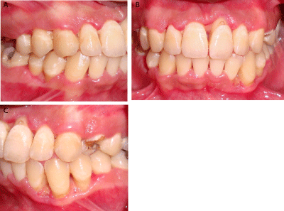
    Figure 4:  Day 7; intra oral view [(a) Wright side, (b) front, (c) left side] great
resolution of inflammation and suppuration.
Clinical Outcomes
The patient was very responsive to the treatment provided. At onemonth recall visit, there was a complete resolution of inflammation and a healing of ulcerated areas with epithelialization of gingival interdental craters (Figure 5).
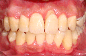
    Figure 5:  Day 30; Physiologic count our and new attachment of gingiva with
epithelialization of interdental craters.
The patient was compliant to treatment and regularly followed its meetings to smoking cessation center. A psychologist was consulted, and the patient was kept on maintenance with instructions of oral hygiene and proper nutrition.
Discussion
Necrotizing periodontitis (NP) is a necrotizing periodontal disease that has an acute onset and requires urgent treatment [1,15].
The pathognomonic clinical characteristics are the typical punched out appearance and interproximal craters which were evident in our patient. Bone sequestration like in our case may occur in extreme cases [6].
Although its clinical signs are easy to identify, much is still not understood regarding the etiology and pathogenesis [16]. It is a known fact that the pathogenic processes involved in any periodontal diseases is initiated by bacterial plaque and modified by predisposing factors [17,18].
The bacterial flora associated NP is dominated by fusiform bacilli and spirochetes. However, it is not clearly established whether this flora is the cause because of their capacity to invade the epithelium and the connective tissue where they release endotoxins or the consequence of the disease and then they may result from a secondary growth since the necrotic tissues are the perfect environment for bacterial colonization and tissue invasion [7]. Recently, researchers have pointed out the possible etiological role of Herpes viruses, especially HCMV and EBV-1, in necrotizing periodontal diseases [7]. Herpes viruses are usually contracted in childhood, and may be reactivated under various conditions such as stress [19]. In the reported case, the microbiological diagnosis was not done, clinical features and the presence of risk factors were sufficient to establish the diagnosis which was confirmed by the clinical improvement 48h after the beginning of the treatment.
Extra oral features such as lymphadenopathy, halitosis, and fever have been reported to occur in NP [16]. However, none of these are pathognomonic since they frequently occur in many other forms of periodontal disease such as primary herpetic gingivostomatitis or mononucleosis [16]. In this case, their presence is notable and may be probably related to the severity of the disease since it is usually observed in advanced cases.
Predisposing systemic factors such as AIDS, diabetes, chemotherapy or leukemia are described in the literature to be associated to most cases of PUN [20-22]. In our patient a systemic disease was rule out since biological analysis were normal and he was otherwise healthy, and his weight loss is argued by pain and general discomfort that might severely compromised food intake.
This case report describes an acute presentation of necrotizing periodontitis associated with poor oral hygiene, malnutrition, lack of sleep, alcohol and tobacco abuse, which are all related to intense psychological stress. Many pathways have been suggested to explain the relationship between NP and psychological stress. Indeed, a recent study evaluated the association between levels of the stress-related steroid hormones and periodontitis and reported that high serum levels of cortisol were associated with periodontitis severity [23]. Host immune response to periodontal pathogens may be reduced by increasing the adrenocortical activity, which lead to altered cytokine profile and affected cells recruitment of macrophages and fibroblasts [24]. Stress also causes reduction of tissue matrix metalloproteinase levels, which leads to impaired tissue turnover [25].
During stress periods, the subject’s behaviors also change and can lead patients to neglect oral hygiene [26].
Malnutrition related to extreme stress has also been reported as a predisposing factor for necrotizing periodontal diseases. The basis for this interaction has been termed ‘protein-energy malnutrition’, implying a marked reduction in antioxidant nutrients and an altered acute-phase response against infection [7].
Among the many harmful habits induced by emotional disturbances, smoking is possibly the most important. Many studies have showed the impact of the nicotine on the periodontal tissues. Indeed, vasoconstriction induced by smoking leads to a lack of nutrients for the periodontal tissues and inhibition of oral neutrophil function [27]. Alcohol consumption has also been associated with the physiological and psychological factors favoring necrotizing periodontal disease [7].
In the light of these considerations, it might be argued that the severity of NP in this case is directly related to extreme stress and consumption of tobacco and alcohol.
The management strategy for NP lesions is divided into two phases: acute and maintenance phase treatment [1]. The aim of the acute phase is to remove local irritating factors, plaque and calculus and necrotic tissues, as was done in this case. At the first consultation, we adopted a conservative approach, with the application of hydrogen peroxide to increase the supply of oxygen to the anaerobic microbes thereby inhibiting their growth. Mouthwash based 0.2% Chlorhexidine was a very effective adjunct to reduce plaque formation over the debrided lesions.
The patient’s rapid and positive response to the intervention within one week could be attributed to several factors: use of systemic metronidazole, which has been described as first antibiotic choice [1] because it is active against strict anaerobes, proper oral hygiene, adequate nutrition and rest.
Other systemic drugs have also been proposed, with acceptable results, including penicillin, tetracyclines, clindamycin, amoxicillin or amoxicillin plus clavulanate [7]. Conversely, topical application of antibiotics is not indicated in the treatment of NPD, because of the large numbers of bacteria present within the tissues, and topical application does not result in sufficient intra lesion concentration of antibiotics [1,7].
Conclusion
A successful outcome of necrotizing periodontal disease depends on a confidential relationship between patient and dentist.
This paper highlights the imminent danger faced by the populations where stress, tobacco, smoking and alcohol are not only frequent, but widespread especially among children and young adults. A prompt periodontal therapy and oral hygiene reinforcement and patient compliance are essential for the necrotizing periodontitis treatment.
References
- Holmstrup P. Necrotizing periodontal disease. In: Lindhe J, Lang NP. Clinical periodontology and implant dentistry. 6th ed. Oxford: Wiley-Blackwell; 2015.
- Horning GM, Cohen ME. Necrotizing ulcerative gingivitis, periodontitis, and stomatitis: clinical staging and predisposing factors. J Periodontol. 1995; 66: 990-998.
- Murayama Y, Kurihara H, Nagai A, Dompkowski D, Van Dyke TE. Acute necrotizing ulcerative gingivitis: Risk factors involving host defense mechanisms. Periodontol 2000 1994; 6: 116-124.
- Magan-Fernandez A, O’Valle F, Pozo E, Liebana J, Mesa F. Two cases of an atypical presentation of necrotizing stomatitis. J Periodontal Implant Sci. 2015; 45: 252-256.
- Corbet EF. Diagnosis of acute periodontal lesions. Periodontol 2000. 2004; 34: 204-216.
- Albandar JM, Tinoco EM. Global epidemiology of periodontal diseases in children and young persons. Periodontol 2000. 2002; 29: 153-176.
- Herrera D, Alonso B, de Arriba L, Santa Cruz I, Serrano C, Sanz M. Acute periodontal lesions. Periodontol 2000. 2014; 65: 149-177.
- Novak MJ. Necrotizing ulcerative periodontitis. Ann Periodontol. 1999; 4: 74-78.
- Paster BJ, Russell MK, Alpagot T, Lee AM, Boches SK, Galvin JL, et al. Bacterial diversity in necrotizing ulcerative periodontitis in HIV-positive subjects. Ann Periodontol. 2002; 7: 8-16.
- Tappuni AR, Fleming GJ. The effect of antiretroviral therapy on the prevalence of oral manifestations in HIV-infected patients: a UK study. Oral Surg Oral Med Oral Pathol Oral Radiol Endod. 2001; 92: 623-628.
- Zia A, Mukhtar-Un-Nisar Andrabi S, Qadri S, Bey A. Necrotizing periodontitis in a heavy smoker and tobacco chewer - A case report. Singapore Dent J. 2015; 36: 35-38.
- Dannewitz B, Eickholz P, Kohl A, Komposch G, Tomakidi P. Molecular changes in the gingival epithelium associated with necrotizing ulcerative periodontitis: a case report. Int J Periodontics Restorative Dent. 2006; 26: 191-196.
- Umeizudike KA, Savage KO, Ayanbadejo PO, Akanmu SA. Severe presentation of necrotizing ulcerative periodontitis in a Nigerian HIV-positive patient: a case report. Med Princ Pract. 2011; 20: 374-376.
- Bermejo-Fenoll A, Sánchez-Pérez A. Necrotising periodontal diseases. Med Oral Patol Oral Cir Bucal. 2004; 9 Suppl: 114-119.
- Armitage GC. Development of a classification system for periodontal diseases and conditions. Ann Periodontol. 1999; 4: 1-6.
- Rowland RW. Necrotizing ulcerative gingivitis. Ann Periodontol. 1999; 4: 65-73.
- Güntsch A, Erler M, Preshaw PM, Sigusch BW, Klinger G, Glockmann E. Effect of smoking on crevicular polymorphonuclear neutrophil function in periodontally healthy subjects. J Periodontal Res. 2006; 41: 184-8.
- Ishikawa I. Host responses in periodontal diseases: a preview. Periodontol 2000; 2007; 43: 9-13.
- Cappuyns I, Gugerli P, Mombelli A. Viruses in periodontal disease - a review. Oral Dis. 2005; 11: 219-229.
- Williams CA, Winkler JR, Grassi M, Murray PA. HIV-associated periodontitis complicated by necrotizing stomatitis. Oral Surg Oral Med Oral Pathol. 1990; 69: 351-355.
- Santos FA, Pochapski MT, Pilatti GL, Kozlowski VA, Goiris FA, Groppo FC. Severe necrotizing stomatitis and osteomyelitis after chemotherapy for acute leukaemia. Aust Dent J. 2009; 54: 262-265.
- Nakano H, Ota Y, Yura Y. Calcifying epithelial odontogenic tumor of the maxilla with ulcerative stomatitis: a case report. Br J Oral Maxillofac Surg. 2009; 47: 222-224.
- Ishisaka A, Ansai T, Soh I, Awano S, Yoshida A, Hamasaki T et al. Association of cortisol and dehydroepiandrosteronesulphate levels in serum with periodontal status in older Japanese adults. J Periodontol. 2008; 78: 1767-1773.
- Deinzer R, Kottmann W, Förster P, Herforth A, Stiller-Winkler R, Idel H. After-effects of stress on crevicular interleukin-1beta. J Clin Periodontol. 2000; 27: 74-77.
- Knuutinen A, Kokkonen N, Risteli J, Vähäkangas K, Kallioinen M, Salo T, et al. Smoking affects collagen synthesis and extracellular matrix turnover in human skin. Br J Dermatol. 2002; 146: 588-594.
- da Silva AM, Newman HN, Oakley DA. Psychosocial factors in inflammatory periodontal diseases. A review. J Clin Periodontol. 1995; 22: 516-526.
- Johnson GK, Guthmiller JM. The impact of cigarette smoking on periodontal disease and treatment. Periodontol 2000. 2007; 44: 178-194.