
Case Report
Austin J Dent. 2016; 3(5): 1047.
Revascularization of Permanent Premolars; Report of Three Cases with Clinical and Radiographic Outcomes
Hatipoğlu Z*, Atabek D and Akal N
Department of Pediatric Dentistry, Gazi University, Turkey
*Corresponding author: Hatipoğlu Z, Department of Pediatric Dentistry, Gazi University, Gazi University Faculty of Dentistry No: 4 Emek, Ankara, Turkey
Received: September 29, 2016; Accepted: October 15, 2016; Published: October 17, 2016
Abstract
Objective: Revascularization is a treatment in immature necrotic teeth which allows the continuation of root development. The aim of the present report was to introduce the successful regenerative endodontic treatment of three immature premolar teeth.
Study Design: The root canals were irrigated with 2.5% sodium hypochlorite with minimal instrumentation, followed by tri-antibiotic paste dressing (metronidazole, ciprofloxacin, amoxicillin). Instead of minocycline that is principally recommended for regenerative procedures, amoxicillin was used in order to avoid the discoloration of the crown. At a subsequent visit, the antibiotic paste was removed. A blood clot aroused in the canal by irritating periapical tissues and the root canals were sealed with MTA or BioAggregate, glass ionomer cement, and composite resin.
Conclusion: After 18, 24, and 30 months of radiographic and clinical followups respectively, the teeth were asymptomatic without discoloration, and the root developments were continued. It can be suggested that amoxicillin can be used successfully instead of minocycline in order to eliminate discoloration for regenerative endodontic treatment procedures.
Keywords: Antibiotics; Revascularization; Endodontics; Tri-antibiotic paste
Case Presentation
Treatment of necrotic immature teeth has always been a challenge in comparison to that of mature teeth [1]. The lack of an adequate apical barrier for condensation of gutta percha has traditionally required an apexification procedure to close the apex, enabling subsequent canal obturation. Apexification techniques originally involved induction of a calcific barrier at the apex with calcium hydroxide (Ca(OH) 2). Long-term use of calcium hydroxide inside the root canal space has several disadvantages such as multiple visits over a period of 9 to 20 months, a shortened root with thin walls, increased the likelihood of root fractures, poor crown-root ratio, probable recontamination of the root canal system, and increased dentin fragility [2].
Placing barrier material such as mineral trioxide aggregate (MTA) in the apical third of the canal is an alternative technique for apexification with calcium hydroxide, and MTA apexification success has been reported [3]. It has a high pH and antibacterial qualities similar to Ca(OH)2 and induces hard tissue formation. Its biocompatibility, an excellent sealing ability in the presence of moisture and efficient mechanical properties as an apical sealing material have also been reported. Especially, the shorter chair time seems to be an important advantage of the MTA apexification [4]. However, either apexification or the barrier technique doesn’t allow the continued root maturation that leads to weak root structure [ 5].
Regenerative endodontic treatment, which is a paradigm shift in the treatment of immature, necrotic teeth allows continued root development of the dentinal walls and apical closure [6]. The continued deposition of dentin occurs throughout the length of the root, providing greater strength and resistance to fracture. It is based on the presence stem and progenitor cells that have the ability to generate osteoid and odontoid structures from the pulp and /or periodontium [7]. The treatment offers regeneration of the stem cells, presence of a protein scaffold, and continuation of root development. These stem cells are dental pulp stem cells (DPSCs), which are more populated in the central-cell-rich zone of the pulp, bone marrow stem cells (BMSCs), stem cells from human exfoliated deciduous teeth (SHED), and stem cells from apical papilla (SCAPs) [8].
Regenerative procedures generally advocate that the placement of a tri- antibiotic paste is for eliminating intra-radicular infection from the root canal system [9]. The tri-antibiotic paste consists of a combination of ciprofloxacin, metronidazole (bactericidal), and minocycline (bacteriostatic), which was found sufficiently potent to eradicate the bacteria whereas the respective drugs used alone only reduced the bacteria [10]. However, crown discoloration, presumably due to minocycline, was reported as a disadvantage of this technique [11]. An alternative approach to discoloration was taken by Thomson and Kahler [12]. where amoxicillin was used instead of minocycline and a successful regenerative endodontic procedure without discoloration of a case was reported.
The other most important stage that affects the success of regenerative treatment occurs when removing the antibioticpaste formation of a blood clot and sealing of the canals with a biocompatible material that allows for the regeneration of new tissue adjacent to it [13]. Although the successful use of MTA as a sealing material is suggested, crown discoloration, depending on the presence of MTA, in the cervical portion of the canal is also reported [14]. In this context, researchers are looking for new, alternative materials. BioAggregate is a relatively new product, and its clinical applications include apexification, repair of root perforation and resorption, and vital pulp therapy [15]. Although MTA and BioAggregate have similar compositions and uses, BioAggregate does not create a risk of discoloration due to aluminum-free content [16].
This paper presents three case reports with successful regenerative endodontic treatment of three immature premolars using triantibiotic paste containing ciprofloxacin, metronidazole, amoxicillin, and BioAggregate as the sealing material.
Case 1
A healthy 10-year-old girl was referred to our clinic with complaints about spontaneous pain and swelling in the right side of her lower jaw. Patient’s medical history revealed no significant medical ailment. Clinical examinations revealed the mandibular right second premolar (LR5) tooth to be intact with no signs of caries and without any history of trauma; however, the developed abscess and the mobility were recognized. After radiographic examination, incomplete root formation (1/3 root formation) and a diffuse periapical radiolucency were diagnosed. A thin dentinal wall at the apex of the root that exhibited a wide- open apex and intracoronal radiolucent lesion was noted.
Considering the outcome of the clinical and radiographic examinations and the immaturity of the tooth, the diagnosis was pulpal necrosis with apical abscess, and the treatment option was revascularization.
After a complete explanation of the treatment procedure, its risks, benefits, and experimental nature, an informed consent was obtained from the patient’s parents. Firstly, local anesthesia with 3% plain mepivacaine (Septodont, Cedex, France) and rubber dam isolation was performed. An access cavity on tooth LR5 was prepared by using a diamond-coated fissure bur (Diatech, Heerbrugg, Switzerland) and a high-speed handpiece. The tooth was irrigated with 2.5% sodium hypochlorite for 10 minutes with minimal instrumentation. The canals were dried with paper points( Dentplus,Korea).The triantibiotic paste consisted of a powder of 20 mg of metronidazole (Flagyl, Eczacibasi, Turkey), ciprofloxacin (Cipro, Biofarma, Turkey), and amoxicillin (Largopen, Bilim, Turkey) each, mixed with 1 mL of sterile water. The paste was introduced into the canal with a lentulo and filled to the level of just below the cemento-enamel junction, and the tooth was restored temporarily with cavite (I-PRO, EU) and glass ionomer cement (Kavitan PRO, SpofaDental, Czech Republic) After three weeks, the patient was recalled, reporting that the tooth had been asymptomatic for that entire time. Moreover, the tooth was not sensitive to percussion and palpation. The temporary restorations were removed, the tooth was irrigated with 2.5% sodium hypochlorite, and the antibiotic paste was removed. The 25 K-file (Antaeos, Germany) was over-extended beyond the root length of the tooth several times in order to irritate the apical tissue until bleeding occurred apically in the root canal space and rose to the crown. By doing this, a biological scaffold for the regenerative process was created. After waiting 10 minutes, BioAggregate (Innovative BioCeramix Inc., Vancouver, British Columbia, Canada) was prepared according to the manufacturer’s directions, and 3 mm of BioAggregate was placed with the aid of a plugger and adapted softly to dentinal walls via a cotton pellet. The access cavity was sealed with glass ionomer cement and the acid etched composite resin (Tetric N-Ceram, Ivoclar Vivadent, Asia).
The patient was recalled at 3, 6, 12, and 30 months after treatment to evaluate the outcome. In clinical examination, the tooth was asymptomatic and functional with a normal periodontal condition. Also, the tooth was not tender to percussion or palpation and there was no presence of swelling. Radiographically, osseous healing of the periapical lesion was evident as well as the thickening of the dentinal wall and root maturation (Figure 1A-C). The appearance of the tooth showed no obvious change in shade or color (Figure 1D).
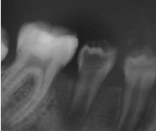
    Figure 1a:  Periapical radiograph. It llustrates a periapical radiolucency
associated with the mandibular right second premolar with a wide open apex.
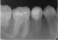
    Figure 1b:  Periapical radiograph. Radiographic image taken at the 12-month
follow-up appointment.
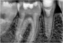
    Figure 1c:  Periapical radiograph. Radiographic image taken at the 24-month
follow-up appointment.
    Figure 1d:  Intraoral photograph. Clinical photograph of the tooth at the
24-month follow-up appointment.
Case 2
A 12-year-old male was referred to our clinic with complaint about pain upon biting on the tooth (UL5) and intraoral swelling at the buccal alveolar mucosa of the tooth. Patient’s medical history revealed no significant medical ailment. Clinical examination revealed slight caries on occlusal surface of tooth UL5, localized swelling of the buccal mocosa and also sensitivity to percussion. After radiographic examination, incomplete root formation (1/2 root formation) and a diffuse periapical radiolucency extending to the apex of tooth UL4 were diagnosed.
On the basis of the results of the clinical and radiographic examinations, the diagnosis of the tooth UL5 was pulpal necrosis with apical abscess.
The patient and his parents were informed of regenerative endodontic treatment and consent was obtained from them. After local anesthesia with 3% plain mepivacaine, the tooth was isolated with rubber dam. An access cavity on tooth UL5 was prepared by using a diamond-coated fissure bur and a high-speed handpiece. After irrigation of canals by using the same technique described in case 1, canals were dried, the tri-antibiotic paste (20 mg of each metronidazole, ciprofloxacin, and amoxicillin, mixed with 1 mL of sterile water) placed inside the canal. The tooth was then temporized with cavite and glass ionomer cement.
At the following appointment, 30 days later, the patient was recalled, reporting that the tooth had been asymptomatic for that entire time. Moreover, the tooth was not sensitive to percussion and swelling were resolved. The temporary restorations were removed, the tooth was irrigated with 2.5% sodium hypochlorite, and the antibiotic paste was removed and the apical tissues in the canal were irritated by using the same technique as described in case 1. After 10 minutes, subsequent the formation of blood clot in the canal, BioAggregate was placed over blood clot with the hand plugger and adapted softly to dentinal walls via a moist cotton pellet. An approximately 2-mm-thick layer of glass ionomer cement base was placed over the BioAggregate and the tooth was restored permanently with composite.
The patient was recalled at 6, 12, and 18 months after treatment. In clinical examinations, the tooth was asymptomatic and functional with disappeared of periradicular radiolucency. Also, the tooth was not tender to percussion or palpation and there was no presence of swelling. Radiographically, osseous healing of the periapical lesion was evident as well as the thickening of the dentinal wall. However, apical one-third of the root canal showed only thickening of the wall, but not fully developed (Figure 2A-2C). The appearance of the tooth showed no obvious change in shade or color (Figure 2D).
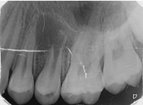
    Figure 2:  Periapical radiograph. It llustrates incomplete root formation (1/2
root formation) and a diffuse periapical radiolucency extending to the apex
of tooth 24#
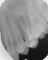
    Figure 2b:  Periapical radiograph. Radiographic image taken at the 12-month
follow-up appointment.
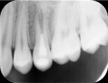
    Figure 2c:  Periapical radiograph. Radiographic image taken at the 18-month
follow-up appointment.
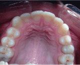
    Figure 2d:  Intraoral photograph. Clinical photograph of the tooth at the
18-month follow-up appointment.
Case 3
An 8-year old girl was referred to our clinic as an emergency patient with a chief complain of pain on her right side of the maxilla in the area of first premolar. Her medical history was noncontributory. Clinical examination showed extensive caries in the tooth UR4 and sensitivity to percussion. Radiographic examination showed almost no root formation and a radiolucent periapical lesion adjacent to the tooth UR4.
After a complete explanation of the treatment procedure, its risks, benefits, and experimental nature, an informed consent was obtained from the patient’s parents. Firstly, local anesthesia with 3% plain mepivacaine and rubber dam isolation was performed. An access cavity on tooth UR4 was prepared by using a diamond-coated fissure bur and a high-speed handpiece. After irrigation of canals by using the same technique described in case 1, canals were dried, the tri-antibiotic paste (20 mg of each metronidazole, ciprofloxacin, and amoxicillin, mixed with 1 mL of sterile water) placed inside the canal. The tooth was then temporized with cavite and glass ionomer cement.
At the following appointment in 2 weeks, the tooth was asymptomatic with no sensitive to percussion. The temporary restorations were removed, the tooth was irrigated with 2.5% sodium hypochlorite, and the antibiotic paste was removed. The 25 K-file was over-extended beyond the root length of the tooth several times in order to irritate the apical tissue until bleeding occurred apically in the root canal space and rose to the crown. By doing this, a biological scaffold for the regenerative process was created. After waiting 10 minutes, MTA (Angelus Soluçes Odontológicas, Londrina PR, Brazil) was prepared according to the manufacturer’s directions, placed over blood clot with the hand plugger and adapted softly to dentinal walls via a moist cotton pellet. An approximately 2-mmthick layer of glass ionomer cement base was placed over the MTA and was the tooth was restored permanently with composite.
The patient was recalled at 3, 6, 12, and 24 months after treatment. Clinical examination showed the tooth was asymptomatic with no sensitive to percussion and functional. Radiographically, osseous healing of the periapical lesion and thickening of the dentinal wall was evident as well as the fully development of the root (Figure 3AC). Unfortunately the appearance of the tooth showed an obvious change in color (Figure 3D).
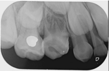
    Figure 3:  Periapical radiograph. It llustrates almost no root formation and a
radiolucent periapical lesion adjacent to the tooth #14.
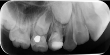
    Figure 3b:  Periapical radiograph. Radiographic image taken at the 12-month
follow-up appointment.
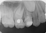
    Figure 3c:  Periapical radiograph. Radiographic image taken at the 24-month
follow-up appointment.
Discussion
In endodontics, treatment of necrotic immature teeth is very challenging because of the root anatomic structure, difficulty of the apical seal, and susceptibility to fracture. To eliminate these complexities and obtain a better prognosis, regenerative endodontic treatment or pulp revascularization is improved as an alternative method for the treatment of necrotic immature teeth. The regenerative endodontic treatment technique was first applied by Nygaard-Ostby and Hjortdal in 1971 and many case-based studies have been done about this approach until recently [6,17]. Basically, regenerative endodontic treatment is described as the stem-cell therapy that includes pulp revascularization, apexification, apexogenesis, and tissue engineering [18]. It has been proposed that stem cells from the apical papilla could be introduced into the canal because of the disorganization of the apical papilla tissue with an endodontic file and carried into the canal space by the forming blood clot [19]. The histological evaluations of tooth tissues that had been regenerated using revascularization clearly show that inside the root canal space, dentinal walls were covered with a layer of cementum, a neo-ligament, and an osteoid structure that appeared to be of periodontal origin [20]. On the other hand, some authors suggest that there is approximately a 30% chance of pulp tissue re-entering the pulp space [21]. Current case-based studies, published reports show a success of regenerative endodontic treatment [22]. Kontakiotis, et al. [22] reported that antibiotic combination paste was used as the intracanal medicament in 80% of the clinical articles. Also, as filling the root canal space with vital biological tissue has many biological advantages, the regenerative endodontics will likely be the treatment choice for those cases. [22].
In these 3 cases, revascularization treatment include continuation of root development, and the thickening of dentin wall was used instead of apexification with calcium hydroxide or the artificial barrier technique.
Elimination of microorganisms and necrotic tissues from the root canal system is an important point in a successful revascularization. The treatment procedure began with chemical disinfection by irrigating the root canal space with NaOCl. When considering the type and period used for disinfection of the root canal, it is important to balance the need to eliminate bacteria with the need to maintain vitality to stem cells. Although NaOCl has favorable antimicrobial actions, its cytotoxic effect to stem cells is also known. In studies, the concentration of NaOCl ranges between 1% and 6%, and the irrigation period varies from between 1 to 20 minutes [23]. In this regard, lower concentrations of NaOCl are recommended, so in these cases, 2.5% NaOCl was used for 10 minutes, followed by 3 weeks of triple antibiotic paste dressing. The length of antibiotic paste dressing time has also varied from 1 week [17]. to several months [ 5]. In accordance with the case reports in which clinical success was reported for 2 to 4 weeks, in the present case, 2-4 weeks of tri-antibiotic paste dressing was applied [17].
It is claimed a mixture of metronidazole, ciprofloxacin and minocycline as a tri- antibiotic paste is effective against endodontic pathogens that disinfected even the deep layers of dentin in infected teeth [13]. Although different reports show successful outcomes for revascularization treatment by using this tri-antibiotic paste there have been a few published reports address the discoloration caused by the minocycline [24,25]. It binds to calcium ions via chelation to form an insoluble complex. Hence, the minocycline incorporated into the tooth matrix causes discoloration. In the study of Sato, et al. [24] a 6-weeks tri-antibiotic paste dressing period can also explain the crown discoloration. To avoid discoloration, Reynolds, et al. [11] first etched the crown of the teeth with 35% phosphoric acid and sealed with SingleBond and flowable composite before placing the tri-antibiotic paste. Thus the dentinal tubules of the crown didn’t contact the tri-antibiotic paste. In conclusion, they suggest minocycline should be limited to the root canal or should be used suitable techniques for preventing contact with the coronal dentin despite the biologic success of minocycline. Also, they reported grey MTA indicated discoloration, while white MTA didn’t indicate any discoloration. In addition, Kim, et al. [25] reported that dentine bonding agents decreased the tooth discoloration but did not prevent it completely. Sato, et al. [26] found that amoxicillin used as the third antibiotic was as effective as minocycline when used in combination with metronidazole and ciprofloxacin eliminating bacteria. A modified tri-antibiotic paste in which amoxicillin was used instead of minocycline was developed by Hoshino, et al. [10]. In a study by Thomson, et al. [12] no observable discoloration was reported over an 18-month follow-up to successful regenerative treatment in which amoxicillin was used instead of minocycline. In all cases mentioned above, like Thomson, et al. [12] amoxicillin was used as the third antibiotic and in case 1 and case 2 no crown discolouration was observed during the 30 and 18 months, respectively. However tooth UR4 in case 3 revealed discolouration possibly related to the use of grey MTA unlike using Bioaggregate in case 1 and 2.
Three weeks after the initial appointment, in the absence of symptoms, the intracanal medicament was removed and the formation of a blood clot in the canal space was introduced. A clinical study revealed failure to induce the blood clot after disinfection might be one of the reasons for treatment failures in revascularization [13]. It was also important to induce a bacteria-tight coronal seal for successful revascularization. Although various materials have been used to induce a bacteria-tight coronal seal, MTA was the first choice because of its sealing ability and biocompatibility [13]. Because of the cementogenic and osteogenic properties of MTA, the formation of a cemental bridge was reported under MTA. However, presence of MTA in the cervical part of the canal is also stated to cause tooth discoloration [13,27]. Thus, as an alternative sealing material BioAggregate was used in case 1 and case 2. Tuna, et al. [28] stated that BioAggregate could be a promising material for the endodontic treatment of teeth with immature apexes because it provides the highest fracture resistance. There are studies demonstrating significantly better inflammatory and foreign body reaction for BioAggregate compared to MTA. So it was concluded that BioAggregate is a biocompatible material [28-30].
Although regenerative endodontic treatment is still not clear, the success of this approach was reported for necrotic, immature teeth with pulp regeneration inside the root canal space and continued root growth. Present cases reported successful regenerative endodontic treatment that utilized a slight variation of the tri-antibiotic paste, as amoxicillin was used instead of minocycline. In addition, these are the first cases in which BioAggregate was used as the sealing material in regenerative procedures, and a 30 and 18-month followup was exhibited. Future studies are needed to develop and clarify the procedure.
Conclusion
Present cases reported on successful regenerative endodontic procedure that utilizes a slight variation of the tri-antibiotic paste, using amoxicillin instead of minocycline to reduce the risk of discoloration of the tooth. In addition, clinical success of the new water-based cement BioAggregate, which has similar compositions and uses to MTA as a sealing material, has been reported.
References
- Waterhouse P, Whitworth J, Camp J, Fuks A. Pediatric endodontics: Endodontic treatment for the primary and young permanent dentition. In: Hargreaves K, Cohen S, eds. Pathways of the pulp. 10th ed. St Louis: Mosby Elsevier. 2011.
- Andreasen J, Farik B, Munksgaard E. Long-term calcium hydroxide as a root canal dressing may increase risk of root fracture. Dent Traumatol. 2002; 18: 134-137.
- Simon S, Rilliard F, Berdal A, Machtou P. The use of mineral trioxide aggregate in one-visit apexification treatment: a prospective study. Int Endod J. 2007; 40: 186-197.
- Steinig TH, Regan JD, Gutmann JL. The use and predictable placement of mineral trioxide aggregate in one-visit apexification cases. Aust Endod J. 2003; 29: 34-42.
- Chueh LH, Ho YC, Kuo TC, Lai WH, Chen YH, Chiang CP. Regenerative endodontic treatment necrotic immature permanent teeth. J Endod. 2009; 35: 160-164.
- Hargreaves K, Law A. Regenerative endodontics. In: Hargreaves K, Cohen S, eds. Pathways of the pulp. St Louis: Mosby Elsevier. 2011; 602-619.
- Hargreaves KM, Diogenes A, Teixeira FB. Treatment options: biological basis of regenerative endodontic procedures. J Endod. 2013; 39: 30-43.
- Morsezeck C, Schmalz G, Reichert TE, Völlner F, Galler K, Driemel O. Somatic stem cells for regenerative dentistry. Clin Oral Invest. 2008; 12: 113- 118.
- Thibodeau B, Trope M. Pulp revascularization of a necrotic infected immature permanent tooth: case report and review of the literature. Pediatr Dent. 2007; 29: 47-50.
- Hoshino E, Kurihara-Ando N, Sato I, Uematsu H, Sato M, Kota K. In-vitro antibacterial susceptibility of bacteria taken from infected root dentine to a mixture of ciprofloxacin, metronidazole and minocycline. Int Endod J.1996; 29: 125-130.
- Reynolds K, Johnson JD, Cohenca N. Pulp revascularization of necrotic bilateral bicuspids using a modified novel technique to eliminate potential coronal discoloration: a case report. Int Endod J. 2009; 42: 84-92.
- Thomson A, Kahler B. Regenerative endodontics - biologically-based treatment for immature permanent teeth: a case report and review of the literature. Aust Endod J. 2010; 55: 446-452.
- Nosrat A, Seifi A, Asgary S. Regenerative endodontic treatment (revascularization) for necrotic immature permanent molars: a review and report of two cases with a new biomaterial. J Endod. 2011; 37: 562-567.
- Chen MY, Chen KL, Chen CA, Tayebaty F, Rosenberg PA, Lin LM. Responses of immature permanent teeth with infected necrotic pulp tissue and apical periodontitis/ abscess to revascularization procedures. Int Endod J. 2012; 45: 294-305.
- Park JW, Hong SH, Kim JH, Lee SJ, Shin SJ. X-Ray diffraction analysis of white ProRoot MTA and Diadent BioAggregate. Oral Surg Oral Med Oral Pathol Oral Radiol Endod. 2010; 109: 155-158.
- Yan P, Yuan Z, Jiang H, Peng B, Bian Z. Effect of bioaggregate on differentiation of human periodontal ligament fibroblasts. Int Endod J. 2010; 43: 1116-1121.
- Law AS. Considerations for regeneration procedures. J Endod. 2013; 39: 44- 56.
- Garcia-Godoy F, Murray P. Recommendations for using regenerative endodontic procedures in permanent immature traumatized teeth. Dent Traumatol. 2012; 28: 33-41.
- Simon SR, Tomson PL, Berdal A. Regenerative Endodontics: regeneration or repair? J Endod. 2014; 40: 70–75.
- Shimizu E, Ricucci D, Albert J, Alobaid AS, Gibbs JL, Huang GT, et al. Clinical, radiographic, and histological observation of a human immature permanent tooth with chronic apical abscess after revitalization treatment. J Endod. 2013; 39: 1-6.
- Ritter AL, Ritter AV, Murrah V, Sigurdsson A, Trope M. Pulp revascularization of replanted immature dog teeth after treatment with minocycline and doxycycline assessed by laser Doppler flowmetry, radiography and histology. Dent Traumatol. 2004; 20: 75-84.
- Kontakiotis EG, Filippatos CG, Tzanetakis GN, Agrafioti A. Regenerative endodontic therapy: a data analysis of clinical protocols. J Endod. 2015; 41:146-154.
- Jung IY, Lee SJ, Hargreaves KM. Biologically based treatment of immature permanent teeth with pulpal necrosis: a case series. J Endod. 2008; 34: 876- 887.
- Nosrat A, Seifi A, Asgary S. Regenerative endodontic treatment (revascularization) for necrotic immature permanent molars: a review and report of two cases with a new biomaterial. J Endod. 2011; 37: 562-567.
- Kim JH, Kim Y, Shin SJ, Park JW, Jung IY. Tooth discoloration of immature permanent incisor associated with triple antibiotic therapy: a case report. J Endod. 2010; 36: 1086-1091.
- Sato I, Ando-Kurihara N, Kota K, Iwaku M, Hoshino E. Int Endod J. 1996; 29: 118-124.
- Sato T, Hoshino E, Uematsu H, Noda T. In vitro antimicrobial susceptibility to combination of drugs on bacteria from carious and endodontic lesions of human deciduous teeth. Oral Microbiol Immunol. 1993; 8: 172-176.
- Tuna EB, Dinç ME, Gençay K, Oya KE. Fracture resistance of immature teeth filled with BioAggregate, mineral trioxide aggregate and calcium hydroxide. Dent Traumatol. 2011; 27: 174-178.
- Kim J, Song YS, Min KS, Kim SH, Koh JT, Lee BN, et al. Evaluation of reparative dentin formation of ProRoot MTA, Biodentine and BioAggregate using micro-CT and immunohistochemistry. Restor Dent Endod. 2016; 41: 29-36.
- Chang SW, Lee SY, Kum KY, Kim EC. Effects of ProRoot MTA, Bioaggregate, and Micromega MTA on odontoblastic differentiation in human dental pulp cells. J Endod. 2014; 40: 113-118.