Case Report
Austin J Dent. 2016; 3(5): 1048.
The Debatable Dilemma- A Case of Anaesthetic Dental Trauma in a Syndromic Patient: Unavoidable or Negligence?
Pauly G*, Rao PK, Kini R, Kashyap RR and Bhandarkar GP
Department of Oral Medicine and Radiology, AJ Institute of Dental Sciences, India
*Corresponding author: Geon Pauly, Department of Oral Medicine and Radiology, AJ Institute of Dental Sciences, Kuntikana, NH-66, Mangaluru, PIN– 575004, Karnataka, India
Received: September 29, 2016; Accepted: October 24, 2016; Published: October 26, 2016
Abstract
Perioperative tissue damage is one of the most common anaesthesia-related adverse events and is responsible for the greatest number of malpractice claims against anaesthesiologists. Damage to the teeth during general anaesthesia is a frequent cause of morbidity for patients and a source of litigation against anaesthetists. Mucopolysaccharidoses (MPSs) are a group of uncommon genetic disease of connective tissue metabolism. Hunter syndrome (HS), or mucopolysaccharidosis II (MPS II), is a lysosomal storage disease caused by a deficient or absent enzyme- iduronate-2-sulfatase (I2S). It is well established that the elective treatment of subjects affected by MPS is multidisciplinary and must be carried out by experienced personnel in highly specialist centres. A thorough evaluation may necessitate a dentist’s help, requiring that anaesthesiologists receive more formal training regarding oral and dental anatomy, and enables performing the treatments necessary to minimize the risks of dental injuries. Various attempts to produce guidelines have been made for MPS. We want to provide a summary of anaesthetic management for these high-risk patients, who require surgical procedures and diagnostic examinations under sedation with a higher frequency than the general population.
Keywords: Anaesthetic trauma; Mucopolysaccharidoses; Hunter syndrome
Introduction
‘Iatrogenic injury’, it is a rather broad term that may be defined as ‘harm, hurt, damage or impairment that results from the activities of a doctor [1]. This includes physical injuries, surgical mishaps, adverse drug reactions, drug errors and adverse outcomes associated with equipment failure. Some causes of iatrogenic injury are difficult to avoid, in particular unexpected adverse drug reactions such as anaphylaxis. However, many are the result of human error and may be avoided through anticipation and high standards of practise [2]. Hunter syndrome or MPS II, an X-linked recessive disorder is a serious genetic disorder that primarily affects males, It interferes with the body’s ability to break down and recycle specific mucopolysaccharides; also known as glycosaminoglycan (GAG).
Hunter syndrome is one of several related lysosomal storage diseases called the MPS diseases [3]. The symptoms of HS are generally not apparent at birth, but usually start to become noticeable after the first year of life. Often, the first symptoms of Hunter syndrome may include ear infections, runny noses, colds and abdominal hernias [4]. In case of abdominal hernias, surgical intervention is indicated. Here we report a case of a 9 year old boy with Hunter syndrome.
Case Presentation
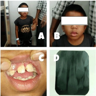
A 9 year old boy was referred from the hospital to our out-patient department, for general dental evaluation. Additionally, the patient’s parents gave a complaint of mobile teeth in upper front teeth region since one week. The patient was short in stature and was of asthenic built and moderately nourished (Figure 1A). The patient was a known case of hunter syndrome with a family history of having a sibling with the same condition. The patient had undergone surgery for umbilical hernia one week prior. On clinical examination, the shape of the head was acrocephalic (Figure 1B). The patient was not fully cooperative for intra-oral examination. On local examination, the left upper central incisor was slightly extruded, had distinct proclination, exhibited grade II mobility and was tender on palpation (Figure 1C). An intra-oral periapical radiograph was taken, which revealed complete crown formation with incomplete root formation (Figure 1D). The patient was further advised for splinting of maxillary left central incisor after suitable pulpal therapy, and further referred to department of pedodontics for the same. The patient has undergone oral prophylaxis, splinting has not been done yet ailing to patient’s non co-operation and is currently undergoing endodontic treatment.

    Figure 1:  A & B –Straight Facial Profile. C – Rotated Central Maxillary
Incisors After Surgery.
D – Intra-Oral Periapical Radiograph of 11, 21.
Discussion
The estimated incidence of Hunter syndrome is between 0.69 and 1.19 per 100,000 live births [5,6]. Although this rare disorder is X-linked, thus occurring almost exclusively in males, Hunter syndrome has also been reported in a small group of female patients, manifesting with equal severity [7].
Patients with Hunter syndrome experience a wide spectrum of progressive, multi-systemic clinical symptoms. The clinical diagnosis of Hunter syndrome requires in the first instance a thorough patient medical and family history. Pediatricians are likely to be the first clinicians to encounter a patient with Hunter syndrome, and there are a number of very early signs and symptoms that should arouse clinical suspicion, for example, lumbar gibbus, recurrent ear infections, hernia, myocarditis, or progressive hepatosplenomegaly may occur before the age of 6 months. Other signs and symptoms that are commonly found include the following: [8]
- Facial dysmorphism: coarsening of facial features, broadened nose with flared nostrils, prominent supraorbital ridges, enlarged protruding tongue, large jowls and thickened lips.
- Abdominal symptoms: hernia, abdominal distension due to hepatosplenomegaly.
- Respiratory symptoms: recurrent upper airway infection, particularly affecting the ears; sleep apnoea.
- Skeletal and joint problems: Dysostosis multiplex on radiographic examination, including abnormal bone thickening and irregular epiphyseal ossification in the joints of the hand, shoulder, and elbow; Carpal tunnel syndrome.
- Laryngoscope: Upper incisors can be damaged if used incorrectly; the mechanism of this damage is a substantial force that is applied to the teeth by the laryngoscope blade when the clinician uses the patient’s upper teeth as a fulcrum for levering the laryngoscope blade [14].
- Oropharyngeal airways: Oropharyngeal airways should be used with caution for patients with vulnerable anterior teeth and should not be used as a bite block [15,17].
- Jaw clamping: Use of a jaw clamp during light anaesthesia, particularly when used with an oropharyngeal airway, can put pressure on the teeth.
- Suction devices: Aggressive suctioning the posterior region of the mouth can cause oral injuries; dental injuries can occur when anterior teeth are subjected to extreme lateral forces. Suctioning should be therefore done with great care, preferably using a soft latex catheter [17].
- Dental props and mouth gags: Dental props and mouth gags can damage teeth during insertion or removal or when they are moved from one side of the mouth to the other [11,16].
- All teeth fragments need to be recorded.
- In case of any missing fragments, a chest radiography is necessary to exclude aspiration [15]. However, some dental prostheses are not radio-opaque and direct visualization may be required.
- In children, the loss of a primary tooth does not require treatment. In fact, the return of an avulsed primary tooth into its original socket can damage the underlying permanent successor [15,17].
- Although most dental fragments will pass though the gastrointestinal tract without causing harm, large prostheses may cause obstruction and perforation. These patients may therefore require surgical or endoscopic removal [15,18].
- If a permanent tooth is displaced from its socket, it should be stored in cool, fresh milk or normal saline until it can be splinted or fixed back in place [19,20].
- When the patient is sufficiently awake, a full explanation must be given. This should include a clear apology and a description of the events that led up to the damage and the efforts made to minimize any complications. The presence of a relative, a member of the nursing staff, or a patient liaison officer is often very useful in this situation. Similarly, from a junior anaesthesiologist’s point of view, the attendance of a senior colleague is invaluable and should be sought [15].
- All actions and discussions should be clearly documented in the patient’s records.
- It is the responsibility of the anaesthesiologist to organize an urgent dental assessment and arrange subsequent treatment. The patient should not leave hospital without a clear written treatment plan and arrangements for follow-up [15].
- Taylor T. Avoiding iatrogenic injuries in theatre. Br Med J. 1992; 305: 595- 596.
- Jenkins K, Baker A. Consent and anaesthetic risk. Anaesthesia. 2003; 58: 962-984.
- Wraith J, Scarpa M, Beck M, Bodamer O, De Meirleir L, Guffon N, et al. Mucopolysaccharidosis type II (Hunter syndrome): a clinical review and recommendations for treatment in the era of enzyme replacement therapy. European Journal of Pediatrics. 2007; 167: 267-277.
- Schwartz I, Ribeiro M, Mota J, Toralles M, Correia P, Horovitz D, et al. A clinical study of 77 patients with mucopolysaccharidosis type II. Acta Paediatrica. 2007; 96: 63-70.
- Alcalde-Martín C, Muro-Tudelilla J, Cancho-Candela R, Gutiérrez-Solana L, Pintos-Morell G, Martí-Herrero M , et al. First experience of enzyme replacement therapy with idursulfase in Spanish patients with Hunter syndrome under 5 years of age: Case observations from the Hunter Outcome Survey (HOS). European Journal of Medical Genetics. 2010; 53: 371-377.
- Ballabio A Gieselmann V. Lysosomal disorders: From storage to cellular damage. Biochimica et Biophysica Acta (BBA) - Molecular Cell Research. 2009; 1793: 684-696.
- Jurecka A, Krumina Z, Żuber Z, Różdżyńska-Światkowska A, Kloska A, Czartoryska B, et al. Mucopolysaccharidosis type II in females and response to enzyme replacement therapy. Am J Med Genet. 2012; 158A: 450-454.
- Martin R, Beck M, Eng C, Giugliani R, Harmatz P, Munoz V, et al. Recognition and Diagnosis of Mucopolysaccharidosis II (Hunter Syndrome). PEDIATRICS. 2008; 121: e377-e386.
- Santos S, López L, González L, Domínguez M. Hearing Loss and Airway Problems in Children With Mucopolysaccharidoses. Acta Otorrinolaringologica (English Edition). 2011; 62: 411-417.
- Bready L, Dillman D, Noorily S, Peduto V. Anestesiologia. London: Elsevier Health Sciences. Italy. 2011.
- Givol N, Gershtansky Y, Halamish-Shani T, Taicher S, Perel A, Segal E. Perianesthetic dental injuries: analysis of incident reports. Journal of Clinical Anesthesia. 2004; 6: 173-176.
- Pea M, Aujla PK, Choi SS, Zalzal GH Acute airway distress from endotracheal intubation injury in the pediatric aerodigestive tract. Otolaryngology - Head and Neck Surgery. 2004; 130: 575-578.
- Vallejo M, Best M, Phelps A, O’Donnell J, Sah N, Kidwell R, et al. Perioperative dental injury at a tertiary care health system: An eight-year audit of 816,690 anesthetics. J of Healthcare Risk Mgmt. 2012; 31: 25-32.
- Kim H, Lee J, Bahk J. Assisted head extension minimizes the frequency of dental contact with laryngoscopic blade during tracheal intubation. The American Journal of Emergency Medicine. 2013; 31: 1629-1633.
- Windsor J,Lockie J. Anaesthesia and dental trauma. Anaesthesia & Intensive Care Medicine. 2008; 9: 355-357.
- Rosenberg MB. Anesthesia-induced dental injury. International Anesthesiology Clinics. 1989; 27: 120-125.
- Yasny JS. Perioperative dental considerations for the anesthesiologist. Anesth Analg. 2009; 108:1564-1573.
- Skeie A, Schwartz O: Traumatic injuries of the teeth in connection with general anaesthesia and the effect of use of mouthguards. Endod Dent Traumatol. 1999; 15: 33-36.
- Newland MC, Ellis SJ, Reed Peters K, Simonson JA, Durham TM, Ullrich FA, et al. Dental injury associated with anaesthesia: a report of 161,687 anaesthetics given over 14 years. J Clin Anesth. 2007; 19: 339-345.
- Givol N, Gerhtansky Y, Halamish-Shani T, Taicher S, Perel A, Segal E, et al. Peri-anaesthetic dental injuries: analysis of incident reports. J Clin Anaesth. 2004; 16: 173-176.
- Vogel J, Stubinger S, Kaufmann M, Krastl G, Filippi A. Dental injuries resulting from tracheal intubation--a retrospective study. Dental Traumatology. 2009; 25: 73-77.
- Rosa Maria G, Paolo F, Stefania B, Letizia T, Martina A, Massimiliano D, et al. Traumatic dental injuries during anaesthesia: part I: clinical evaluation. Dental Traumatology. 2010; 26: 459-465.
- Bhatnagar N, Lin CJ, Orebaugh SL, Vallejo MC. Regional anesthesia considerations for awake endotracheal intubation and prevention and management of dental injuries. International Anesthesiology Clinics. 2012; 50: 1-12.
Every MPS type is characterized by progressive craniofacial, articular, and skeletal deformities, cardiac involvement, and early death due to pulmonary infections or heart failure, often before adulthood. Patients usually look normal at their birth but progressively develop clinical manifestations according to the kind of syndrome they are affected by [9] .The management of these situations is a challenge for the anaesthesiologist. The anesthetic risk of MPS patients must be considered high for many reasons, including airway abnormalities, orthopaedic deformities, pulmonary predisposition, and cardiac and neurological involvement [2,10].
Dental injury occurs during 1% of general anaesthetics. It is most commonly sustained during laryngoscopy and the teeth most likely to be injured are the upper incisors [2], as seen in this case. While preoperative assessment of dentition may guide anaesthetists as to which patients are at risk of dental injury, the majority of incidents are not associated with predicted difficult intubation [11]. Dental protectors may protect the teeth from injury; however, they may cause dental damage themselves and make laryngoscopy more difficult. A small proportion of dental injuries occur at the time of extubating, often in patients who have occluded their tracheal tube through biting, again which was a distinct feature in this case. This situation can be avoided by inserting a Guedel airway or bite-block. If a tooth is accidentally avulsed, it should be re-implanted in its socket and a dental surgeon consulted at the earliest opportunity. Injuries to lips and oral mucous membrane are easily caused if adequate care is not taken during laryngoscopy, insertion of laryngeal masks or manual maintenance of the airway using a face mask. Most injuries heal uneventfully, but may be quite distressing after operation. Soft paraffin ointment should be applied to any cuts on the lips to minimize postoperative discomfort [12].
Several anaesthetic equipment can cause dental damage, particularly rigid equipment if used inappropriately [13]. The following devices are often associated with damage to teeth; therefore, extra care must be taken when they are used: [11,14-17].
Another viable option is the use of mouthguards. The main indications for its use during surgical operations with intubation is the presence in the mouth of fixed prosthetic restorations such as crowns and bridges, especially those made of porcelain, which are very fragile. The risk of dental damage, especially to the frontal part of dental arches significantly increases in cases of endodontictreated teeth. Mixed dentition in children between 5 and 10 years and teeth with III and IV class dental fillings are also characterized by a lower resistance to damage. The use of elastic occlusal splints is also recommended in cases of patients with toothless gums, in whom they could offer stable support for an inserted laryngoscope, protecting the soft tissues of the maxillary alveolar processes against damage. Another option is use of protective occlusal splints during intubation, but should be based on an assessment of the conditions and potential difficulties during an operation [18]. Where possible, the anaesthesiologist should discuss the choice of equipment with the patient prior to the procedure, outlining the benefits and risks of each equipment, and ensure that the patient freely consents to the use of a particular piece of equipment as well as record details of this discussion in the medical records [19,20].
Anaesthetic departments should develop protocols, which should include the following: [15,17,21-23].
However, even with experienced anaesthesiologists who were adequately trained in the use of devices and aware of the potential damage while using excessive forces, some unexpected difficulties may lead to injuries. The damage to the teeth and oral structures may occur even in the absence of negligence.
Conclusion
MPS children are high anaesthetic risk patients because of airway narrowing, bone dystrophy, cardiac illness, and neurological impairment. They require the cooperation of counsellors with different professional competences. The dentist too plays a pivotal role in ensuring risk free surgery by thoroughly evaluating the dental status of the patient. The best management we can pertain is by creation of a communicative multidisciplinary team, available to cooperate with other professionals worldwide. Any surgical procedures if indicated should be carried out only after consulting the anaesthesiologist, who has the obligation to discuss risks and benefits with the parents. Also, the administration of anaesthesia should be carried out only in specialized centres by experienced anaesthesiologists and trained personnel. All of this is principally aimed at improving the quality of life of both patients and families. However, despite all necessary steps taken, at times it is extremely difficult to avoid a possible injury to the dental tissues especially pertaining to syndromic patients where in the risk probabilities are much higher due to various constrains. Despite the challenges, utmost care and precautions should be instilled to ensure minimal discomfort to the patients. Because, the bottom line is – Health care reforms are about the patients, and not about the physicians.
References