Clinical Image
Austin J Dent. 2016; 3(6): 1053.
Healing Herpes Labialis
Peter M*, Rao JPK, Kini R, Bandarkar GP, Kashyap RR and Shetty DN
Department of Oral Medicine and Radiology, AJ Institute of Dental Sciences, India
*Corresponding author: Mathew Peter, Department of Oral Medicine and Radiology, AJ Institute of Dental Sciences, Kuntikana, Mangalore, PIN–575004, Karnataka, India
Received: October 28, 2016; Accepted: November 16, 2016; Published: November 18, 2016
Clinical Image
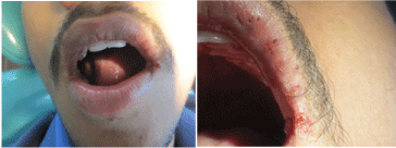
A 17 year old male patient visited the dental outpatient department for routine dental check. Family history, past dental and personal history was non-contributory. Medical history revealed that the patient had taken medication for fever since two days. Extraoral examination revealed multiple vesicles of varying sizes ranging from pin point size to 0.5 cm were seen on the left side of the upper lip and the left side commissure areas which were in the healing stage and appeared erythematous. On palpation there was tenderness present without any bleeding. Based on history and clinical examination provisional diagnosis of Herpes labialis in relation to the left side of the lip was given. Herpex ointment was prescribed daily four times for the patient for two weeks. Patient was recalled after two weeks to check for resolution of symptoms.
Herpes Labialis is a recurrent Herpes infection caused by herpes simplex virus. Around 15 to 30% of the community is affected by episodes of herpes labialis. Common colds, influenza, fever, UV exposure, menstruation, emotional upset, stress and anxiety predispose the patient to recurrent infection, as these cause reactivation of the virus, which subsequently migrates along one of the sensory divisions of the trigeminal nerve. The lesions are most often seen at the muco cutaneous junction of the lip or peri-oral skin.

Figure 1:
A burning sensation usually precedes the development of a small cluster of vesicles. These vesicles enlarge, coalesce, ulcerate and become crusted before healing within 10 days [1]. Preventative therapy, such as using sun block, is the management of choice for herpes labialis, while antiviral drugs such as acyclovir are only useful if applied at the prodromal stage before the development of vesicles [2]. Patients must be discouraged from touching the lesions in order to reduce the risk of spreading the infection to other sites [1].
References