
Clinical Image
Austin J Dent. 2016; 3(6): 1054.
Bifid Condyle
Shetty US*, Burde KN, Rao PK, Kini R, Naikmasur V and Rao D
Department of Oral Medicine and Radiology, A J Institue of Dental Science and Hospital, India
*Corresponding author: Ujwala Shivarama Shetty, Department of Oral Medicine and Radiology, A J Institue of Dental Science and Hospital, Mangalore, Karnataka –575004, India
Received: October 28, 2016; Accepted: November 16, 2016; Published: November 24, 2016
Clinical Image
A 26 year old male patient reported with a chief complaint of missing teeth in the upper left back tooth region. Past medical history dental and personal history was non-contributory. On intra oral examination, there was missing 26, 27 teeth. Patient was subjected for radiological investigations to carry out further treatment. Cone beam computed tomography (CBCT) was taken to assess the height and width of bone in the missing area region for implant placement. The CBCT image showed change in the morphology of condyle suggestive of bifid condyle which is a rare anatomic variation of mandibular condyle (Figure 1). It can be symptomatic or asymptomatic. In our case it was a symptomatic and was diagnosed incidentally on radiographic examination.
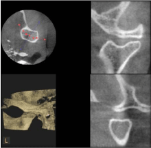
Figure 1: Morphology of condyle suggestive of bifid condyle.