Clinical Image
Austin J Dent. 2016; 3(7): 1057.
Amelogenesis Imperfecta: Hypomaturation Type
Kulkarni AV¹*, Kini R², Rao PK³, Bandarkar GP4 and Kashyap RR4
Department of Oral Medicine and Radiology, AJ Institute of Dental Sciences, India
*Corresponding author: Arati V Kulkarni, Department of Oral Medicine and Radiology, AJ Institute of Dental Sciences, Kuntikana, Mangalore, PIN-575004, Karnataka, India
Received: December 06, 2016; Accepted: December 26, 2016; Published: December 30, 2016
Clinical Image
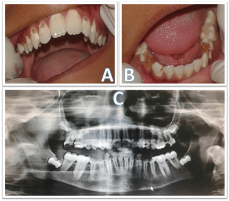
A 13 year old medically fit male patient reported with a chief complaint of decay in the upper and lower back teeth which was noticed first as a yellowish brown discoloration six years back. History of food lodgement and pain occasionally. Past dental history of decay in the primary teeth for which restorations had been done was recorded. Intraoral local examination revealed brownish discoloration with cavitation in the labio cervical, palatocervical, linguocervical and occlusal aspect of maxillary and mandibular teeth (Figure A and B). Generalised chipping of enamel on the occlusal and cervical aspect was noted. Orthopantamograph was advised which showed the presence of all unerupted third molars with crown formation complete, as well as a seemingly normal pattern and timing of eruption of teeth (Figure C). Thin enamel with normal pulp chamber and root canal spaces with no signs of obliteration seen. Radiolucency involving enamel ,dentin and pulp seen in few teeth. Based on history and clinical examination a Diagnosis of Amelogenesis Imperfecta – Hypomaturation Type was given.

    Figure 1:  A and B: Brownish discoloration with cavitation seen on the
maxillary and mandibular teeth C: Orthopantamograph showing thin enamel
with no obliteration of pulp chamber.
Amelogenesis imperfecta (AI) is a diverse collection of inherited diseases that exhibit quantitative or qualitative tooth enamel defects in the absence of systemic manifestations. Synonyms include Hereditary enamel dysplasia, Hereditary brown enamel and Hereditary brown opalescent teeth. Four types of AI include Hypoplastic AI, Hypo maturation AI, Hypocalcified AI and Hypo maturation and Hypo calcified AI types [1]. The Present case represented the Hypomaturation type where in enamel matrix is laid down appropriately and begins to mineralize, but there is defective maturation of enamel’s crystal structure leading to normal shape but abnormal mottled, opaque white-brown color of enamel. Enamel can be pierced by an explorer point under firm pressure and can be lost by chipping away from the underlying normal appearing dentin. Snow capped pattern exhibits a zone of white opaque enamel on the incisal or Occlusal third of the crown [2]. Radiographic features may vary as in enamel may appear totally absent or when present may appear as a thin layer , chiefly over the tips of the cusps and on the interproximal surfaces. Treatment ranges from preventive care using sealants and bonding for esthetics to extensive removable and fixed prosthetic reconstruction [3].
Oral Diagnostician has to diagnose the condition as early as possible to offer early intervention and balance the decision with long-term survival of the restorations to avoid social distress due to esthetic problems in patients.
References