
Clinical Image
Austin J Dent. 2016; 3(7): 1059.
Vascular Lesion: A Challenge to Diagnosis
Kalkur C*, Lakshman AR and Halim N
Department of Oral Medicine & Radiology, Century International Institute of Dental Science & Research Centre, India
*Corresponding author: Kalkur C, Department of Oral Medicine &Radiology, International Institute of Dental Science & Research Centre, Kasargod, Kerala, India
Received: December 08, 2016; Accepted: December 26, 2016; Published: December 30, 2016
Clinical Image
A 25 year old female patient presented to the department with chief complaint of swelling in the right cheek region since 15 years. On General and extra oral examination no abnormality was detected. On intraoral examination there was a large solitary purple coloured swelling on the right buccal mucosa. The swelling was sessile with broad base and was located at the line of occlusion. It measured about 2x2 cm, it had waxed and waned in size, surface was smooth, rubbery on palpation (Figure 1). Diascopy test gives negative result as there is no blanching on pressure over the lesion. Provisional diagnosis of low flow vascular anomaly and differential diagnosis of Capillary Hemangioma, Intraoral varix, lymphangioma and Arteriovenous malformation was made. Patient advised for surgical opinion but patient didn’t follow up later.
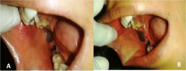
    Figure 1: (A): A Solitaty raised lesion on right buccal mucosa. (B): No
blanching on diascopy test.