Clinical Image
Austin J Dent. 2016; 3(7): 1060.
A Case of Intra-oral Hemangioma- The Blue Blemish of the Oral Cavity
Pauly G*, Kashyap RR, Kini R, Rao PK and Bhandarkar GP
Department of Oral Medicine and Radiology, AJ Institute of Dental Sciences, India
*Corresponding author: Geon Pauly N, Department of Oral Medicine and Radiology, AJ Institute of Dental Sciences, Kuntikana, NH-66, Mangaluru, PIN- 575004, Karnataka, India
Received: December 10, 2016; Accepted: December 29, 2016; Published: December 30, 2016
Clinical Image
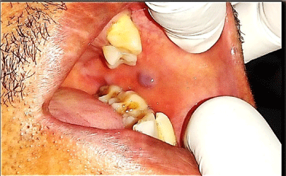
A 56 year old male patient came with a chief complaint of missing teeth and wanted replacement for the same. His past medical and dental history was non-contributory. Intra-oral examination revealed an asymptomatic bluish dome shaped raised growth on the left buccal mucosa located about 1 cm above the mandibular occlusal plane in the region of mandibular second molar. It appeared smooth, was circular in shape and had less distinct borders. It measured less than 1 cm in size and was soft, smooth, compressible and non-tender on palpation (Figure 1). A diascopy test was undertaken which showed blanching of the lesion. A provisional diagnosis of hemangioma of left buccal mucosa was given, and a biopsy further confirmed the diagnosis. The patient was further referred to department of prosthodontics for the needful. As the lesion was asymptomatic no treatment was advised for the lesion and patient was kept under observation and recalled after one, three and six months for periodic follow-ups still revealing the asymptomatic lesion with no growth nor regression.
Hemangiomas are tumors identified by rapid endothelial cell proliferation in early infancy, followed by involution over time. Hemangiomas of head and neck are common, but are rarely seen in the oral cavity, especially in the oral soft tissues [1]. Therefore, are not commonly encountered by dental professionals. About 85% of childhood onset hemangiomas regress after puberty, where in older individuals, once formed it does not regress. Majority are congenital, but some are acquired later in life [2]. They are associated with syndromes such as: Rendu-Osler-Weber syndrome, Sturge-Weber- Dimitri syndrome, Kasabach-Merritt syndrome, Maffucci syndrome, Klippel-Trenaunay-Weber syndrome and PHACE syndrome [3]. Concerning the treatment, most true hemangioma require no intervention; as they undergo spontaneous regression. Merely, 10- 20% require treatment because of the location, size or behavior. Various therapeutic procedures available include microembolization, cryotherapy, sclerosing agents, radiation, corticosteroids and LASER therapy. However, complete surgical excision if possible, offers the best chance of cure [4].

Figure 1:
References
- Rachappa MM, Triveni MN. Capillary hemangioma or pyogenic granuloma: A diagnostic dialemma. Contemp Clin Dent. 2010; 1: 1-4.
- Bayrak S, Dalsi K, Hamza T. Capillary hemangioma of the palatal mucosa: report of unusual case. SÜ Dishek Fak Derg. 2010; 19: 87-89.
- Acýkgöz A, Sakallioglu U, Ozdamar S, Uysal A. Rare benign tumours of oral cavity- capillary hemangioma of palatal mucosa: a case report. Int J Paediatr Dent. 2000; 10: 161-165.
- Brannan S, Reuser TQ, Crocker J. Acquired capillary haemangioma of the eyelid in an adult treated with cutting diathermy. Br J Ophthalmol. 2000; 84:1322.