
Clinical Image
Austin J Dent. 2017; 4(2): 1069.
Pregnant Tooth
Santannavar S*, Bhandarkar GP, Vidya Holla A, Prasanna Kumar Rao J, Kini R and Kashyap RR
Department of Oral Medicine and Radiology, A J Institute of Dental Sciences, India
*Corresponding author: Sunmith Santannavar, Department of Oral Medicine and Radiology, A J Institute of Dental Sciences, NH-66, Mangaluru, Kuntikana, India
Received: February 13, 2017; Accepted: February 28, 2017; Published: March 06, 2017
Clinical Image
A 30-year-old female patient visited the dental outpatient department with a complaint of pain in upper left front tooth region. Family history,medical history and dental history was non–contributory. Patient chews betelnut since 2 years after meals. Intraoral examination revealed edematous gingiva on maxillary left lateral incisor and canine region with generalized calculus and stains. Peg shaped maxillary left lateral incisor was tender on palpation and percussion (Figure A). Intraoral periapical radiograph revealedexamination peg shaped tooth with well-definedradiopacity around the pulp not extending to the root with root resorptionwith well-defined radiolucency measuring about less than 1cm around the apex suggestive of periapical granuloma ( Figure B). After Correlating the findings clinically and radiographically a final diagnosis of Dens in dente with periapical granuloma in relation to maxillary left lateral incisor was given. Later, patient was sent to department of oral and maxillofacial surgery for the extraction in relation to maxillary left lateral incisor.
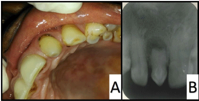
    Figure 1: A) Maxillary left peg shaped lateral. B) Intra oral periapical
radiographs suggestive of Dens Invaginatus with periapical granuloma.
Dens in dente refers to a developmental anomaly associated with an abnormal infolding of the inner enamel epithelium into the dental papilla during tooth development and gives rise to a possible communication between the pulp and the oral environment increasing the susceptibility of the tooth to caries, pulpitis and pulpal necrosis [1]. Dens in dente is observed in 0.25%-5% of individuals [2] and is most commonly associated with maxillary lateral incisors (1.7%-38.5%) [1]. Dilated composite odontome, gestantanomaly, and dens telescope are the terms commonly used synonyms of dens in dente [3]. In our case the affected tooth is maxillary left lateral incisor and it is more frequent in maxilla than mandible [4] and is seen most often in Caucasians and Asians [1]. It allows entry of irritants into an area which is separated from pulpal tissue by only a thin layer of enamel and dentine and presentsa predisposition for the development of dental caries.Therefore pulp necrosis often occurs rather early,within a few years of eruption. Sometimes even before root end closure [5]. Dens invaginatus is coincident with other dental anomalies,malformations and even dental or medical syndromes like Williams syndrome,Ekman-Westborg-Julin syndrome and Nance Huran syndrome [6-7]. In most cases Dens in dente is detected by radiograph. Clinically, as ususual crown morphology or a deep foramen coecum may be important hints. As maxillary lateral incisors are the teeth most susceptible to coronal invaginations these teeth should be investigated thoroughly clinically and radiographically, with special considerations to cases with a deep pit at the foramen coecum. Treatment plan includes preventive and restorative treatment, root canal treatment, surgical treatment and extraction [4].
- Sisodia N, Manjunath MK. Double Dens In Dente: A rare anomaly- Case Report. SADJ. 2016; 71: 410-411.
- Pindborg JJ. Pathology of the dental hard tissues. Philadelphia: WB Saunders;1970.
- Alaini A, Bishop K. Dens invaginatus. Part 1: classification; prevalence, and etiology. Int Endod J. 2008; 41: 1123-1136.
- Oehlers FA. Dens Invaginatus.variations of the invagination process and associated anterior crown form. Oral Surg Oral Med Oral Pathol. 1957; 10: 1204-1218.
- Hulsmann M. Dens invaginatus. aetiology, classification, prevalence, diagnosis, and treatment considerations. Int Endod J. 1997; 30: 79-90.
- Serrano J. Triple Dens in invaginatus in a mesidens. Oral Surg Oral Med Oral Pathol Oral Radiol Endod. 1991; 648-649.
- Mupparapu M, Singer SR, Pisano D. Diagnosis and clinical significance of dens invaginatus to practicing dentists. NY State Dent J. 2006; 72: 42-46.