Short Communication
Austin J Dent. 2017; 4(4): 1080.
History of Dental Implantology
Xavier Riaud*
History of Sciences and Techniques, Laureate and Associate Member of the National Academy of Dental Surgery, Free Member of the National Academy of Surgery, France
*Corresponding author: Xavier Riaud, History of Sciences and Techniques, Laureate and Associate Member of the National Academy of Dental Surgery, Free Member of the National Academy of Surgery ; 145, route de Vannes, 44800 Saint Herblain, 0240766488, France
Received: May 01, 2017; Accepted: June 01, 2017; Published: June 12, 2017
Short Communication
Faïd Souar II was discovered by G. Laplace (1954). His age was between 18 and 25 years old.
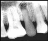
7000 years B.C. It was a Prehistoric osteoimplanted dental prosthesis. Indeed, tooth #15 had been replaced by a dental pseudoelement which was neither a human tooth, nor an animal tooth. It was neither made in ivory, nor in wood but in bone. It was perfectly still, without any trace of bonding (20 mm long, 8 mm long in the bone). It was a small-sized bone such as the bone of a hand, a human foot or a foot of a small mammal in which this tooth was carved, then placed in the mouth. However, it was impossible to know the reasons why it was there. The texture of the bone tissue was the same as that in these mammals, only the morphology had changed. The radiographs that we carried out confirmed it (Figure 1).

Figure 1:
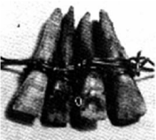
In the Antiquity, in Phoenicia exactly, in a necropolis, Doctor Gaillardot (1841) found a piece whose two canines were connected with gold threads (4th century B.C). The two incisors belonged to another individual. This item can be seen in the Louvre Museum in Paris. In Greece, in the case of maxillary fractures, Hippocrates (c. 460-c. 370 BC) recommended to reinsert the displaced teeth and to ligate them (Figure 2).

Figure 2:
The Etruscans replaced prosthesis of 2 central incisors. A goldplated band, that was approximately 5 mm wide, had been modelled in order to hug the labial and lingual surfaces of teeth #32, 42 and 43, as well as the distal surfaces of teeth #32 and 43. Some strips, which were oriented in the labio-lingual direction, had been welded on the inside of the main band, at the interdental space of teeth #42, 43, at the mesial area of teeth #32 and 42, and at the interdental space of teeth #31 and 41. Two edentulous paces delineating teeth #31 and 41 were to be replaced. Teeth of an animal or of a patient (Cornetto museum, Italy >>calf (4th century B.C.)). Once adjusted in their respective spaces, they had been riveted in the labio-lingual direction (7th century B.C./came from Tarquinia).
In the middle Ages, Abulcasis (936-1013) was an Arab surgeon from Spain. He was caliph Abdel-Rahman III’s surgeon, in Córdoba, the capital city of the Islamic empire of Spain. He wrote some scientific and medical study entitled The Practice. This 1500-page piece of work was divided into 30 books. It was translated in Latin and published in 1497 in Venise, 1541 in Basel and in 1778 in Oxford.
He made numerous dental instruments for dentists who used them to clean the teeth or to extract them. He also made prosthetics with oxe bones. Abulcasis used the method of ligating with a gold or silver thread to replace or to fix teeth when they needed to be transplanted.
In the Pre-Columbian era - era which covered the period from the appearance of the primitive man to the first traces of the European man in America-, in 1893, Andrews reported that a black stone was replacing a left lateral incisor from Copan’s excavation (Honduras, 1931). On this subject, M. Jeanneret underlined that this 1000-yearold stone, “set” in the tooth socket without any support, was placed while the carrier was still alive, as it was proved by a significant layer of sediment. This tooth (female adult) can be seen at the Peabody Museum of Harvard University. At that time, the primitive men attempted to model the teeth which were filed in stone [1-4].
In the Renaissance, in 1545, R.W. Hermann, a surgeon from Frankfurt, made one of the first statements on the methods and techniques about implants. In 1557, M. de Cartillo filled a cleft palate with a gold plate. Under the reign of Francis the 1st of France and Henry the 2nd, Riolon wrote: “Teeth are bones of their own kind”. In 1582, Foret, a Dutch dentist, recommended vigorous dental subluxation equivalent to the reimplantation of natural teeth after dental avulsion to soothe dental pains. In 1560, Ambroise PARE described the use of transplant and reimplantation in one of his treatises. In 1575, Ambroise Paré talked about transplants and quoted: “A princess who had had a tooth pulled, immediately received another from another young woman. The tooth grew and became solid as before”. In 1582, Ambroise Paré (1517-1590) published De plusieurs indispositions qui adviennent aux dents (About many dental discomforts). He was the greatest barber surgeon of Europe. He was successively the official surgeon of Henry II (1559), Francis II (1559), Charles IX (1562) and of Henry III (1575).
In the XVIIth Century, in 1615, in Le miroir de la beauté et de la santé du corps (The mirror of beauty and the body health), L. Guyon advised that dentists should immediately put a tooth back in its tooth socket and bind it to the neighboring tooth with a small thread. In 1633, Dupont, Louis XIII’s dentist published De la greffe dentaire (About dental transplant) and recommended the extraction and immediate reimplantation of teeth in the case of toothache treatment. He was the first to carry out the operation of a dental avulsion and the reimplantation in the case of the treatment of a pathological tooth. In 1649, Schearmmer filled teeth before reimplanting them.
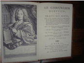
In the XVIIIth Century, Pierre Fauchard (1678-1761) recommended: “Never to pull out and to throw away a loose tooth”. He added: “First, one should extract it, then slightly file its root, fill it with lead or gold leaves and put it back in its socket, making sure to ligate them to the neighbouring teeth with silver or gold metal threads” (Figure 3).

Figure 3:
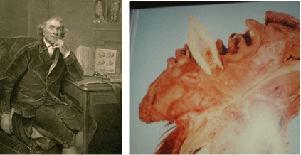
In 1771, John Hunter (1728-1793) in The Natural History of the Human Teeth (1771) transplanted a living person’s healthy tooth on the comb of a rooster. A few months later, the rooster was killed and John HUNTER noticed that the tooth was fixed to the tissue and blood flow. This practitioner had always considered the tooth as a bone planted in another bone. In 1780, John Hunter carried out dental reimplantations and transplantations (Figure 4).

Figure 4:
In the XIXth Century, in 1856, J. Younger from San Francisco carried out the first real implantation: an operation which consisted in digging a tooth socket in the jawbone: either where a tooth had disappeared, or where the space was edentulous in the first place. He used natural teeth. He transplanted an upper tooth respecting the impacted position. In 1865, Magitot presented a dissertation on reimplantation and restitution transplant. In 1882, he failed 8 times out of 117 cases of reimplantation. In 1875, John Younger was the first to open an artificial tooth socket with a drill. In 1879, E. Magitot (1833-1897) published De la greffe chirurgicale dans ses applications à la thérapeutique des lésions de l’appareil dentaire (From surgical graft in its applications to the therapeutic of lesions caused by a dental appliance). From then on, dental implantations were usually referred to as “Magitot’s operations”. In 1886, L. Fredel studied dental transplantation.
That’s all for dental reimplantations and transplantations.
Let’s see what about implantations of dental implants. In 1807, when his book Manuel de l’art du dentist (Handbook on the dentist’s art) was published, Maggiolo described the implantation of a tooth made of 18-carat gold alloy. We see also:
- In 1804, Ch. Bell studied the tissue reactions that various metals could cause.
- In 1885, Waisser carried out implantations with porcelain roots.
- In 1886, Hausman reduced the risk of new fractures or immobilised fractures, thanks to a stainless steel plate and fixing screws.
- In 1887, Harris implanted porcelain tooth on a root made of plaster and covered with lead.
- In 1889, Frantzen invented celluloid roots. He was followed by Franck in his clinical trials.
- In 1891, Znamenski implanted porcelain and rubber on a dog. It was a great success. However, what was applicable to dogs was not for men.
In 1891, Hillischer (1850-1926) made platinum and gold implants that he immediately put in a tooth socket after dental avulsion to increase retention. From 1891 to 1894, Lewis, D’Ewardt and Bonwill used platinum and gold.
In the XXth Century, around the year 1900, following the trend which was characteristic of that period, Berr was trying to implant teeth whose roots were made of precious metals. In 1901, Payne presented a communication proposing the implementation of gold roots on a full prosthesis. In 1906, Hentze had implanted a porcelain root for 3 months and a root made of rubber for 2 years.
In 1913, E. Greenfield (US Patent Office Serial n°478 360 of December 14th 1909) used an artificial root in the shape of a small windowed cage made of platinum-iridium alloy metal lattice. The tooth socket was not dug but the drill hollowed out a circular slit in the middle of which the bone remained undamaged. This set was made up of 4 ladders. The tooth was chiseled in a space which had the shape of a swallow. These experiments lasted 7 years before being presented to the Academy of Stomatology, in Philadelphia.
In 1920, Bricke screwed a root made of ivory onto a tooth socket that he drilled himself. In 1928, Meissner “inserted” artificial teeth in holes dug in the sockets. In 1933, Dag indicated the existence of a bridge on the lower maxillary which was made up of 5 elements. Its abutment was a wood screw-shaped root in 22-carat gold. The bridge had been worn for 10 years.
In 1937, in a substantial amount of time, Wuhrmann wanted to exclude the device incorporated in the bone tissue from the action of the buccal medium.
The year 1939 saw the use of stainless steels and stellites in dental surgery. The Strock brothers from Boston used vitallium screws (worn during 17 years). In 1943, Alvin E. Strock (1911-1996) and Moses Strock from the Surgical Laboratory of Harvard Medical School were the pioneers of this new method. The same year saw the Dr Gustav Dahl’s juxta-osseous implant. It was a simple bar of Stellite with four rods (two canine rods and two molar rods piercing the gum and supporting a full prosthesis).
Following an extraction, Formiggini left iodoform gauze in the tooth socket. A few weeks later, he found it difficult to extract the gauze. The bone had reformed around the gauze. He created an original screw made of stainless steel and which was internally empty. On February 27th 1947, he held his first conference in Milan. The handcrafted method of his screw and the tapping were impossible to reproduce. He carried out his first surgical procedure in 1943. After the war, he lost most of his customers.
In 1955, Palfer-Sollier and Chercheve described transalveolar transfixion. This implant was made up of three nuts screwed on the symphyseal surface and positioned after a chin incision, of three rods springing up on the gum. In 1962, Chercheve made his spiral implant. It was a double spiral helical implant: connective tissue formed inside the propeller. It was first in chrome cobalt, then in titanium. In 1960, Orlay codified a technique which required to increase the radicular length of the tooth and thus, to lower its center of gravity. The same year, Sandhaus made a prefabricated implant in platinum iridium, made up of an extra gingival abutment mounted on a subperiosteal anchor equipped with four claws. It was put in the space previously dug in the bone which then received a coaxial band and implant. The vestibular and oral portions of the anchor were folded, modeled on the bone and tightened thanks to specially-designed pliers. The intraprosthetic element was shaped with a telescope which was passing through each extragingival post. The two notches on the canine surface received a full prosthesis. In 1961, Lehmans created an arch implant. It was made up of a threaded tantalum axis at one end and a shoulder at about one third of its length. A one-millimeter-wide tantalum strip, which had a hole in its center and on its both ends, was shaped like a ring, so that the further holes came on top of one of the poles of the ring. Thus the centre hole of the strip was opposite to the other pole of the ring. Threaded on this axis, the shoulder supported this ring with its two superimposed holes.
The Revolution will come soon.
1952: the Swedish professor Per-Ingvar Brånemark conducted an experimental research during which he used a titanium implant for a better understanding of blood circulation at bone level. He made an optic chamber allowing to film the phenomena of bone repair and of revascularisation in vivo. This chamber, made in titanium, was placed in the tibia of some rabbits. At the end of the study, when professor Brånemark wanted to recover this optic chamber, he realised that it was knitted to the bone of the sacrificed rabbit (Figure 5).

Figure 5:
Osteointegration was born. In 1965, Brånemark treated his first patient, Gösta Larson. In 1986, Brånemark’s technique was published in France for the first time in l’Information dentaire.
But nothing was possible without related specialities:
- Anaesthesia with Horace Wells (1815-1848) and the discovery of the Nitrous oxide in 1844 and with William Thomas Green Morton (1819-1868) and the discovery of the Sulphuric ester in 1846. In 1884, local anaesthesia with cocaine hydrochlorate was introduced by Carl Köller (1857-1944), an Austrian ophthalmologist. And in 1885, truncular anesthesia and rubber gloves were introduced by William S. Halsted (1852-1922), an American surgeon.
- Discovery of penicillin (September 3rd 1928) by Fleming. Production of 100 mg of penicillin in 1940 by Florey and Chain. December 10th 1945: Nobel Prize for Medicine awarded to Fleming, Florey and Chain.
- Bone grafts
Bones are the most transplanted human tissues. Bone graft is the oldest graft of tissues. Th e patron saints of surgeons, Saint Come and Damien, carried out the first leg allograft in 5th century AD. Van Meekeren (1611-1666), a surgeon in Amsterdam, related another anecdote: that of a Russian soldier whose skull was partially reconstructed with that of a dog (xenotransplantation). As the Church tthreatened him with excommunication, he demanded its removal and died shortly afterwards, but in a church. Percy, a military surgeon (1754-1825), carried out the first xenotransplantation (bovine) around 1800. Albee, an American surgeon, popularised autograft and published a book on the techniques of bone autograft in 1815. In 1820, the first autograft was attributed to von Walther (1782-1849), a German ophthalmologist and surgeon. McEwen carried out the first allograft on a child in Glasgow in 1879, and Poncet, a French military surgeon, carried out the first allograft on an adult in Paris in 1887.
- Radiography
December 22nd 1895: the first radiography in history was carried out on Anna Bertha Röntgen’s hand, the wife of the discoverer of X-Rays, Wilhelm Conrad Röntgen (1845-1923) (1st publication on December 28th 1895). Two weeks later (January 5th 1896), Dr Otto Walkhoof (1860-1934) carried out the first dental radiography on himself in Braunschweig. It took up to 25 minutes of exposure. He used a photographic glass plate, covered with black paper and a rubber dyke.
References
- Gaulthier Robert, Histoire de l’implantologie (History of Implantology), in anatomieartistique.com, sans date.
- Granat Jean, Heim Jean-Louis. Prothèse dentaire préhistorique ostéoimplantée (Préhistoric Dental Prosthesis), in Actes de la Société française d’histoire de l’art dentaire, Marseille, 2000.
- Riaud Xavier. La première fois en implantologie (First time in Implantology), in Indépendentaire, n°144, janvier. 2017; 89-90.
- Ruel-Kellerman Micheline. Quatre siècles de greffes dentaires et invention de la première racine artificielle (Four centuries of dental grafts and invention of the first artificial root), in Actes de la Société française d’histoire de l’art dentaire, Paris. 2009; 14: 51-55.