
Case Report
Austin J Dent.2017; 4(5): 1085.
Subciliary Incision with a Frost Suture: A Case Report Along with Review
Sunny Deshmukh*
Department of Oral and Maxillofacial Surgery, D J College of Dental Sciences and Research, India
*Corresponding author: Sunny Deshmukh, Department of Oral and Maxillofacial Surgery, D J College of Dental Sciences and Research, Modinagar, UP, India
Received: May 30, 2017; Accepted: June 30, 2017; Published: July 11, 2017
Abstract
Injuries in the orbital region have profound functional as well as aesthetic implications. Treatment of orbital fractures remains one of the most controversial issues in maxillofacial trauma with regard to the classification, diagnosis, surgical approach and treatment. The purpose of the article is to discuss a case report of orbital rim fracture operated using subciliary incision with a frost suture and review pertinent literature.
Keywords: Subciliary incision; Inferior orbital rim fracture; Ectropion
Introduction
Orbital fractures represent one of the more common conditions encountered after motor vehicle accidents resulting in loss of an aesthetically pleasing appearance [1]. The management of orbital floor fractures has changed significantly over past decades with introduction of new materials and methods of reconstruction there are several surgical interventions recommended based on clinical examination and radiographic examination but the consequences of inadequate repair of orbital floor fractures can lead to significant facial asymmetry and visual defects. Conventional approach to the infraorbital rim/orbital floor has been by cutaneous infraciliary incisions namely the subciliary, mid lower eyelid or subtarsal and infraorbital incisions. These approaches leave behind a scar which may be cosmetically disfiguring at times [2]. The Frost suture is a novel technique to oppose downward tension vectors and to preserve or restore to position of lower eyelid. It was first originally described for the placement of a tarsorrhaphy suture through the upper and lower lid tarsal plates to correct ptosis and then this has been evolved for both the preventive and corrective procedures [3]. The purpose of this case report is to highlight the advantages of subciliary incision with a frost suture (stabilizing suture) as it offers good visualization of anterior fractures and would result in good outcomes in cases of orbital rim and floor fractures, prevents obnoxious scar, ectropion formation.
Case Presentation
A 22 year old young female patient reported to our unit with a complaint of pain and mild swelling over the right eye region. Patient had given a history of fall from motorcycle two days back when he reported to our institution. Patient on examination presented with subconjunctival hemorrhage, periorbital edema, mild enopthalmos and tenderness over the right infraorbital region (Figure 1) and there are no signs of any derangement of globe, any visual loss, impaired papillary reflex or any abnormality in Diplopia test. Basic radiographic examination on Paranasal sinus view revealed fracture of infraorbital rim on the right side (Figure 2), although C.T. scan is gold standard for orbital fractures and patient is not comfortable with it.. Based on the above findings, open reduction and internal fixation of the right infraorbital rim was performed after tarsorrhaphy (Figure 3) and prior to the fixation the entrapment of the muscle is released from the fracture site with subciliary incision (Figure 4). Even though the transconjuctival incision leaves no visible scar but it has its own group of complications like minor granuloma and cyst of lower eyelid. The subciliary incision placed 2mm caudal to cilial line and it gives very nice results as we can observer in postoperative views (Figure 5A and B).
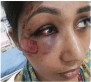
Figure 1: Preoperative view.
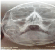
Figure 2: Paranasal sinus view.
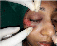
Figure 3: Tarsorrhaphy suture.
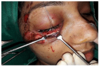
Figure 4: Exposure of surgical site.
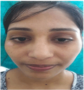
Figure 5A: 1 Month postoperative view.
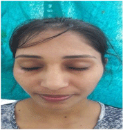
Figure 5B: 1 Month postoperative view.
The frost suture technique is used as a stabilizing suture performed in the literature quite a few times, for the placement of the frost suture we need an adequate anaesthesia for that we have to place a needle lateral and angled slightly inferior direction to tarsal plate to minimize the risk of injury to the globe. Slow infiltration both subcutaneously and subconjunctivally is performed. A 4-0 or 5-0 polypropylene, nylon, or silk suture on 3/8 circle arc needle is passed through the tarsal plate at the gray line or through the pretarsal skin, orbicularis oculi, and tarsus, taking care to remain anterior to the palpebral conjunctiva. We used 6-0 prolene suture for our case to achieve more superior results. The sutures can be placed at any location along the horizontal axis of lid margin to create tension vector directly opposing the area of greatest downward pull. Frost sutures may be removed as early as 3 to 5 days after placement [4]. However, removal is most commonly performed 7 days postoperatively. This time frame may be extended to 10 to 12 days for lager defects or for repair of ectropion.
Potential limitations and alternatives to frost suture
There are several limitations of frost suture like inability to open the affected eye while suture is in place, examination of visual acuity or papillary function is not possible. There is a theoretical risk of corneal abrasion and superficial skin erosion of upper lid or brow from suture, also it will not provide durable position of the lower lid margin, and excessive downward tension vectors from suboptimal reconstructive design carry a high risk of ectropion after removal of frost suture. The alternatives to frost suture are eyelid splinting and supportive tape, the main advantage of these techniques are preservation of the visual field and ability to inspect the eye while these support measures in place. Decreased risk of corneal abrasion is another theoretical advantage [5]. A barbed suture technique for suspension of lower lid after Mohs surgery also used as an alternative to frost suture [6].
Discussion
The evolution of treatment involving the orbital skeleton has undergone substantial change in the past century. Closed reduction, external fixation, antral packing, and kirschner wires were all used until open reduction with internal wire fixation was introduced in the 1940s becoming widely adapted by the 1950s [7]. A variety of surgical approaches to the orbital floor and infraorbital rim exist and can be conveniently categorized as either transcutaneous or transconjunctival. These approaches are widely used for exposure, evaluation, and treatment of orbital trauma, pathology, and cosmesis.
According to the literature, the indications for surgery are increased orbital pressure, persisting diplopia, enophthalmos, visual impairment, and hypoanaesthesia of the infraorbital nerve. Management of orbital fractures is controversial because of the difficulty in evaluating the anatomy of the defect area, and the amount of soft tissue herniation [8]. Selection of suitable incision for orbital fractures is one of the challenging problems. The lower eyelid skinmuscle flap is now widely used for cosmetic blepharoplasty, primarily because of the ease and speed of dissection it offers [9]. The subciliary, or blepharoplasty incision, is made approximately 2 mm inferior to, and parallel with, the superior free margin of the lower lid. It extends from the medial canthal region into or parallel to one of the resting skin tension lines located along the lateral aspect of the orbit, which usually turn slightly inferiorly. With the skin-only technique the dissection is entirely between the skin and orbicularis muscle to the level of the infraorbital rim. The skin-muscle technique differs in that the flap is made by dissecting through the orbicularis muscle either initially, or in a stepped manner, first dissecting the skin for several millimeters before penetrating the orbicularis oculi muscle. Both of these approaches preserve the position of the pretarsal orbicularis oculi muscle. However, when the skin and orbicularis muscle are incised coincidently, no orbicularis oculi muscle is left attached to the inferior tarsus. With either method, once the orbital rim is reached, an incision through the periosteum is made and subperiosteal dissection exposes the orbital region of interest.
Lower eyelid ectropion is a commonly acquired eyelid malposition affecting the population. Local eye symptoms include discomfort, itching redness, and tearing frequently associated with coexistent trichiasis. Severe cases may cause chronic ocular surface irritation, punctate keratopathy, and corneal pannus formation with decreased visual acuity. The clinical follow-up was performed at 3 months, 6 month and one year after the operation. The results were assessed from aesthetic and functional aspects by evaluating the followings: the presence of a visible or hypertrophic scar. The position of the lower lid in contrast to the globe on the treated side than on the other side. It may include rounding of the lateral canthal angle, lower eyelid retraction with inferior scleral show or frank ectropion.
Conclusion
Proper management of orbital fractures can be complex, mainly due to the variability of injuries, the proximity of eyes and brain, and the position that the eyes and thus the orbits occupy as the focal point in the middle of the face. Most of the potential important adverse sequele associated with orbital trauma reconstruction occur as a result of optic neuropathy, ocular dysmotility, ocular injury, or undesirable appearance. Careful attention to a thorough preoperative assessment, individualized surgical plan, honest appreciation of one’s own limitations, meticulous attention to surgical technique with a clear understanding of the relevant anatomy, and a thoughtful respect for the local essential structures will, as in most surgery, steer us clear of most complications.
References
- Ansari MH. Maxillofacial fractures in Hamedan province, Iran: a retrospective study (1987-2001). J Craniomaxillofac Surg. 2004; 32: 28-34.
- Subramanian B, Krishnamurthy S, Suresh Kumar P, Saravanan B, Padhmanabhan M. Comparison of various approaches for exposure of infraorbital rim fractures of zygoma. J Maxillofac Oral Surg. 2009; 8: 99-102.
- Frost A. Supporting suture in ptosis operations. Am J Ophthalmol. 1934; 17: 633.
- Desciak EB, Eliezri YD. Surgical Pearl: Temporary suspension suture (Frost suture) to help prevent ectropion after infraorbital reconstruction. J Am Acad Dermatol. 2003; 49: 1107-1108.
- Jothi S, Moe KS. Lower eyelid splinting: An alternative to the Frost suture. Laryngoscope. 2007; 117: 63-66.
- Kim JH, Yi SM, Kim S, Lee Kg. Berbed suture suspension technique for preservation of lower eyelid ectropion after Mohs microscopic surgery. Ophthal Plast Reconstr Surg. 2011; 27: e79-81.
- Rohrich RJ, Hollier LH, Watumull D. Optimizing the management of orbitozygomatic fractures. Clin Plast Surg. 1992; 19: 149-165.
- Farwell DG, Strong EB. Endoscopic repair of orbital floor fractures. Otolaryngol Clin North Am. 2007; 40: 319-328.
- Whitaker LA. Selective alteration of palpebral fissure form by lateral canthopexy. Plast Reconstr Surg. 1984; 74: 611-619.