Case Report
Austin J Dent. 2018; 5(3): 1107.
Expansile Haematomas in a Patient with Vascular Ehlers- Danlos Syndrome following Routine Dental Treatment
Keat RM¹*, Siddique I², Albuquerque R¹ and Mohammed-Ali R²
¹Birmingham Dental School/School of Dentistry, University of Birmingham, UK
²Sheffield Teaching Hospitals NHS Foundation Trust
*Corresponding author: Ross M. Keat, Birmingham Dental School/School of Dentistry, University of Birmingham, UK
Received: January 02, 2018; Accepted: February 07, 2018; Published: February 16, 2018
Abstract
We report a case of a 17-year-old male suffering from Vascular Ehlers- Danlos syndrome. He presented to the Oral and Maxillofacial Surgery department with multiple facial haematomas following dental extractions under general aesthetic. He was treated conservatively and haematomas controlled.
Poor surgical outcomes are well documented in individuals with Vascular Ehlers-Danlos syndrome, but there is no robust evidence about complications following intra-oral surgery.
We described a successful management of this condition with conservative measures, recognising the novel effect dental surgery can have on these patients. We highlight the need for rapid referral to the correct secondary care setting, where these patients can receive the multidisciplinary treatment they require.
Keywords: Vascular Ehlers-Danlos syndrome; Routine dental treatment; Multidisciplinary treatment
Introduction
Vascular Ehlers-Danlos syndrome (Type IV, vEDS) is a rare hereditary connective tissue disorder resulting from mutations in the COL3A1 gene [1]. The resultant defect in type III procollagen synthesis causes blood vessel wall fragility. Normal Ehlers-Danlos characteristics such as hypermobility of joints and hyper-extensibility of skin are not typical in the vascular type. The incidence of vEDS is approximately 1:200,000. Poor surgical outcomes and spontaneous aneurysm leading to increased mortality are well documented [2,3]. There is no cure with management focused upon preventing and treating associated complications. Patients often have a characteristic facial appearance including a thin nose and protruding eyes [4]. vEDS is also associated with morphological changes to the dentition posing challenges to oral health care, with sufferers experiencing abnormal tooth morphology including high cusps and deep fissures along with readily bleeding gingivae [5].
There is a paucity of case reports to date which have linked the development of post-operative complications following dental surgery in sufferers of vEDS. The condition is notoriously difficult to diagnose with many sufferers are erroneously believed to have another form of Ehlers-Danlos syndrome [1]. We believe it is therefore important for health care professionals in both primary and secondary care environments to have their awareness of, and potential complications associated with, vEDS. Recognising the novel effect dental surgery can have on these patients can facilitate rapid referral to the correct secondary care setting, where these patients can receive the multidisciplinary treatment they require. The article also raises awareness of medical conditions that may present with a morphologically affected dentition alongside dental considerations that should be made for patients on a calorie rich diet.
Case Presentation
A male patient, aged seventeen years, with Vascular Ehlers- Danlos syndrome was previously seen at a teaching hospital with substantial pain and multiple areas of infection. He was listed for elective treatment within the same hospital.
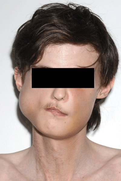
He underwent twelve routine dental extractions under General Anaesthetic (GA) (11, 14, 15, 25, 26, 27, 34, 36, 43, 44, 45, 46). The operation was uneventful. No significant bleeding was noted, haemostasis was achieved and the patient was discharged on the day of the procedure. He presented to the Emergency Department at a local district hospital as an outpatient department seven days postoperatively with pain and mild swelling associated with his right body of mandible and left infraorbital region. He was diagnosed with an infection associated with his extraction sites and discharged on oral antibiotics. He re-presented three days later to the Emergency Department with significantly larger facial swellings at the same sites (Figure 1).

Figure 1: Showing the extensive haematomas experienced by our patient.
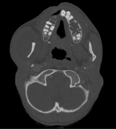
On examination, the patient was apyrexial with a firm, tender swelling overlying his right body of mandible. Cranial nerve and remaining head and neck examination was normal. No neck lymphadenopathy was palpable. A Computed Tomography scan showed a collection encasing the right mandibular ramus extending down onto the body and buccal space, displacing the masseter laterally. A lingual collection was also reported (Figure 2). He was transferred to the initial teaching hospital, where a multidisciplinary assessment involving an oral and maxillofacial surgeon, head and neck radiologist and rheumatologist was initiated. A conservative approach was undertaken including close inpatient observation to monitor for on-going expansion of the haematomas, ensuring these presented no risk a to his airway. The haematomas reduced in size over seven days and our patient was discharged with an appropriate outpatient appointment.

    Figure 2:  Computed Tomography scan slice showing the extent of our
patient’s swelling.
Discussion
Vascular Ehlers Danlos syndrome typically transmitted as an autosomal dominant trait [6]. However, in the case of our patient there was no known familial history of vEDS, making this a sporadic instance of the condition. Diagnosis of the condition was made at a young age, where the primary presenting complaint was multiple bruises to the legs and arms. The patient had been an inpatient frequently throughout the course of his life and, from his past experiences, was very keen to avoid peripheral venous access where possible. The act of placing a cannula was very painful for the patient and resulted in profound bruising, a finding which is not uncommon in individuals who suffer from this condition [7].
In this clinical case, the patient was non-compliant with regular dental attendance due to his dental anxiety. Although there is no documented association between vEDS and low Body Mass Index (BMI), our patient had a BMI of 14 on admission. Due to this, his dieticians recommended a high calorie, carbohydrate rich diet. Morphological changes to the teeth were evident, and he complained of bleeding gums when tooth brushing. These risk factors are likely to have contributed to our patient’s poor oral health and resultant need for dental extractions.
A potentially co-causative factor in our patient’s poor dental health are reports of deposits fibrinoids, which produce vasculitis, in the gingival tissues suggesting that the intra-oral mucosa of patients with vEDS is more prone to inflammation [8]. The condition is linked with early periodontitis, mucosal ulceration, gingival bleeding, mobility and loose teeth, enamel hypoplasia and irregular dentine structures, further highlighting the increased dental needs in this cohort of patients [8]. Our patient would therefore have benefitted from focused oral health preventative measures including specific dietary advice with oral health surveillance and management of caries by a general dental practitioner.
Our patient had previously undergone closure of his unilateral incomplete cleft lip and palate. Cleft palate is not commonly associated with vEDS, however Okamura noted this link in a 6-yearold [9]. Further research into this topic may be beneficial, as there is potentially a mechanistic relationship between connective tissue disorders and failure of fusion at embryonic nasal prominences, but this is poorly understood [10].
Delayed healing and spontaneous haematoma following surgery is a known complication of vEDS due to vascular wall fragility [11]. In other surgical specialties, the incidence of post-operative bleeding in these patients is deemed ‘significant,’ and it seems that the late presentation of significant haematoma in our patient is concurrent with previous findings in other specialties [12]. Vascular complications in individuals suffering from vEDS have been noted in all anatomical areas, with surgical repair of major vessels often difficult due to vessel wall fragility [4].
In this case, the subsequent haematoma was not managed invasively. Simple measures such as pressure and coolant therapy may have reduced the timescale of the acute episode. Steroids or medicinal leech use may also have reduced recovery time [13]. Use of suction drainage as a treatment modality for the patient was discussed and is frequently used to treat haematomas within the maxillofacial region [14]. However, in this instance, it was decided that such an intervention would be too traumatic and could result in further complications.
Conclusion
Patients with vEDS require particular attention with regards to oral health prevention and management. This is crucial in order to reduce dental disease, which may require surgical management resulting in postoperative bleeding and an increased risk of morbidity and mortality. We believe this is the first reported case of a bleeding complication associated with routine oral surgery in a patient with vEDS. A multidisciplinary approach to the patient’s management resulted in a favourable outcome and complete resolution of the haematomas without further surgical intervention.
References
- Pepin M, Schwarze U, Superti-Furga A, Byers PH. Clinical and genetic features of Ehlers–Danlos syndrome type IV, the vascular type. N Engl J Med. 2000; 342: 673-680.
- Mattar SG, Kumar AG, Lumsden AB. Vascular complications in Ehlers- Danlos syndrome. The American Surgeon. 1994; 60: 827-831.
- Jindal R, Choong A, Arul D, Dhanjil S, Chataway J, Cheshire NJ. Vascular Manifestations of Type IV Ehlers–Danlos Syndrome. EJVES Extra. 2005; 9: 135-138.
- Germain DP. Ehlers-Danlos syndrome type IV. Orphanet J Rare Dis. 2007; 19; 2: 32.
- Paepe AD, Malfait F. Bleeding and bruising in patients with Ehlers–Danlos syndrome and other collagen vascular disorders. Br J Haematol. 2004; 127: 491-500.
- Germain DP. Clinical and genetic features of vascular Ehlers-Danlos syndrome. Ann Vasc Surg. 2002; 16: 391-397.
- Parfitt J, Chalmers RT, Wolfe JH. Visceral aneurysms in Ehlers-Danlos syndrome: case report and review of the literature. J Vasc Surg. 2000; 31: 1248-1251.
- Mao J, Bristow J. The Ehlers-Danlos Syndrome: Beyond collagens. J Clin Invest. 2001; 107: 1063-1069.
- Okamura H, Matsumoto Y. A case of Ehlers-Danlos syndrome associated with cleft lip and palate. J Laryngol Otology. 1984; 98: 311-316.
- Tan PW, Song C, Lalonde D. Ehlers-Danlos syndrome associated with cleft lip and palate. Can J Plast Surg. 2009; 17: e24.
- Kawabata Y, Watanabe A, Yamaguchi S, Aoshima M, Shiraki A, Hatamochi A, et al. Pleuropulmonary pathology of vascular Ehlers-Danlos syndrome: spontaneous laceration, haematoma and fibrous nodules. Histopathology. 2010; 56: 944-950.
- Oderich GS, Panneton JM, Bower TC, Lindor NM, Cherry KJ, Noel AA, et al. The spectrum, management and clinical outcome of Ehlers-Danlos syndrome type IV: a 30-year experience. J Vasc Surg. 2005; 42: 98-106.
- Ramzan M, Droog W, Sleeswijk V, van Roessel EW, Meynaar IA. Leech got your tongue? Haematoma of the tongue treated with medicinal leeches: a case report. Neth J Crit Care. 2010; 14: 268.
- Bull PD, Lancer JM. Treatment of auricular haematoma by suction drainage. Clinical Otolaryngology. 1984; 9: 355-360.