Special Article - Amelogenesis Imperfecta
Austin J Dent. 2018; 5(5): 1117.
Hereditary Brown Teeth - A Case Report
Kavitha PT¹, Pradeep L¹, Mohankumar M¹, Santannavar S²*, Bhandarkar GP³, Rao PK4, Kini R5, Kashyap RR3, Shetty D3
¹House surgeon, Department of Oral Medicine and Radiology, AJ Institute of Dental Sciences, Kuntikana, Mangalore, Karnataka, India
²Postgraduate Student, Department of Oral Medicine and Radiology, AJ Institute of Dental Sciences, Kuntikana, Mangalore, Karnataka, India
³Reader, Department of Oral Medicine and Radiology, AJ Institute of Dental Sciences, Kuntikana, Mangalore, Karnataka, India
4Professor, Department of Oral Medicine and Radiology, AJ Institute of Dental Sciences, Kuntikana, Mangalore, Karnataka, India
5Professor and Head, Department of Oral Medicine and Radiology, AJ Institute of Dental Sciences, Kuntikana, Mangalore, Karnataka, India
*Corresponding author: Sunmith Santannavar, Department of Oral Medicine & Radiology, AJ Institute of Dental Sciences, Kuntikana, Mangaluru, Karnataka, India
Received: August 07, 2018; Accepted: September 05, 2018; Published: September 12, 2018
Abstract
Amelogenesis imperfecta encompasses a complicated group of conditions that demonstrate developmental alterations in the structure of the enamel in the absence of a systemic disorder or syndrome. It may be differentiated into three main groups; Hypoplastic (HP), Hypocalcified (HC), Hypomatured (HM). It may show autosomal dominant, autosomal recessive, sex-linked or sporadic pattern. It is necessary to diagnose this disorder and provide durable, functional and esthetic management of these patients to improve the quality of their lives.
Keywords: Amelogenesis Imperfect; Enamel, Dental; Genetic; Inherited
Introduction
AI represents a group of conditions, genomic in origin, which affect the structure and clinical appearance of the enamel of all or nearly all the teeth in a more or less equal manner, and which may be associated with morphologic or biochemical changes elsewhere in the body [1].
The prevalence of Amelogenesis imperfecta has been estimated to range from (1 in 718) to (1 in 14000), depending on the population study. Hypoplastic AI represents 60-73 % of all cases, hypomaturation AI represents 20-40 % and hypocalcification represents 7%. Disorders of enamel epithelium also can cause alterations in the eruption mechanism, resulting in anterior open bite [2]. Clinical researchers usually classify AI into four main types of which 17 subtypes are recognized. The main types are based on clinical appearance, radiographic appearance and enamel thickness, and the subtypes are based on mode of inheritance and gene mutation [3].
Witkop and Sauk listed the varieties of AI, divided according to whether the abnormality lay in a reduced amount of enamel (hypoplasia), deficient calcification (hypocalcification), or imperfect maturation of the enamel (hypomaturation), and also recognized the combined defects [4].
Tooth enamel is the most highly mineralized structure in the human body, with 85% of its volume occupied by hydroxyapatite crystals. The physical properties and physiological function of enamel are directly related to the composition, orientation, disposition, and morphology of the mineral components within the tissue. During organogenesis, the enamel transitions from a soft and pliable tissue to its final form, which is almost entirely devoid of protein. The final composition of enamel is a reflection of the unique molecular and cellular activities that take place during its genesis. Deviation from this pattern may lead to amelogenesis imperfect [5].
Case Presentation
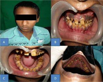
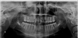
A 13year old male patient came to department of oral medicine and radiology with chief complaint of discoloration of teeth in upper and lower jaws since childhood. Patient also gave the history of discoloration of deciduous teeth which had been exfoliated 3years back. Patient’s mother and sister had the same discoloration with all teeth and underwent full mouth FPD. On inspection there was generalized yellowish brown discoloration seen on buccal, lingual or palatal surface of teeth with reduced height and with of the crown, evidence of open contact points with posterior and anterior teeth, also there was black pinpoint pits in relation to upper anteriors which suggests that there was a total enamel loss (Figure 1). On palpation, the surface was rough on all aspects, surface doesn’t abrade on scraping with the help of probe/explorer. A clinical diagnosis of Amelogenesis Imperfecta (hypoplastic type-Pitted type) was made with differential diagnosis of dentinogenesis imperfecta and molarincisor hypoplasia. Orthopantamograph showed impacted upper right canine, upper left canine with retained upper left deciduous canine, generalized radiopaque enamel which was less in density as compared to dentin or equivalent to it with decreased enamel height (Figure 2). So based on radiographic examination final diagnosis of amelogenesis imprefecta (Hypoplastic type) was made.

    Figure 1:  1A: Show a 13yr old male patient, 1B: Loss of enamel and
showing a mild pinpoint pits on facial surface of maxillary and mandibular
teeth, 1C: Discoloured and eroded mandibular posterior teeth, 1D: Showing
discolouration of teeth on palatal surface of maxillary anteriors.

    Figure 2:  Orthopantamogram shows a generalized radiopaque enamel which
is less in density as compared to dentin or equivalent to it.
Discussion
Amelogenesis imperfecta is a developmental, often inherited disorder, affecting dental enamel. It usually occurs in the absence of systemic features and comprises of diverse phenotypic entities.
The predominant clinical manifestations of affected individuals are enamel hypoplasia (enamel is seemingly correctly mineralized, but thin), hypomineralization (subdivided into hypomaturation and hypocalcification), or a combined phenotype, which is seen in most cases. The trait of AI can be transmitted by an autosomaldominant, autosomal-recessive, or X-linked mode of inheritance. The distribution of AI types is known to vary among different populations. The amelogenin gene is a tooth-specific gene expressed in preameloblasts, ameloblasts, and in the epithelial root sheath remnants; while a low-expression of amelogenin mRNAs has been recently shown in odontoblasts. To date, there are 14 AMELX-associated AI mutations.
The enamelin (ENAM) gene is a tooth-specific gene expressed predominantly by the enamel organ, and, at a low level, in odontoblasts. Enamel phenotypes of ENAM mutations may be dosedependent, with generalized hypoplastic AI segregating as a recessive trait and localized enamel pitting segregating as a dominant trait. The ameloblastin (AMBN) gene is expressed at high levels by ameloblasts and at low levels by odontoblasts and pre-odontoblasts, while moderate expression is also observed in Hertwig’s epithelial root sheath, and in odontogenic tumors, such as in ameloblastomas. The amelotin gene (AMELOTIN 4q13) has been reported by Iwasaki et al., 2005, to play a role in the molecular interaction during amelogenesis. However, so far no mutation of the amelotin gene has been related to AI5. An additional locus for autosomal dominant AI has been found recently on chromosome 8q24.3 [6]. Clinically, there is deficiency of enamel matrix with subsequent normal mineralization. The enamel doesn’t develop to normal thickness; at places the enamel is so thin that crowns do not meet at contact points. The decreased crown height leads to anterior open bite (in about 50% of cases).Enamel matrix defects vary from pinpoint to pinhead size pits arranged in rows or column on labial or buccal surface of the permanent teeth. Sometimes pits and grooves of hypoplastic enamel are seen in a horizontal fashion across the middle-third of teeth. Enamel agenesis is seen in Type IG where the tooth has a rough granular surface and has no contact with adjacent teeth [7].
Types of Amelogenesis imperfecta as follows:
I. Hypoplastic type: The enamel has not formed to full normal thickness on newly erupted developing teeth.
II. Hypocalcified type: The enamel is so soft that it can be removed by a prophylaxis instrument.
III. Hypomaturation type: The enamel can be pierced by an explorer point under firm pressure and can be lost by chipping away from the underlying normal- appearing dentin [8].
IV. Hypomaturation – hypoplastic with taurodontism
Diagnosis clinically through family history, pedigree plotting, clinical observation and meticulous recording form the backbone of diagnosis in this, as in any potentially inherited condition. Extra-oral radiographs may reveal the presence of unerupted and sometimes spontaneously resorbing teeth. Intra-oral radiographs will reveal the relative contrast between enamel and dentine in cases where mineralisation may have been affected. The same films in conjunction with clinical observation will provide information on the degree of any enamel hypoplasia and genetic diagnosis through laboratory genetic diagnostic research tool. Extrinsic disorders of tooth formation, chronological disorders of tooth formation and localised disorders of tooth formation should be considered in the differential diagnosis. The commonest differential diagnosis is dental fluorosis, dentinogenesis imperfecta, tricho-dento-osseous syndrome, molarincisor hypoplasia. The variability of this condition, from mild white “flecking” of the enamel to profoundly dense white colouration with random, disfiguring areas of staining and hypoplasia, requires careful questioning to distinguish from AI [9]. In treating amelogenesis imperfecta, the patient’s appearance and restoration of occlusion are of prime importance. Modern methods and materials have widened the range of available treatment [10]. The three principles of management of patients with AI involve; Alleviation of pain and anxiety. Restoration and maintenance of the remaining dentition with regards to esthetics and maintenance/restoration of the occlusal vertical height. The management of young patients with AI is preferably done in three phases. The temporary phase, undertaken during the primary or mixed dentition. The transitional phase, when all permanent teeth have erupted; this phase continues till adulthood. The permanent phase, which lasts through adulthood7. The varying etiology of AI conjures a wide array of clinical features whose restorative management poses a challenge for dentists. As both esthetics and function are compromised in these patients, their management usually involves complete oral rehabilitation by way of full coverage crowns, direct and indirect veneers, and bonded esthetic restorations, depending on the condition of the individual tooth and the age of the patient. If the treatment is delayed, a loss of usable crown length occurs. In those patients without sufficient crown lengths, full dentures (overdentures in some cases); often become the only satisfactory approach. The other types of AI demonstrate less rapid tooth loss, and the esthetic appearance is the prime consideration. In some cases a lack of good enamel bonding of veneers occurs and does not result in a durable restoration. The use of glass ionomer cements with dentinal adhesives often overcomes this weakness [5].
Conclusion
The restoration of the defects created by amelogenesis imperfecta improves the esthics and functional concern of the patient. Treatment planning of these case involves an interdisciplinary approach to evaluate, diagnose, and to resolve esthetic problems using a combination of periodontal, prosthodontic, and restorative treatment. Dental practitioners should consider the social implications for these patients and intervene to relieve their suffering. Thus, this article is an attempt to improve the clinician’s knowledge about the clinical diagnosis as well as intervention required for such a condition.
References
- Hemagaran G, Aravind M. Amelogenesis imperfecta - Literature review. Journal of dental and medical science 2014; 13: 48-51.
- Shafer, Hine, Levy. Textbook oral pathology. 7th Ed. Elsevier. 2012: 3-80.
- Amelogenesis Imperfecta-NORD.
- Aldred MJ, Savarirayan R, Crawford PJ. Amelogenesis imperfecta: A classification and catalogue for the 21st century. Oral Diseases. 2003; 9: 19- 23.
- Chaudhary M, Dixit S, Singh A, Kunte S. Amelogenesis Imperfecta: Report of a case and review of literature. J Oral maxillofac Pathol. 2009: 13: 70-77.
- Mendoza G, Pemberton TJ, Lee K, Scarel-Caminaga R, Mehrian-Shai R, Gonzalez-Quevedo C, et al. A new locus for autosomal dominant amelogenesis imperfecta on chromosome 8q24.3. Hum Genet. 2007; 120: 653-662.
- Ongole R, Praveen. Textbook of oral medicine, oral diagnosis and oral radiology. Elsevier. 2013: 11-60.
- Shafer, Hine, Levy. Textbook oral pathology. 4th Ed. Saunders. 1983: 2-85.
- Crawford P, Michael A, Bloch-Zupan A. Amelogenesis Imperfecta - review. Orphanet Journal of rare diseases. 2007; 2: 17.
- Lamb D J. Treatment of amelogenesis Imperfecta. The journal of prosthetic dentistry. 1976; 36: 286-291.